ABSTRACT
Bone remodeling at microscrew interface near
extraction site in the beagle dog
mandible-histologic and immunohistochemical analyses
Guangxi WEI1,2,3, Yun HU2,3,4, Leilei ZHENG1,2, Jinfeng HUO1,2, Tian TANG5, Feng DENG1,2,3
1- Department of Orthodontics, the Afiliated Hospital of Stomatology, Chongqing Medical University, Chongqing, P. R. China.
2- Chongqing Research Center for Oral Diseases and Biomedical Science, College of Stomatology, Chongqing Medical University, Chongqing, P. R. China. 3- College of Biomedical Engineering, Chongqing Medical University, Chongqing, P. R. China.
4- Department of Pediatric Dentistry, the Afiliated Hospital of Stomatology, Chongqing Medical University, Chongqing, P. R. China.
5- Department of Orthodontics, West China College of Stomatology, Sichuan University, Chengdu, P. R. China.
Corresponding address: LeiLei Zheng - Chongqing Research Center for Oral Diseases and Biomedical Science - College of Stomatology - Chongqing Medical University - 426#, Songshibei Road - Yubei District - Chongqing - 401147- P. R. China - Phone: 86-23-88860107 - Fax: +86 23 8886 0222 - e-mail: [email protected]
Submitted: February 8, 2013 - Modiied: July 26, 2013 - Accepted: August 6, 2013
e
xtraction is often used as part of orthodontic therapy, and good control of anchorage is a key step after extraction. Although microscrews can be implanted close to the extraction site in order to achieve orthodontic support, the eficiency of bone remodeling at the implant-bone interface near the extraction region is dubious. Objective: The purpose of this study was to investigate bone remodeling of the bone-microscrew interface near the tooth extraction site, in the absence of loading. Material and Methods: Third and fourth premolars were extracted from the mandibles of beagle dogs, followed by placement of test microscrews near the extraction sites. Control microscrews were placed further away from the extraction site. All samples were collected after 1, 3, 8, or 12 weeks of healing following extraction. The bone remodeling process at the interface was evaluated using histologic and immunohistochemical analyses. Results: Initially, a large number of inlammatory cells were aggregated at the interface. The expression levels of core binding factor (Cbfa1), osteocalcin (OC) and transforming growth factor beta (TGF-β) were inconspicuous in both groups, whereas tumor necrosis factor alpha (TNF-α) was strongly expressed, especially in the test groups (P<0.05). Subsequently, the expression levels of Cbfa1, OC and TGF-β were found to increase signiicantly, and active osteogenesis was observed. Conclusions: During week 1, inlammatory reaction is a major concern at the bone-microscrew interface near the extraction site. However, with healing, the inluence of extraction on the remodeling of bone surrounding the microscrews decreases, thus facilitating successful treatment.Keywords: Bone remodeling. Core binding factor. Osteocalcin. Transforming growth factor alpha. Transforming growth factor beta.
INTRODUCTION
Anchorage control is a challenge to nearly every orthodontist. Since the first successful
attempt to move teeth against a ixed screw by
Linkow12 (1969), the application of microscrews
in orthodontic treatment has become a mainstay in contemporary orthodontics7,14.Owing to its
miniature size, the interradicular site is the most common choice for microscrews in orthodontic clinical treatment9. Moreover, palatal implants,
the implant system for orthodontic anchorage, have shown promising results in recent years by achieving maximal intraoral orthodontic anchorage purposes1. However, orthodontists are
often faced with complicated challenges, such as: 1) the low-order maxillary sinus that hinders implantation in the molar area, 2) the maxillary tubercle and external oblique line where implanting microscrews is a greater challenge compared to
the lat areas in the jaw bone, 3) the complicated
4) too many missing teeth or rampant caries/ periodontitis within the same quadrant. These
factors make it very dificult to identify the ideal
location for implants. Although microscrews can be implanted close to the extraction site in order to achieve orthodontic support, the stability of the
microscrews and the eficiency of bone remodeling
at the implant-bone interface near the extraction region is dubious. To our knowledge, this issue has not been resolved.
Literature reports11,17 show that extraction
undoubtedly leads to a decrease of bone density in the extraction vicinity. Miyawaki and his colleagues14 (2003) proved that the decrease in
bone density increases the risk of non-integration at the implant-bone interface. Zheng’s examination indicated that the risk of loosening of microscrews near extraction site was the most severe in
the irst week following implant placement21.In
addition, there are several factors associated with the stability of microscrews, such as the diameter of the miniscrew, proximity with dental
roots and inlammation within peri-implant tissue.
To understand and overcome these challenges, numerous studies have been conducted, with the aim of promoting bone tissue remodeling at the implant-bone interface to increase stability of microscrews under diverse conditions13,19,21.
However, the various animal and clinical studies have focused on the stability of microscrews under loading in order to mimic the actual clinical process as closely as possible. This leads to the evaluation
of microscrew stability under the inluence of
iatrogenic factors such as intention, direction and occasion of loading.
In order to determine the ideal implantation strategy for loading, clinical orthodontists certainly need to understand the state of the bone-device interface during the healing process. In accordance with the change rule of bone healing15,the bone
remodeling process at the interface was evaluated at 1, 3, 8 or 12 weeks following implantation. In this study, we hypothesized that tooth extraction will inluence bone tissue remodeling near the microscrews, and we evaluated this effect via histologic and immunohistochemical analyses.
MATERIAL AND METhODS
Animals and surgical procedures
Twelve male beagles meeting the following criteria were selected: 18 months of age, 10 kg in weight, presence of fourth premolars on mandible, and healthy with no malocclusion and periodontal diseases. They were handled according to the experimental protocol approved by the Bioethics Committee of Sichuan University, China (Number of permit: SYXIC111 2009-045, China).
The animal model was described as our previous study21. All the third and the forth premolars were
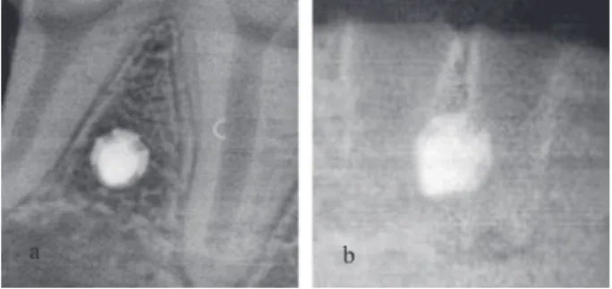
surgically extracted from the mandibles. Ninety-six microscrews (diameter 1.6 mm, length 6 mm) (Medicon Company, Tuttlingen, Germany), were placed between the mesial and distal roots of P2, P3, P4, and M1, on the buccal side of the mandible, 6 mm beneath the top of the alveolar crest (Figures 1a and 1b). The test implants (48 microscrews) were placed near the extraction sites (between the mesial and distal roots of P3 and P4), and the control implants (48 microscrews) were placed at the normal sites (between the mesial and distal roots of P2 and M1) (Figures 2a and 2b). Microscrews in both the test and control groups experienced no loading. The beagles were fed liquid diet in order to avoid the impact of hard
food on the microscrews, and were inally executed
at 1, 3, 8 or 12 weeks following implantation.
histologic analysis
The mandibles were removed from the executed animals and carefully sectioned into small tissue blocks (10x10x6 mm). each tissue block contained one microscrew surrounded by at least 4 mm-thick bone tissue. The blocks were
ixed in cold buffered formalin, pH 7.0, for 3–6 days and then demineralized in 20% tetrasodium
ethylenediaminetetraacetic acid (eDTA, pH 7.0) for about 4 to 6 months until the microscrews could be easily removed without breaking the implant-bone
interface. All parafin-embedded blocks were cut
into 4 μm-thick slices. Some tissue sections were
stained with Masson’s Trichrome for descriptive analysis and determination of neutrophil and osteoblast densities. For the determination of cell
density, 5 histological ields of the implant-bone
interface were randomly selected and the number of neutrophils and osteoblast was counted by
manual method using 200× magniication coupled
to Nikon e600 microscope (Nikon Instruments Inc, Melville, USA). The cell-density was calculated by the mean of cell number per ield.
Immunohistochemistry analysis
Following parafin removal from the tissue sections, they were hydrated by incubation in 95%, 90%, 80%, and 70% ethanol for 5 minutes. After
antigen retrieval with TRIS eDTA (pH 9.0) solution for 30 min, sections were immersed in PBS-H202
(0.01 ml, pH 7.0, PBS 99 ml + 30% H202 1 ml)
for 20–30 minutes, at room temperature (23°C) to eliminate the endogenous peroxidase. The
sections were irst washed in distilled water for 5
min, and then washed in phosphate-buffered saline
(PBS) for another 5 minutes. Before incubation in primary antibody, sections were immersed in
non-immune serum (5% bovine serum albumin,
BSA) and diluted 1:5–1:20 for 30 minutes without wash. Following the above step, the sections were incubated with mouse anti-dog OC antibodies (1:75, R&D Systems, Minneapolis, MN), rabbit
anti-dog TGF-β antibodies (1:100, Santa Cruz
Biotechnologies, Inc, Santa Cruz, CA, USA) and
caprine anti-dog TNF-α antibodies (1:200, R&D
Systems, Minneapolis, MN, USA) at 4°C overnight. Next, the sections were sequentially incubated with secondary biotinylated goat anti-mouse antibodies (Santa Cruz Biotechnologies, Inc, Santa Cruz, CA, USA), mouse anti-rabbit antibodies (1:200, R&D Systems, Minneapolis, MN, USA) and pig anti-caprine antibodies (1:100, R&D Systems, Minneapolis, MN, USA) for 30 minutes at 23°C. The
sections were washed and the speciic antibody binding reaction was ampliied using streptavidin
peroxidase. Diaminobenzidine (DAB 0.5 mg/ml) staining and counterstaining with hematoxylin were performed to provide enhanced orientation of the tissue topography. Finally, the sections were dehydrated in an ethanol gradient and mounted for microscopic observation. As negative controls,
slides were incubated with PBS 1% instead of primary speciic antibodies. The images were acquired at 200× magniication using a Nikon
e600 microscope (Nikon Instruments Inc, Melville,
USA). All iles were saved in tagged-image ile
format (TIFF). The integral optical density (IOD) of the target protein was measured with Image-Pro Plus 5.0 (Media Cybernetics, Rockville, MD, USA). In the process of measurement, the value was
deined irstly by determining the positive staining
of control sections, and was used to automatically analyze images of all samples that were stained Figure 2- Radiographs illustrating implanted microscrews.
(a) Radiograph of microscrew implanted between the
roots of irst molar. (b) Radiograph of a microscrew
implanted near an extraction site
Figure 3- Histologic analysis at the implant-bone interface in the test group (lower panel) and control group (upper panel) using Masson staining. (a) In the test group, a large number of neutrophils were aggregated at the interface at week 1 while
a large amount of ibrous tissue was aggregated in the control group. (b) the new bone layer in the test group at week 3. (c)
A large-scale dematrix bone (DB) excreted by osteoblasts around the microscrew was observed in both groups at week 8.
(d) A mass of mature lamellar bone in the implant-bone interface in both groups at week 12 (Masson stain). Magniication:
Figure 4- Graphs showing the changes in the density of neutrophil and osteoblast in both groups at 1, 3, 8 and 12-weeks
healing time. * indicates statistically signiicant differences
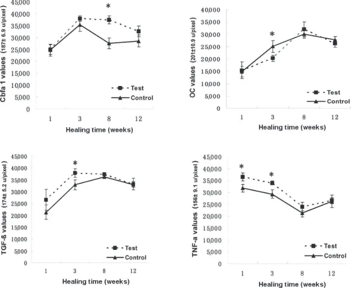
Figure 5- Graphs showing the changes in expression of Cbfa1, osteocalcin (OC), TGF-β and TNF-α, in both groups at 1, 3, 8 and 12-week healing time. * indicates statistically signiicant differences. (a) At week 8, the expression in the test group was signiicantly higher than in the control group (p<0.01). (b) The expression of OC reached peak values at week 8 and subsequently decreased. Signiicant differences were observed at week 3 and values of OC in the control group were signiicantly higher than in the test group (p<0.01). (c). The peak of TGF-β in the control group appeared at week 8, and subsequently went down to the level as in week 3. At week 3, signiicant differences were observed between both groups, but the expression of TGF-β in test groups was stronger than in control groups (p<0.01). (d) Signiicant differences
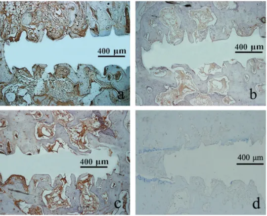
Figure 6- Immunohistochemical staining of Cbfa1 in sections from beagle mandible from test (a and c) and control groups (b and d), during weeks 3 and 8. The expression of Cbfa1 in both groups reached peak values at week 3, but there were
no statistically signiicant differences between test group (a) and control group (b). At week 8, signiicant differences were
observed between both groups, and the expression of Cbfa1 in test group (c) was stronger than in control group (d).
Magniication: 200x
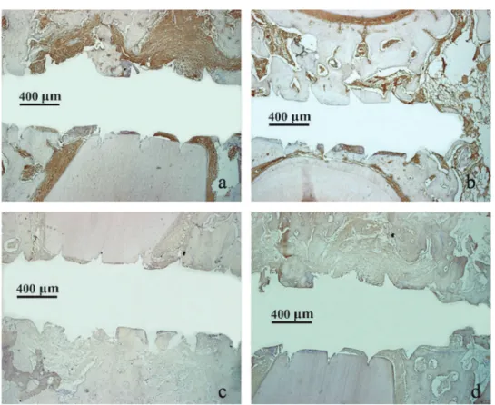
Figure 7- Immunohistochemical staining of osteocalcin (OC) in beagle mandible sections from test (a and c) and control groups (b and d), during weeks 3 and 8. The expression of OC in control group (b) was stronger than in test group (a)
under identical conditions.
In situ hybridization
In situ hybridization for Cbfa1 was performed using digoxigenin-labeled riboprobes. Before unsealing, the Cbfa1 probes were briefly centrifuged and immersed in ddH2O. These probes were then stored at -20°C until needed. Deparaffinage, hydration and deactivation of
endogenous enzymes in the parafin sections
were performed as mentioned in the previous section. Tissue sections were dropped in pepsin
diluted 3% citric acid for 30 min at 37°C, ixed for 10 min (1% paraformaldehyde (0.1 M PBS, PH 7.0–7.6)), and washed in distilled water 3 times. The sections were pre-hybridized for 2 hours at
37–42°C using 20 μL pre-hybridization solution per sections, and then they were hybridized with the
probe (2 μg/ml) diluted in hybridization buffer and
in 2×SSC (standard saline citrate) for 16–18 h at 38–42°C. The sections were washed sequentially in 0.2×SSC, blocked with blocking solution and then incubated with anti-mouse antibody for 1 h at 37°C, washed in PBS. These sections were then exposed to SABC (Strept Avidin-Biotin Complex) and Biotin peroxidase for 30 min at 37°C, and washed again in PBS. Finally, sections were
stained, counterstained, dehydrated and mounted.
The expression of cbfa1 was quantiied using the
same methodology for immunohistochemical analysis.
Statistical analysis
All statistical analyses were performed with SPSS software (SPSS, Chicago, Ill). Student’s t-test was used to determine statistical differences in the values between the test groups and the control groups. Data were presented as means with standard deviations. A difference of P<0.05
was accepted statistically signiicant.
RESULTS
Histologic and immunohistochemical sections from the samples are shown in Figures 4–9. Twelve male beagles received 93 samples. Three samples were omitted due to the loss of microscrews in the test group at week 1.
Histologic indings
At week 1, a large number of neutrophil were aggregated at the bone-screw interface (Figure 3a). In the test group, the neutrophil density was higher (p<0.01) while the osteoblast density was
Figure 8- Immunohistochemical staining of TGF-β in sections from beagle mandible from test (a and b) and control group (c and d), during weeks 3 and 8. The expression of TGF-β in test group reached the peak values at week 3 (a). The
expression in control groups was low at week 1 (d) and reached the peak values at week 8 (c). There were statistically
Figure 9- Immunohistochemical staining of TNF-α in beagle mandible sections from test (a and c) and control group (b and d), during weeks 3 and 8. Statistically signiicant differences were observed between the two groups at week 3. The TNF-α values in test group (a) were higher than in control group (b). The expression in test group (c) and control group (d) presented a downward trend at week 8. Magniication: 40x
lower (p<0.01) in relation to control group (Figure 4). There were new bone layers in the test group at week 3 (Figure 3b). By week 8, many active osteoblasts gathered along the interface and excreted a large-scale bone matrix around the microscrew (Figure 3c). By week 12, there was a mass of mature lamellar bone in the
implant-bone interface, calciied to a degree close to that
of normal bone tissue (Figure 3d). However, the amount of dematrix bone in the control group was greater than in the test group at week 3 and week 8.
Immunohistochemistry analysis
The expression of Cbfa1 in both groups reached a peak at week 3 (Figure 5). At week 8, the
expression in the test group was higher signiicantly
than in the control group (Figure 6) (p<0.01). The expression of OC reached peak values at week 8
and subsequently decreased (Figure 5). Signiicant
differences were observed at week 3 and values of
OC in the control group were signiicantly higher
than in the test group (Figure 7) (p<0.01). The
TGF-β values in test groups reached a peak at week 3. At week 3, signiicant differences were
observed between both groups, but the expression
of TGF-β in test groups was stronger than in control
groups (Figure 8) (p<0.01). The mean levels of
TNF-α in both groups were high in the irst three weeks after implantation. Signiicant differences
in expression between two groups were observed at week 1 and week 3, and the values in the test group were higher than those in the control group (Figure 9) (p<0.01).
DISCUSSION
Owing to its miniature size and simple surgical placement, miniscrews are easy to place in the maxillae and mandibles, with the aim of providing skeletal anchorage for orthodontic patients. However, in face of the variety of oral conditions seen clinically, orthodontists often need to choose the most suitable miniscrew site, and at present, interradicular sites are the most common choice. In this study, all miniscrews were placed between the mesial and distal roots of P2, P3, P4 and M1 at the buccal side of the mandible of beagles. In order to avoid damaging the roots of neighboring teeth, as reported in a study by Asscherickx, et al.2 (2005),
radiographs of the beagle mandibles were taken
to conirm that the furcation angles of the roots
accurate, and these sites had not interfered with the roots of neighboring teeth or other important structures of the mandible.
In this study, histologic indings from the test
group revealed that, at week 1, the original bone was destroyed, with aggregation of a large number
of inlammatory cells at the screw-bone interface.
Active osteoblasts were gathered around the new bone by week 3. Moreover, by week 8, osteoblasts had secreted a large-scale bone matrix around the microscrew. Literature had reported that, during
the irst week, pull-out strength of the miniscrews was signiicantly lower near the extraction site
than it was at a distance away from it, followed by an increase in strength during weeks 3 and
8. This indicates that inlammatory reaction and
bone resorption at the implant-bone interface were 2 major initial events following implantation. This likely explains why 3 microscrews in the test group failed at this stage. However, subsequent to longer healing time and formation of new bone, the risks surroundings the stability of microscrews decreased signiicantly, as can be conirmed by the ensuing molecular regulation of osteogenesis around the miniscrews.
At week 1, the expression of Cbfa1, OC and
TGF-β was inconspicuous in control and test groups. In contrast, TNF-α expression in both groups was
most robust following implantation. This suggests that there emerged a mass of macrophages
and osteoclast mediated by TNF-α3,10, which
aggravated directly the damage of interface bone, especially in the test group, which likely caused 3 microscrews to fail in this group. However, due
to the low-expression of Cbfa1, OC and TGF-β,
bone formation and bone mineralization triggered by osteoblasts were still inconspicuous during this stage4,5,8. In addition, literature reports have
suggested that extraction leads to a decrease in bone density in the surrounding vicinity, which increases the risk of non-integration at the implant-bone interface11,14,17. Thus, it is safe to
assume that iniltration of numerous inlammatory
cells had reduced the stability of the microscrews at week 1, and that the area near the extraction site was not suitable for implantation, even without loading.
Tu, et al.16 (2007) discovered that alveolar bone
defects were largely illed with ibrous connective
tissues 3 weeks after surgery in normal mice. In contrast, wound healing was dramatically delayed in Cbfa1-deficient mice. Therefore, with the increasing level of Cbfa1, the most active period of bone remodeling possibly occurred at week 3 post-implantation. Although the high-intensity
expression of Cbfa1 was not signiicantly different
at week 3 between the two groups in our study, it maintained its intensity until week 8 in the test
groups, and decreased signiicantly in the control
groups. esposito, et al.6 (2010) showed that the
most active period of bone remodeling following extraction was week 8, which may explain why the level of Cbfa1 was higher in test groups at
week 8. Likewise, the expression of TGF-β and
OC was high from week 3 to week 8. Thus, active osteoblasts and large-scale new bone were formed at this stage. Osteogenesis was observed at the implant-bone interface during this stage, and the
expression of TNF-α, as well as the inlammation mediated by it, began to decline signiicantly.
On the other hand, the expression of OC in the control group was higher than in the test group
at week 3. TNF-α can likely inhibit the expression
of matrix protein genes at week 310. However,
by week 8, the expression level of TGF-β and OC
was the same in both the test and control groups. Literature21 reported that values of microscrew
pull-out strength were similar between the test and
the control groups at week 8. Thus, these indings
suggest that, with a longer period of healing, the risk of microscrew instability decreased
signiicantly, and that, by week 8, the remodeling
of the interface bone, both in test and control groups, tended to be similar.
As for week 12, there was a large amount of mature lamellar bone at the implant-bone interface,
calciied to a degree that was similar to that of normal bone tissue. The expression of TGF-β,
OC and Cbfa1 began to decline, which illustrated
a decline in bone tissue remodeling. TNF-α
expression had begun to rebound, which suggests that lack of corresponding bone stimulation aggravates bone resorption20.
CONCLUSIONS
After investigating the remodeling of the bone-microscrew interface near extraction sites via histologic and immunohistochemical analysis, we conclude that:
In the early days, the bone remodeling of extraction will affect stability of microscrew near extraction;
Subsequent to a longer healing period, the influence of extraction on the remodeling of interface bone surrounding microscrews decreases;
Irrespective of the location of the interface, near or away from an extraction site, microscrews are suitable for implantation.
ACKNOWLEDgMENTS
Science (cstc2012jja10053); Foundation of the Chongqing Municipal Health Bureau (2012-2-126); Foundation of the Chongqing Municipal Commission of education (kj120329).
REFERENCES
1- Asscherickx K, Vannet BV, Bottenberg P, Wehrbein H, Sabzevar MM. Clinical observations and success rates of palatal implants. Am J Orthod Dentofacial Orthop. 2010;137:114-22.
2- Asscherickx K, Vannet BV, Wehrbein H, Sabzevar M. Root repair after injury from mini-screw. Clin Oral Implants Res. 2005;16:575-8.
3- Azuma Y, Kaji K, Katogi R, Takeshita S, Kudo A. Tumor necrosis factor-alpha induces differentiation of and bone resorption by osteoclasts. J Biol Chem. 2000;275:4858-64.
4- Devescovi V, Leonardi e, Ciapetti G, Cenni e. Growth factors in bone repair. Chir Organi Mov. 2008;92:161-8.
5- Dowd TL, Rosen JF, Mints L, Gundberg CM. The effect of Pb(2+) on the structure and hydroxyapatite binding properties of osteocalcin. Biochim Biophys Acta. 2011;1535:153-63.
6- esposito M, Grusovin MG, Polyzos IP, Felice P, Worthington HV. Timing of implant placement after tooth extraction: immediate, immediate-delayed or delayed implants? eur J Orthod. 2010;3:189-205.
7- Huang LH, Shotwell JL, Wang HL. Dental implants for orthodontic anchorage. Am J Orthod Dentofacial Orthop. 2005;127:713-22. 8- Janssens K, ten Dijke P, Janssens S, Van Hul W. Transforming
growth factor-β1 to the bone. Endocr Rev. 2005;26:743-74.
9- Kim SH, Yoon HG, Choi YS, Hwang eH, Kook YA, Nelson G. evaluation of interdental space of the maxillary posterior area for orthodontic mini-implants with cone-beam computed tomography. Am J Orthod Dentofacial Orthop. 2009;135:635-41.
10- Kobayashi K, Takahashi N, Jimi e, Udagawa N, Takami M, Kotake S, et al. Tumor necrosis factor alpha stimulates osteoclast differentiation by a mechanism independent of the ODF/RANKL-RANK interaction. J exp Med. 2000;191:275-86.
11- Lindskog-Stokland B, Wennström J, Nyman S, Thilander B. Orthodontic tooth movement into edentulous areas with reduced bone height: an experimental study in the dog. eur J Orthod. 1993;15:89-96.
12- Linkow LI. The endosseous blade implant and its use in orthodontics. Int J Orthod. 1969;7:149-54.
13- Marquezan M, Souza MM, Araújo MT, Nojima LI, Nojima MC.
Is miniscrew primary stability inluenced by bone density? Braz
Oral Res. 2011;25:427-32.
14- Miyawaki S, Koyama I, Inoue M, Mishima K, Sugahara T, Takano-Yamamoto T. Factors associated with the stability of titanium screws placed in the posterior region for orthodontic anchorage. Am J Orthod Dentofacial Orthop. 2003;124:373-8. 15- Tarantino U, Cerocchi I, Scialdoni A, Saturnino L, Feola M, Celi M. Bone healing and osteoporosis. Aging Clin exp Res. 2011;23:62-4.
16- Tu Q, Zhang J, James L, Dickson J, Tang J, Yang P, et al.
Cbfa1/Runx2-deiciency delays bone wound healing and locally
delivered Cbfa1/Runx2 promotes bone repair in animal models. Wound Repair Regen. 2007;15:404-12.
17- Wehrbein H, Bauer W, Diedrich P. Gingival invagination area after space closure: a histologic study. Am J Orthod Dentofacial Orthop. 1995;108:593-8.
18- Wexler A, Tzadok S, Casap N. Computerized navigation surgery for the safe placement of palatal implants. Am J Orthod Dentofacial Orthop. 2007;131:S100-5.
19- Wu X, Deng F, Wang Z, Zhao Z, Wang J. Biomechanical and histomorphometric analyses of the osseointegration of microscrews with different surgical techniques in beagle dogs. Oral Surg Oral Med Oral Pathol Oral Radiol endod. 2008;106:644-50. 20- Zhao B, Ivashkiv LB. Negative regulation of osteoclastogenesis and bone resorption by cytokines and transcriptional repressors. Arthritis Res Ther. 2011;13:234.
21- Zheng L, Tang T, Deng F, Zhao Z. The inluence of extraction on