O
h
r
c
i
r
g
a
in
e
a
s
l
R
e
Aynur Özen1, İlhan Karacan2, M. Akif Sarıyıldız2, Muharrem Cidem3
1Department of Nuclear Medicine, Vakif Gureba Training and Research Hospital, 2Department of Physical Medicine and Rehabilitation, Vakif Gureba Training and Research Hospital, 3Department of Physical Medicine and Rehabilitation, Bagcilar Training and Research Hospital, Istanbul, Turkey
Hip Strentgh Analysis in Nephrolithiasis Patients
Hip Strentgh Analysis By Dual Energy X-Ray
Absorbtiometry in Nephrolithiasis Patients
Böbrek Taşına Sahip Hastalarda Çit Enerjili X-Işını
Absorbsiyometriyle Kalça Dayanıklılık Analizi
DOI: 10.4328/JCAM.2431 Received: 24.03.2014 Accepted: 16.04.2014 Printed: 01.01.2016 J Clin Anal Med 2016;7(1): 1-5 Corresponding Author: Aynur Özen, İnönü Mah. Ütade Sok. No:37/6 Elmadağ, Şişli, İstanbul, Turkey.
T.: +90 2124404000/3266 F.: +90 2124404013 E-Mail:[email protected] Özet
Amaç: Bu çalışmanın amacı, böbrek taşına sahip hastalarda çit enerjili X-ışını absorbsiyometri ile ölçülen kalça geometric parametrelerini değerlendirmek ve normal popülasyon ile karşılaştırmaktır. Gereç ve Yöntem: Bu prospektif bir çalışmanın verilerinin geriye dönük incelenmesiyle kalça analizi uygulanan çalışma böbrek taşına sahip 72 hastayı (29 kadın, 43 erkek) ve 94 kontrol ol-gusunu (31 kadın, 63 erkek) kapsamaktadır. Kemik mineral yoğunluğu ve kal-ça aks uzunluğu, kesitsel alan, kesitsel eylemsizlik momenti, femur dayanık-lılık indeksi ve femurun kesit modülü gibi yapısal parametreler her bir grup-ta ölçüldü. Bulgular: Hasgrup-talar ve kontrol olguları arasında antropometrik ola-rak fark saptanmadı (P>0.05). Cinsiyetin etkisi dikkate alındığında, gruplar arasında kalça aks uzunluğu, kesitsel alan, kesitsel eylemsizlik momenti, fe-mur dayanıklılık indeksi ve fefe-murun kesit modülü için istatiksel anlamlılık yok-tu (P>0.05). Böbrek taşı varlığının, kemik mineral yoğunluğu, kalça aks uzun-luğu, kesitsel eylemsizlik momenti, femur dayanıklılık indeksi, femurun kesit modülü ve kesitsel alan üzerine prediktif etkisi olmadığı saptandı. Kadın ol-manın kemik mineral yoğunluğu, kalça aks uzunluğu, kesitsel eylemsizlik mo-menti ve kesit modülü üzerine negatif bir etkisi vardı. Tartışma: Sonuç olarak, 20-50 yaş grubunda böbrek taşına sahip hastalar ile kontrol olguları arasın-da kemik mineral yoğunluğu ve kalça yapısal parametrelerinde herhangi bir farklılık tespit edilmedi. Böbrek taşına sahip hastalarda özellikle ileri yaş gru-bunda önceki çalışmalarda yüksek kırık riski tespit edilmiş olup bu hastalarda kemiğin kırığına neden olacak metabolik ve geometrik değişikliklerinin diğer kemik kaybı nedenleriyle (örneğin postmenapozal osteroporoz) birlikte yaş-lanma sürecinde başladığı ve hızlandığı düşünüldü.
Anahtar Kelimeler
Böbrek Taşı; Kemik Mineral Yoğunluğu; Kalça Dayanıklılık Analizi; Kalça Yapı-sal Analizi; Çit Enerjili X-Işını Absorbsiyometri
Abstract
Aim: The aim of this study was to assess the hip geometric parameters mea-sured by dual-energy X-ray absorptiometry in patients with kidney stones and to compare normal population. Material and Method: This study is retro-spectively evaluation and performed hip structural analysis of another pro-spective study data, included 72 patients with kidney stones (29 female, 43 male) and 94 control subjects (31 women, 63 men). Bone mineral density and structural parameters such as hip axis length, sectional area, cross-sectional moment of inertia, femur strength index and section modulus of femur neck have been measured in each groups. Results: The patients and control subjects were anthropometrically identical (P>0.05). There were no statistical diference for hip axis length, cross-sectional area, cross-sectional moment of inertia and section modulus between of groups when take into consideration of gender efect (P>0.05). The presence of nephrolithiasis was determined that there was not predictive efects on femur neck bone mineral density, hip axis length, cross-sectional moment of inertia, section modulus, femur strength index and cross-sectional area. To be female gender was a negatif efect on bone mineral density, hip axis length, cross-sectional mo-ment of inertia and section modulus. Discussion: As a conclusion, we did not found any diferences on bone mineral density and hip structural parameters measured with hip strength analysis program between nephrolithiasis pa-tients and normal subjects at 20-50 ages. We thought that in these papa-tients had high fracture rates determined previous studies especially in older ages, bone metabolic and geometric changes may start or/and fast with aging together other cause of loss of bone mineral (e.g. postmenopausal osteo-porosis).
Keywords
Nephrolithiasis; Bone Mineral Density; Hip Strength Analysis; Hip Structural Analysis; Dual-Energy X-Ray Absorptiometry
Introduction
The patients with nephrolithiasis, particularly presenting with hypercalciuria, may have reduced bone mass. This is based on an increase in bone remodeling [1-5]. The most frequent form of kidney stone is calcium lithiasis, predominantly formed by calcium oxalate stones. The hypercalciuria is present in about 50% of the patients with calcium lithiasis, but it is considered idiopathic since an underlying metabolic impairment is not found [6]. Although increased protein and sodium intake and reduced potassium increase calcium urinary excretion, these do not seem to primarily account for hypercalciuria [7].
The measurement of bone mineral density can be using by photon or X-ray. Single or dual photon and dual-energy X-ray absorptiometry (DXA) images are two-dimensional (2D) pro-jection. These techniques convert pixel values to mineral area density (g/cm2) by using sotware. Although, quantitative com-puted tomograhy is three-dimensional (3D) projection of bone as a gold standard technique. It overcomes limitations of 2D projection. A majority of the previous studies to evaluate bone strength have measured primarily bone mineral density (BMD) relect bone material. Whereas, bone strength be impressed by both material and structural parameters. Therefore, to measure of BMD is inadequate to predict of fracture risk, which exceed-ing stress of bone strength. An important determinant of frac-ture risk is age-related changes in the structural geometry of bone tissue. New sotwares in bone density measurement, cal-culating femoral structural variables that can end hip fracture, have been developed. Cross-sectional bone mineral absorption curves were produced using single photon absorptiometry from Martin and et al. [8] Aterwards, this method has been altered for DXA called hip strength analysis (HSA) by some research-ers [9-11]. Femoral size, shape, and strength can easily assess with this program. The structural variables such as the cross-sectional area (CSA), femoral neck cross-cross-sectional moment of inertia (CSMI), hip axis length (HAL), and femoral neck shat angle can be measured. Besides, the femur strength index (FSI), an amount of the ability of a hip to resist a fall on the greater trochanter, have been calculated union these parameters with age, height, and weight [11]. Crabtree and et al suggested that HSA indicates substantially prospect as a means to enhance the diagnostic precision of predicting hip fracture [12].
The aim of this retrospective study was to assess the hip geo-metric parameters measured by DXA in patients with kidney stones and to compare normal population.
Material and Method Study populations
The database of DXA in our center was searched for 3 years. This is retrospectively evaluation and performed hip structural analysis of another prospective study data, approved by local ethics committee, included 72 patients with kidney stones (29 female, 43 male) and 94 control subjects (31 women, 63 men). The subjects divided two groups as presence of nephrolithiasis. Group A was included patients with kidney stones and Group B was control subjects.
The patients used thiazide diuretics and drug including calci-um or had disease that changes bone mineral such as hyper-parathyroidism, bowel disease, systemic rheumatic disease, as
well as immobilization, or prolonged corticosteroid therapy (>3 months) were excluded. Daily dietary intake of calcium could not be questioned in a clear way, because of the patients did not performed with a regular diet. Patients younger than age of 21 were excluded because of the very low prevalence of bone disease and the lack of reference values for bone mark-ers in this population. Also subject above the age of 50 were not included to avoid postmenopausal osteoporosis in female. Pregnant women were excluded due to change in bone mineral physiology and to avoid radiation efects of X-ray. All subjects were Caucasian.
Anthropometrical data
Just before the densitometric study, the body weight (kg) and body height (cm) were measured using mechanic medical weighing. The body mass index (BMI) was calculated using this formula.
BMI (kg/m2) = Body weight (kg) / Body height2 (m2)
Laboratory tests
In patients with nephrolithiasis, serum creatinine, blood urea nitrogen (BUN), calcium and phosphorus levels and urinary cal-cium excretion in 24-hour urine measured by spectrophotomet-ric method (Abbott Architect c8000) were recorded. Time be-tween laboratory tests and densitometric study were maximum 2 months.
Structural variables and BMD
The BMD (g/cm2) of femoral neck measurements with DXA method were done to all patients in our hospital by using a Lu-nar DPX machine (GE, LuLu-nar Corp., Madison, WI, USA). Structural parameters measuring from DXA X-ray absorption curves were automatically obtained using the manufacturer’s HSA program [8;11]. These variables included (Figure 1):
1. Femoral neck angle (θ, degrees)
around the neck axis necessary to compute resistance to bend-ing, calculated this formula.
CSMI= π / 4 [ ( W / 2 )4 – p ( ED / 2)4 ]
W is the femur neck periosteal diameter. The p is the trabecular porosity. ED is the estimated endocortical diameter [13] 3. CSA, mm2, of the minimum CSMI section within the neck ROI CSA= BMD*W / ρm
ρm is the tissue mineral density [13]
4. d1 (mm), distance from the center of the femoral head to the minimum CSMI
5. y (mm), distance from the center of material to the superior neck margin for the section of minimum CSMI
6. FSI, the rate of approximate compressive yield strength of the femoral neck to the anticipated compressive stress of a fall on the greater trochanter
7. HAL, the distance measured along the neck axis from the base of the greater trochanter to the inner pelvic rim.
8. Section modulus (Z modulus, cm2), a prediction of the ability of the femoral neck to resist bending forces, and it was calcu-lated as CSMI divided by half the width
of the femoral neck.
Statistical analysis
All data were analyzed for normality of distribution using the Kolmogorov-Smirnov test. Unpaired t test was performed to determine statistically diferences in the ages, body height, body weight and BMI between neph-rolithiasis and control. Pearson Chi-square was used for categorical data. The homogeneity of these variables was evaluated Levene’s test. Continu-ous variables (age, body weight, body
height, BMI, structural variables and BMD) were summarized as arithmetic mean and standard deviation (SD). Structural variables and BMD was analyzed using a 2 × 2 [Group (neph-rolithiasis and control) × Gender (female and male)] two way-ANOVA. To perform multiple linear regression (MLR) analysis, the distributions of data (FSI) were itted to a normal distri-bution using logarithmic (log10) transformation. MLR analysis was performed to detect independent predictors of HAL, FSI, CSA, CSMI and Z modulus and to determine confounding efects between potentially independent predictors. A step wise meth-od was used to construct MLR mmeth-odels. A variable was entered into the model if the probability of its score statistic was less than the entry value (0.05), and it was removed if the probability was greater than the removal value (0.1). Multicollinearity was tested with variance inlation factor (VIF) and condition index (CI); autocorrelation was tested Durbin-Watson statistics. A P value of < 0.05 was considered to indicate statistical signii-cance. The sotware package used for data management was PASW Statistics, version 18.
Results
The ages were 36.4 ± 7.6 years at group A and 35.2 ± 7.5 years at group B (p>0.05). The groups were anthropometrically identi-cal (p>0.05). In group A, body height, body weight and BMI were
165.6 ± 8.6 cm, 78.9 ± 12.9 kg, 28.75 ± 4.34 kg/m2, respec-tively. In group B, these parameters were 167.3 ± 8.8 cm, 77.3 ± 12 kg, 27.76 ± 4.88 kg/m2, respectively. Table 1 was expressed these parameters according to gender.
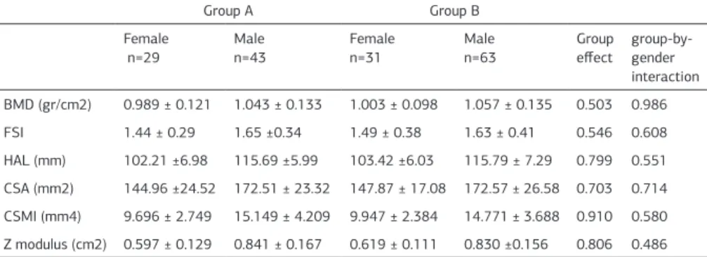
There were no statistical diference for HAL, CSA, CSMI, FSI and Z modulus between of groups when take into consideration of gender efect (p>0.05). Table 2 was expressed these variables according to gender.
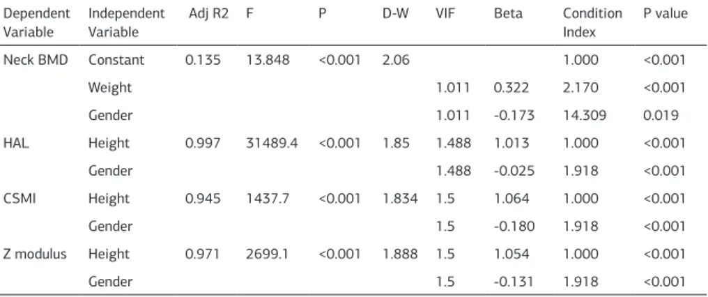
The multipl linear regression analysis was done between depen-dent variables (HAL, FSI, CSMI, Z modulus and CSA) and inde-pendent variables (age, height, weight, BMI, gender, presence of nephrolithiasis). The presence of nephrolithiasis was deter-mined that there was not predictive efects on femur neck BMD, HAL, CSMI, Z modulus, FSI and CSA. To be female gender was a negatif efect on BMD, HAL, CSMI and Z modulus. The predictive factors efected these variables were expressed Table 3 and 4. In none of the patients, increase in BUN, creatinine, calcium and phosphorus were observed. Hypercalciuria was detected in 29 (41.4%) patients with nephrolithiasis. The urinary calcium ex-cretion in 24-hour urine was 154.863 ± 68.339 in normocalciu-ric patients and 417.408 ± 114.738 in hypercalciunormocalciu-ric subjects. There was no statistical diferences for BMD between normo-calciuric and hypernormo-calciuric patients.
Discussion
Bones of nephrolithiasis patients are exposed to eventual frac-ture. Higher fracture rates than normal have been reported in older patients with nephrolithiasis [14]. Until now, HSA, a pre-dictor of fracture risk, was not studied in nephrolithiasis. In this study, we evaluated patients with nephrolithiasis between 20-50 ages. This age group was selected to avoid efects of menopause on bone, and compare structural parameters with
Table 1. Descriptive characteristics of subjects according to gender.
Group A Group B
Female n=29 (40.3%)
Male
n=43 (59.7%) Femalen=31 (33%)
Male
n=63 (67%) Age (years) 36.3 ± 7.5 36.5 ± 7.8 38.7 ± 6.6 33.4 ± 7.3 Height (cm) 158.7 ± 6.6 170.3 ± 6.4 158.9 ± 5.4 171.4 ± 7.1 Weight (kg) 76.3 ± 15.2 80.6 ± 11 76.2 ± 13.6 77.8 ± 11.1 BMI(kg/m2) 30.18 ± 5.26 27.79 ± 3.33 30.25 ± 5.85 26.54 ± 3.81 Data were expressed as Mean ± SD.
BMI: Body mass index.
Table 2. Structural parameters of femur neck in groups and results of Univariate Analysis of Variance
Group A Group B
Female n=29
Male
n=43
Female n=31
Male
n=63
Group efect
group-by-gender
interaction
BMD (gr/cm2) 0.989 ± 0.121 1.043 ± 0.133 1.003 ± 0.098 1.057 ± 0.135 0.503 0.986 FSI 1.44 ± 0.29 1.65 ±0.34 1.49 ± 0.38 1.63 ± 0.41 0.546 0.608 HAL (mm) 102.21 ±6.98 115.69 ±5.99 103.42 ±6.03 115.79 ± 7.29 0.799 0.551 CSA (mm2) 144.96 ±24.52 172.51 ± 23.32 147.87 ± 17.08 172.57 ± 26.58 0.703 0.714 CSMI (mm4) 9.696 ± 2.749 15.149 ± 4.209 9.947 ± 2.384 14.771 ± 3.688 0.910 0.580 Z modulus (cm2) 0.597 ± 0.129 0.841 ± 0.167 0.619 ± 0.111 0.830 ±0.156 0.806 0.486 Data were expressed as Mean ± SD.
normal subjects. Arrabal-Polo and et al. [15] found that patients between 25-60 years of age with calcium lithiasis and severe lithogenic activity in addition to osteopenia/osteoporosis pres-ent with higher levels of hypercalciuria and negative osseous balance. Moyano and et al. [16] studied in nephrolithiasis sub-jects with ages ranging 16-68 years and they found the low Z score than control cases. In a study, decreased BMD value was detected in 10% patients between 20-50 ages when femo-ral and lumbar Z-score are considered [17]. Tsuji and et al. [18] found low BMD values in 30% male (mean age 48.7 ± 13.9 years) and 26.2% female (mean age 52.9 ± 14.9 years) patients. Bones fail when stresses, ascribing forces, exceed the capacity of the structure. If bone material weakens, less force is required to fail. Stresses in loading condition are entirely determined by structural geometry. For this reason, to assess bone strength, both the structural geometry and the features of the material can be quantiied. The long bones are essentially loaded in bend-ing and axial pressure under normal physiologic conditions [19]. Many studies were performed with DXA-based HSA or dual pho-ton absorptiometry. In these clinical studies, gender, age, bone agents and to perdict of fractures risk were studied [20-23]. In a study from Szulc and et al., women with trochanteric hip fractures were examined. In this case control study from the EPIDOS cohort had signiicantly decreased CSMI and section modulus than controls were found [19]. In the conclusion of this study, to predict fracture risk, HSA-derived biomechanical pa-rameters were found to be not superior to BMD. However, in an another study with 60 and older 96 men and women, lower CSMI or section modulus were found as a risk factor for hip fracture even ater adjustment for BMD. On the other hand, the authors concluded that conventional measurement has smaller
superiority to BMD in fracture predic-tion [20]. Melton and et al. in 2005 reached some results in a study with 213 postmenopausal women. They concluded that best risk predictor was femoral neck BMD rather than hip BMD and HSA structural parameters [22]. In a cross-sectional study with 2506 women, FSI and BMD were eval-uated. As a result of the study, FSI pre-dictive value was found to be smaller than BMD in hip fracture patients [24]. In a study performed by Melton et al., observed that the risk of vertebral fracture was greatly increased among men with kidney stones. Although, the risk of fracture at the proximal humer-us, distal forearm, pelvis, and proxi-mal femur was not increased [14]. In a cross-sectional study by using Third National Health and Nutrition Inven-tory Survey (NHANES III), Laudardale et al. found that femoral neck BMD values were lower in men with the his-tory of kidney stones compared to the men without stones [25]. In a study, re-searchers evaluated the efect of hy-percalciuria on bone mineral density in 50 patients with nephrolithiasis. The authors found low lomber and femoral bone density according to T-score in patients with or without hypercalciuria [17].
In a study, researchers evaluated patients with and without re-current calcium lithiasis. They found that patients with recur-rent calcium lithiasis and severe lithogenic activity with loss of BMD (osteopenia/osteoporosis) presented with higher levels of calcium urine excretion, greater elevations of bone resorp-tion markers and lower levels of citrate excreresorp-tion than patients without lithiasis. They suggested that metabolic studies (such as bone remodelling markers (β-CrossLaps, osteocalcin and β-CrossLaps/osteocalcin), calciuria, fasting calcium/creatinine, 24-hour calcium/creatinine, citraturia and calcium/citrate) could be performed in patients with calcium renal stones and BMD loss for accurate therapy [15].
In a study done by Tsuji in 2005, they performed BMD in 310 patients and concluded that there were no diferences in BMD values when both genders were considered however there was a signiicant diference when only female patients were consid-ered [18]. The results of Moyano’s study suggest independent to the presence or absence of hypercalciuric renal calcium lithiasis afect BMD more prominently at the femoral neck. Finally, they concluded that similar distribution was seen between lithiasis patients and controls. Homozygous alleles BB and tt were ob-served less frequently in patients with calcium renal lithiasis [16].
As a conclusion, we did not found any diferences on BMD and hip structural parameters measured with HSA program between nephrolithiasis patients and normal subjects at 20-50 ages. We thought that in these patients had high fracture rates deter-Table 3. Multiple linear regression models for HAL, CMI and Z modulus (n=166).
Dependent
Variable Independent Variable Adj R2 F P D-W VIF Beta
Condition
Index P value Neck BMD Constant 0.135 13.848 <0.001 2.06 1.000 <0.001
Weight 1.011 0.322 2.170 <0.001
Gender 1.011 -0.173 14.309 0.019
HAL Height 0.997 31489.4 <0.001 1.85 1.488 1.013 1.000 <0.001
Gender 1.488 -0.025 1.918 <0.001
CSMI Height 0.945 1437.7 <0.001 1.834 1.5 1.064 1.000 <0.001
Gender 1.5 -0.180 1.918 <0.001
Z modulus Height 0.971 2699.1 <0.001 1.888 1.5 1.054 1.000 <0.001
Gender 1.5 -0.131 1.918 <0.001
D-W: Durbin-Watson coeicient, VIF: Variance inlation factor
Beta: Standardized regression coeicient, Gender: 0 Male, 1 Female
BMD: Bone mineral density, HAL: Hip axis length, CSMI: Cross-sectional moment of inertia, Z modulus: Section modulus
Table 4. Multiple linear regression models for FSI (n=166).
Dependent
variable
Independent Variable
R2 F P D-W VIF B SE Condition
Index
P value
Lg10(FSI) Height 0.770 552.4 <0.001 2.07 1.0 0.001 0.000 1.0 <0.001
CSA Height 0.981 8363.2 <0.001 2.08 1.0 0.981 0.011 1.0 <0.001 Logarithmic (Log) transformation was applied to FSI.
D-W: Durbin-Watson coeicient, VIF: Variance inlation factor B: unstandardized regression coeicient,
mined previous studies especially in older ages, bone metabolic and geometric changes may start or/and fast with aging to-gether other cause of loss of bone mineral (e.g. postmenopaus-al osteoporosis).
Competing interests
The authors declare that they have no competing interests.
References
1. Bataille P, Achard JM, Fournier A, Boudailliez B, Westeel PF, el Esper N, Bergot C, Jans I, Lalau JD, Petit J. Diet, vitamin D and vertebral mineral density in hypercal-ciuric calcium stone formers. Kidney Int 1991;39(6):1193-205.
2. Borghi L, Meschi T, Guerra A, Maninetti L, Pedrazzoni M, Marcato A, Vescovi P, Novarini A. Vertebral mineral content in diet-dependent and diet-independent hypercalciuria. J Urol 1991;146(5):1334-8.
3. Paciici R, Rothstein M, Rifas L, Lau KH, Baylink DJ, Avioli LV, Hruska K. Increased monocyte interleukin-1 activity and decreased vertebral bone density in patients with fasting idiopathic hypercalciuria. J Clin Endocrinol Metab 1990;71(1):138-45. 4. Pietschmann F, Breslau NA, Pak CY. Reduced vertebral bone density in hypercal-ciuric nephrolithiasis. J Bone Miner Res 1992;7(12):1383-8.
5. Weisinger JR, Alonzo E, Bellorín-Font E, Blasini AM, Rodriguez MA, Paz-Martínez V, Martinis R. Possible role of cytokines on the bone mineral loss in idiopathic hypercalciuria. Kidney Int 1996;49(1):244-50.
6. Audran M, Legrand E. Hypercalciuria. Joint Bone Spine 2000;67(6):509-15. 7. Lemann J, Worcester EM, Gray RW. Hypercalciuria and stones. Am J Kidney Dis 1991;17(4):386-91.
8. Martin RB, Burr DB. Non-invasive measurement of long bone cross-sectional moment of inertia by photon absorptiometry. J Biomech 1984;17(3):195-201. 9. Beck TJ, Ruf CB, Warden KE, Scott WW, Rao GU. Predicting femoral neck strength from bone mineral data. A structural approach. Invest Radiol 1990;25(1):6-18. 10. Faulkner KG, Cummings SR, Black D, Palermo L, Glüer CC, Genant HK. Simple measurement of femoral geometry predicts hip fracture: the study of osteopo-rotic fractures. J Bone Miner Res 1993;8(10):1211-7.
11. Yoshikawa T, Turner CH, Peacock M, Slemenda CW, Weaver CM, Teegarden D, Markwardt P, Burr DB. Geometric structure of the femoral neck measured using dual-energy x-ray absorptiometry. J Bone Miner Res 1994;9(7):1053-64. 12. Crabtree NJ, Kroger H, Martin A, Pols HA, Lorenc R, Nijs J, Stepan JJ, Falch JA, Miazgowski T, Grazio S, Raptou P, Adams J, Collings A, Khaw KT, Rushton N, Lunt M, Dixon AK, Reeve J. Improving risk assessment: hip geometry, bone mineral distribution and bone strength in hip fracture cases and controls. The EPOS study. European Prospective Osteoporosis Study. Osteoporos Int 2002;13(1):48-54. 13. Beck TJ, Oreskovic TL, Stone KL, Ruf CB, Ensrud K, Nevitt MC, Genant HK, Cummings SR. Structural adaptation to changing skeletal load in the progres-sion toward hip fragility: the study of osteoporotic fractures. J Bone Miner Res 2001;16(6):1108-19.
14. Melton LJ, Crowson CS, Khosla S, Wilson DM, O’Fallon WM. Fracture risk among patients with urolithiasis: a population-based cohort study. Kidney Int 1998;53(2):459-64.
15. Arrabal-Polo MA, Arrabal-Martin M, Arias-Santiago S, Garrido-Gomez J, De Haro-Muñoz TD, Zuluaga-Gomez A. Metabolic-mineral study in patients with renal calcium lithiasis, severe lithogenic activity and loss of bone mineral density. Sin-gapore Med J 2012;53(12):808-13.
16. Moyano MJ, de Tejada MJG, Lozano RG, Moruno R, Ortega R, Martí V, Palen-cia RS, Miranda MJ, Palma A, Cano RP. Changes in bone mineral metabolism in patients with recurrent urolithiasis and vitamin D receptor gene polymorphisms. Preliminary results. Nefrologia 2007;27(6):694-703.
17. Özen A, Esenyel M, Karacan İ. Efect of hypercalciuria on bone mineral density in patients with nephrolithiasis Turkish Journal of Nuclear Medicine 2010;19(3):132-7.
18. Tsuji H, Umekawa T, Kurita T, Uemura H, Iguchi M, Kin K, Kushida K. Analysis of bone mineral density in urolithiasis patients. Int J Urol 2005;12(4):335-9. 19. Burr DB. Muscle strength, bone mass, and age-related bone loss. J Bone Miner Res 1997;12(10):1547-51.
20. Ahlborg HG, Nguyen ND, Nguyen TV, Center JR, Eisman JA. Contribution of hip strength indices to hip fracture risk in elderly men and women. J Bone Miner Res 2005;20(10):1820-7.
21. Beck TJ, Stone KL, Oreskovic TL, Hochberg MC, Nevitt MC, Genant HK, Cum-mings SR. Efects of current and discontinued estrogen replacement therapy on hip structural geometry: the study of osteoporotic fractures. J Bone Miner Res 2001;16(11):2103-10.
22. Melton LJ, Beck TJ, Amin S, Khosla S, Achenbach SJ, Oberg AL, Riggs BL. Con-tributions of bone density and structure to fracture risk assessment in men and women. Osteoporos Int 2005;16(5):460-7.
23. Szulc P, Duboeuf F, Schott AM, Dargent-Molina P, Meunier PJ, Delmas PD. Structural determinants of hip fracture in elderly women: re-analysis of the data from the EPIDOS study. Osteoporos Int 2006;17(2):231-6.
24. Faulkner KG, Wacker WK, Barden HS, Simonelli C, Burke PK, Ragi S, Del Rio L. Femur strength index predicts hip fracture independent of bone density and hip axis length. Osteoporos Int 2006;17(4):593-9.
25. Lauderdale DS, Thisted RA, Wen M, Favus MJ. Bone mineral density and
frac-ture among prevalent kidney stone cases in the Third National Health and Nutri-tion ExaminaNutri-tion Survey. J Bone Miner Res 2001;16(10):1893-8.
How to cite this article: