ISSN 0104-6632 Printed in Brazil
www.abeq.org.br/bjche
Vol. 26, No. 02, pp. 399 - 405, April - June, 2009
Brazilian Journal
of Chemical
Engineering
DECOLORIZATION OF A TEXTILE DYE,
REACTIVE RED 198 (RR198), BY
Aspergillus
parasiticus
FUNGAL BIOSORBENT
S. Tunali Akar
1*, T. Akar
1and A. Çabuk
21
Department of Chemistry, Faculty of Arts and Science, Phone: + (90) (222) 239 3750 /2862, Fax: + (90) (222) 2393578, Eskişehir Osmangazi University, 26480, Eskişehir, Turkey.
E-mail: [email protected]
2
Department of Biology, Faculty of Arts and Science, Eskişehir Osmangazi University, 26480, Eskişehir, Turkey.
(Submitted: October 7, 2005 ; Revised: January 24, 2006 ; Accepted: February 2, 2006)
Abstract - The decolorization potential of textile dye Reactive Red 198 (RR198) by Aspergillus parasiticus
fungal biosorbent has been investigated as a function of initial pH, contact time, biosorbent and initial dye concentration in a batch system. Maximum dye biosorption capacity 1.03x10⁻⁴ mol g-1 was observed at pH 2.0 and 2.0 g L-1 of biosorbent concentration. Biosorption equilibrium was attained within 50 min. The equilibrium data followed Langmuir, Freundlich and Dubinin-Radushkevich isotherm models at 20, 30, 40 and 50°C. An increase in the biosorption capacity of A. parasiticus with temperature showed that the decolorization process is endothermic. Results indicated that Aspergillus parasiticus was an effective candidate for textile dye RR198 removal from aqueous solutions.
Keywords:Aspergillus parasiticus; Biosorption; Decolorization; Isotherm; Reactive Red 198.
INTRODUCTION
As a result of many industrial activities, the discharge of large quantities of organic pollutants into the receiving waters has caused significant environmental pollution. Synthetic dyes are a group of organic pollutants that are extensively used in textile, paper, printing and dyehauses (Aksu, 2005).
The colored effluents damage the aesthetic quality of water and reduce light penetration and photosynthesis and also some of the dyes are toxic or mutagenic, carcinogenic and allergenic (Aksu and Çağatay, 2006; Kumar et al., 2006; Bakshi et al., 1999; O’Mahony et al., 2002). According to the survey of the Ecological and Toxicological Association of the Dyestuffs Manufacturing Industry (ETAD), over 90% of some 4000 dyes have LD50
values greater than 2000 mg kg-1 (Shore, 1996; Robinson et al., 2001). Hence decolorization of the dye-bearing effluents is of great importance.
The traditional physical or chemical decolorization methods including coagulation, flocculation, ion
exchange, irradiation, precipitation, ozonation and adsorption or a combination of these methods have been used for dye removal from wastewaters (Aksu, 2005; Fu and Viraraghavan, 2001).
However application of these methods is somewhat restricted due to some limitations such as operational costs, formation of hazardous by-products, intensive energy requirement (Padmesh et al., 2005) and limited adaptability to a wide range of effluents (Fu and Viraraghavan, 2001). The use of biological materials as sorbents for the treatment of colored effluents appeared as a potential alternative process to conventional methods (Kumar et al., 2006). Currently, numerous studies have focused on the dye biosorption and biodegradation abilities of microorganisms, including some bacteria, fungi and algae. Live and dead forms of microbial cells can be used in the decolorization process and the main mechanisms of these forms are biodegradation and biosorption, respectively (Fu and Viraraghavan, 2001).
400 S. Tunali Akar, T. Akar and A. Çabuk
which are following; dead organisms are not subject to toxicity limitations, they do not require a continuous supply of nutrients, they can be regenerated simply and reused for many cycles (Akar and Tunali, 2005; Padmesh et al., 2005), biomass can be stored until the use and their operation is easy. Also they can be obtained from industrial sources as a waste product from established fermentation processes (Fu and Viraraghavan, 2001).
Compounds such as chitin, lipids, amino acids and other cellular components present on the cell walls of microorganisms provide active binding sites for dye molecules and biosorption processes based on interactions between the dyes and binding sites in a manner of surface adsorption, ion exchange, complexation, chelatation and microprecipitation (Aksu, 2001).The dye–microorganism interactions depend on different factors varying from the chemical structure of the dye being studied to the type of biosorbent used, as well as operating conditions (Kargi and Ozmıhcı, 2004; Šafaříková et al., 2005).
Currently research is focused on finding optimal microbial biosorbent and reaction conditions in order to develop optimal processes enabling decolorization of large volumes of waters (Šafaříková et al., 2005).
The water-soluble reactive dyes are one of the most important groups of dyes used in the textile dyeing industries due to their highly brilliant colors (Aksu, 2003). Reactive Red 198 (RR198) is also among this group of dyes and it was selected as the model reactive dye for this work.
Aspergillus parasiticus and Aspergillus flavus
fungal strains have only limited parasitic abilities, yet they can successfully colonize and produce aflatoxin in developing host tissue in the field (Brown et al., 1999; Mayer et. al., 2003). However, several studies have been reported in the literature on the biosorption potential of the dead form of A.
flavus (Hafez, et al., 1997; Deepa et al, 2006; Akar
and Tunali, 2006). The commercially available and easily cultivable fungus A. parasiticus was chosen as a biosorbent material because of a lack of
information on its dye sorption ability.
This research aims to investigate the removal of a textile dye RR198 from aqueous solutions by A.
parasiticus cells as an alternative biosorbent. The
effects of various operating parameters were studied including pH, biosorbent dosage and contact time. Biosorption was analyzed by the Freundlich, Langmuir and Dubinin-Radushkevich (D-R) isotherm models at 20, 30, 40 and 50○C.
MATERIALS AND METHODS
Biosorbent Preparation
The filamentous fungus, A. parasiticus (NRRL 502), was obtained from the culture collection of the Biology Department of Eskişehir Osmangazi University. The stock cultures were routinely maintained on agar medium at +4○C and liquid growth conditions for A. parasiticus were described in our previous studies (Kiran, et al., 2005).
The live A. parasiticus cells were separated from the growth medium by filtration after seven days of growth, spread on petri dishes and dried in an oven at 60oC overnight. They were then powdered using a mortar and pestle and sieved to select 150 μm particles for use as a biosorbent.
Dye Solutions
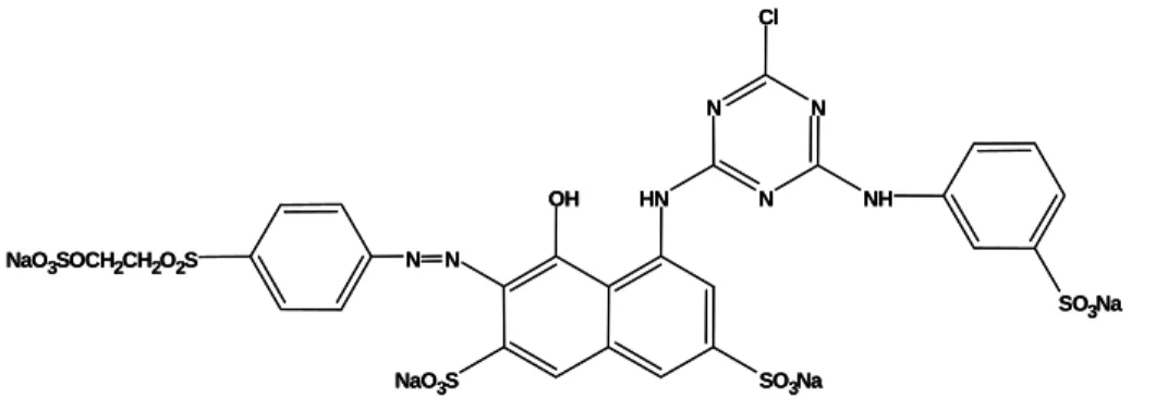
The dye used in this study was Reactive Red 198 (RR198) obtained from a local textile company and used without further purification. Its chemical structure is shown in Fig. 1. A stock solution (1 g L−1) of RR198 was prepared by dissolving an accurate quantity of dye in bidistilled water and other concentrations varying between 100 and 300 mg L−1 were obtained by dilution. Fresh dilutions were used for each biosorption experiment. The pH of the working solutions was adjusted to the required values by 0.1 M HCl or 0.1 M NaOH. All the chemicals used were analytical grade.
OH HN
NaO3S SO3Na
N N NaO3SOCH2CH2O2S
N N N
Cl
NH
SO3Na
OH HN
NaO3S SO3Na
N N NaO3SOCH2CH2O2S
N N N
Cl
NH
SO3Na
Dye Biosorption Experiments
Dye biosorption experiments were performed on a magnetic stirrer. 0.1 g of dried biosorbent of A.
parasiticus was put in a 100 mL beaker and 50 mL
of dye solution (100 mg L−1) was added to it. The mixture was stirred at 200 rpm at 20oC for 1 h to elucidate the optimum conditions of pH, biosorbent dosage and contact time.
The effect of solution pH on dye biosorption capacity of the fungal biosorbent was determined by adjusting the pH values between 1 and 10, adding freshly prepared 0.1 M HCl or 0.1 M NaOH solutions. The optimum pH value was used throughout all biosorption experiments. The effect of biosorbent concentration was investigated by using biosorbent samples ranging from 0.4 to 4.0 g L−1. This was followed by the assessment of the effect of equilibrium time, varied between 10 to 90 min, on the dye biosorption capacity of the biosorbent. The initial dye concentration was varied from 100 to 300 mg L1 and the isotherm appropriateness of the biosorption results was evaluated for the Langmuir, Freundlich and Dubinin-Radushkevich (D-R) isotherm models at temperatures of 20, 30, 40 and 50oC under the optimum conditions.
The biosorption yield (%) and the amount of dye sorbed by the biosorbent were calculated using the following general equations:
Biosorption yield (%) = [(Ci-Ce).100]/Ci (1)
qe = [(Ci-Ce).V]/M (2)
where,
qe: amount of dye biosorbed (mg g-1)
Ci: initial dye concentration in solution (mg L-1)
Ce: final dye concentration in solution (mg L-1)
V: volume of the medium (L)
M: amount of the biosorbent used in the reaction mixture (g).
Biosorbent was separated by centrifugation at
500 rpm for 5 min. The dye concentrations in the supernatants were determined using a spectrophotometer, (UV/Vis, Unicam UV2-100) at
λmax 515 nm. All the experiments were carried out in
triplicate and data presented are the mean values from these independent experiments. Error bars are indicated wherever necessary. All statistical analysis was done using SPSS 9.05 for Windows.
RESULT AND DISCUSSION
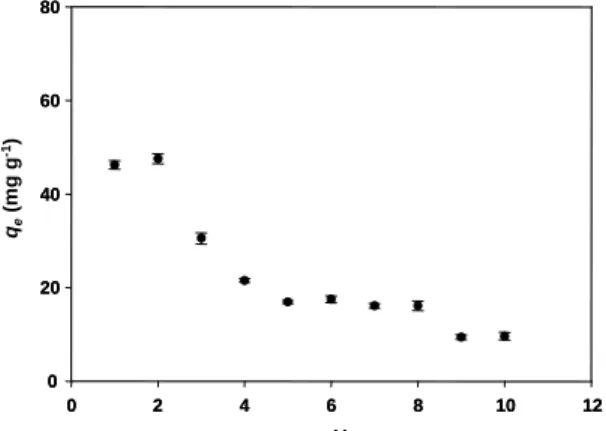
Effect of pH
The pH value of the biosorption medium significantly influences the sorption properties of fungal biosorbents (Aksu and Tezer, 2000). The pH dependence of dye biosorption is related to both protonation or deprotonation of the functional groups of the biopolymers on the biosorbent surface and the ionization potential of complex dye molecules in solution (Maurya et al., 2006). Fig. 2 represents the effect of pH on the biosorption of RR198 onto dried A. parasiticus cells. As seen in this figure, the maximum biosorption of RR198 on
A. parasiticus was found as 47.56 mg g1 at pH 2.0.
Because dye binding sites on the biosorbent were closely associated with H3O+ at lower pH, which
cause an increase in the electrostatic attraction between positively charged biosorbent surface and the negatively charged dye molecules. The biosorption capacity of the dried cells of A.
parasiticus decreased when the pH of the solution
increased from 2.0 to 5.0. Under alkaline conditions biosorption capacity significantly decreased (p<0.05). The decrease of the biosorption capacity with increase in the medium pH can be explained by the dye binding sites on the biosorbent surface becoming negatively charged, restricting the approach of the dye molecules as a result of repulsive forces.
pH
0 2 4 6 8 10 12
0 20 40 60 80
qe
(m
g
g
-1)
pH
0 2 4 6 8 10 12
0 2 4 6 8 10 12
0 20 40 60 80
0 20 40 60 80
qe
(m
g
g
-1)
402 S. Tunali Akar, T. Akar and A. Çabuk
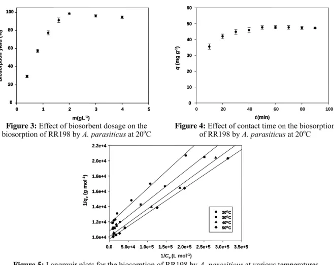
Effect of Biosorbent Dosage
In order to determine the effect of the biosorbent dosage on the biosorption efficiency of RR198, the amounts of biosorbent added into the biosorption medium were varied from 0.4 to 4.0 g L−1 and the results are presented in Fig 3. With increase in the biosorbent dosage, from 0.4 to 2.0 g L−1, the percentage decolorization of RR198 increased from 29.40% to 98.57%, which is the maximum value obtained as the number of possible binding sites increased. Further increase up to 4.0 g L−1 did not change the maximum RR198 biosorption capacity of the biosorbent and it almost stayed constant (p>0.05). This is because, at higher biosorbent concentrations, there is a very fast biosorption onto the biosorbent surface that produces a lower solute concentration in the solution than when the biosorbent concentration is lower (Kumar et al., 2006).
Effect of Contact Time
Fig. 4 indicates the effect of treatment time on the biosorption of RR198 dye onto biosorbent. High biosorption rates were observed at the beginning of
the biosorption and the process reached the plateau values within ca. 50 min. After this period, no considerable change was observed in the RR198 biosorption capacity of the biosorbent (p>0.05) and it was fixed as the optimum contact time.
Biosorption Isotherms
Equilibrium isotherms are plots of the quantity of sorbate retained on the biosorbent (qe) as a function of
sorbate concentration in the liquid phase (Ce).
Biosorption data are commonly represented by an equilibrium isotherm. The equilibrium biosorption isotherm is of fundamental importance for the design and optimization of the biosorption system for the dye decolorization studies (Maurya et al., 2006). Therefore, the equilibrium biosorption isotherms are one of the most important data for understanding the mechanism of the biosorption (Tunali et al., 2006). Several isotherm equations are available and three important isotherm models, namely Langmuir, Freundlich and Dubinin-Radushkevich (D-R) isotherm models were chosen in this study to describe the biosorption equilibrium and the model plots at various temperatures are shown in Figs. 5, 6 and 7.
m(gL-1)
0 1 2 3 4 5
Bi oso rp tio n y ield (% ) 0 20 40 60 80 100
m(gL-1)
0 1 2 3 4 5
Bi oso rp tio n y ield (% ) 0 20 40 60 80 100
m(gL-1)
0 1 2 3 4 5
Bi oso rp tio n y ield (% ) 0 20 40 60 80 100 0 20 40 60 80 100
t (min)
0 20 40 60 80 100
q (mg g -1) 0 10 20 30 40 50 60
t (min)
t (min)
0 20 40 60 80 100
q (mg g -1) 0 10 20 30 40 50 60 0 10 20 30 40 50 60
Figure 3: Effect of biosorbent dosage on the biosorption of RR198 by A. parasiticus at 20oC
Figure 4: Effect of contact time on the biosorption of RR198 by A. parasiticus at 20oC
1/Ce(L mol-1)
0.0 5.0e+4 1.0e+5 1.5e+5 2.0e+5 2.5e+5 3.0e+5 3.5e+5
1/ qe (g mo l -1) 1.0e+4 1.2e+4 1.4e+4 1.6e+4 1.8e+4 2.0e+4 2.2e+4 20 30 40 50 o o o o C C C C
1/Ce(L mol-1)
0.0 5.0e+4 1.0e+5 1.5e+5 2.0e+5 2.5e+5 3.0e+5 3.5e+5 0.0 5.0e+4 1.0e+5 1.5e+5 2.0e+5 2.5e+5 3.0e+5 3.5e+5
1/ qe (g mo l -1) 1.0e+4 1.2e+4 1.4e+4 1.6e+4 1.8e+4 2.0e+4 2.2e+4 1.0e+4 1.2e+4 1.4e+4 1.6e+4 1.8e+4 2.0e+4 2.2e+4 20 30 40 50 20 30 40 50 o o o o o o o o C C C C C C C C
lnCe
-13 -12 -11 -10 -9 -8
ln q e -10.0 -9.8 -9.6 -9.4 -9.2 -9.0 20 30 40 50 o o o o C C C C lnCe
-13 -12 -11 -10 -9 -8
-13 -12 -11 -10 -9 -8
ln q e ln q e -10.0 -9.8 -9.6 -9.4 -9.2 -9.0 -10.0 -9.8 -9.6 -9.4 -9.2 -9.0 20 30 40 50 20 30 40 50 o o o o o o o o C C C C
ε2(J mol-1)2
4.0e+8 5.0e+8 6.0e+8 7.0e+8 8.0e+8 9.0e+8 1.0e+9 1.1e+9 1.2e+9
ln q e -10.0 -9.8 -9.6 -9.4 -9.2 -9.0 o o o 20 30 40 50o C C C C
ε2(J mol-1)2
4.0e+8 5.0e+8 6.0e+8 7.0e+8 8.0e+8 9.0e+8 1.0e+9 1.1e+9 1.2e+9
ln q e -10.0 -9.8 -9.6 -9.4 -9.2 -9.0 o o o 20 30 40 50o C C C C
ε2(J mol-1)2
4.0e+8 5.0e+8 6.0e+8 7.0e+8 8.0e+8 9.0e+8 1.0e+9 1.1e+9 1.2e+9 4.0e+8 5.0e+8 6.0e+8 7.0e+8 8.0e+8 9.0e+8 1.0e+9 1.1e+9 1.2e+9
ln q e ln q e -10.0 -9.8 -9.6 -9.4 -9.2 -9.0 -10.0 -9.8 -9.6 -9.4 -9.2 -9.0 o o o 20 30 40 50 20 30 40 50o C C C C C C C C
Figure 6: Freundlich plots for the biosorption of RR198 by A. parasiticus at various temperatures
Figure 7: D-R plots for the biosorption of RR198
by A. parasiticus at various temperatures
The Langmuir isotherm is represented by the following equation (Langmuir, 1918):
max L e e
L e
q K C
q
1 K C
=
+ (3)
where qe is the biosorbed dye concentration (mol g–
1
), Ce is the residual dye concentration at equilibrium
(mol L–1), qmax is the monolayer biosorption capacity
of the biosorbent (mol g–1), and KL is the Langmuir
biosorption constant (L mol–1).
The Freundlich isotherm involves heterogeneous biosorption and the empirical Freundlich equation is (Freundlich, 1906):
1/n
e F e
q =K C (4)
where KF (L mg−1) and n are Freundlich biosorption
isotherm constants depend on several environmental factors.
The Dubinin-Radushkevich (D-R) isotherm is another less commonly used model. It was applied to distinguish between physical and chemical biosorption (Benhammou et al., 2005) of dye. The D-R isotherm equation (Dubinin and Radushkevich, 1947) is:
2
e m
q =q e−βε (5)
where qm is the theoretical sorption capacity, and ε is
the Polanyi potential; β (mol2 J–2), is the activity coefficient related to mean sorption energy, E (kJ
mol–1). This is the free energy of the transfer per mole of the biosorbate from infinity to the sorbent surface and can be calculated using the following equation (Hobson, 1969; Hasany and Chaudhary, 1996; Dubey and Gupta, 2005):
1/2 1 E
(2 )
=
β (6)
The magnitude of this parameter is useful for information about the type of biosorption process such as chemical ion exchange or physical biosorption. It was found that E values varied from 19.634 to 20.455 kJ mol−1, which are bigger then the energy range of adsorption reactions, 8-16 kJ mol−1. The type of biosorption may be interpreted as chemical biosorption (Helfferich, 1962; Onyango et al., 2004).
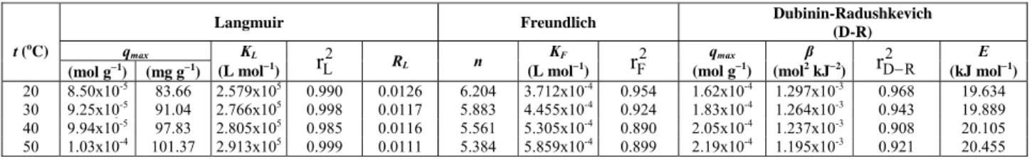
The Langmuir, Freundlich and D-R parameters for the biosorption of RR198 dye onto A. parasiticus are listed in Table 1. The Langmuir isotherm model fits very well for the RR198 biosorption process based on the r2 values in Table 1. Biosorption capacity of A.
parasiticus increased with increasing temperature from
20 to 50oC and was found to be 1.03x10⁻⁴ mol g−1 at 50oC. This observation implies that the biosorption of RR198 by A. parasiticus biosorbent is endothermic nature. The biosorption capacity of A. parasiticus
obtained for RR198 in this study is comparable to and moderately higher than that of many corresponding biosorbents reported in the literature (Table 2).
Table 1: Biosorption isotherm constants for the biosorption of RR198 by A. parasiticus at various temperatures
Langmuir Freundlich Dubinin-Radushkevich
(D-R)
qmax
t (o
C) (mol g−1
) (mg g−1
)
KL
(L mol−1
) 2 L
r RL n
KF
(L mol−1
) 2 F
r qmax
(mol g−1
) β (mol2
kJ−2
) 2 D R
r − (kJ molE −1
) 20 8.50x10⁻⁵
83.66 2.579x10⁵
0.990 0.0126 6.204 3.712x10⁻⁴
0.954 1.62x10⁻⁴
1.297x10⁻³
0.968 19.634 30 9.25x10⁻⁵
91.04 2.766x10⁵
0.998 0.0117 5.883 4.455x10⁻⁴
0.924 1.83x10⁻⁴
1.264x10⁻³
0.943 19.889 40 9.94x10⁻⁵
97.83 2.805x10⁵
0.985 0.0116 5.561 5.305x10⁻⁴
0.890 2.05x10⁻⁴
1.237x10⁻³
0.908 20.105 50 1.03x10⁻⁴
101.37 2.913x10⁵
0.999 0.0111 5.384 5.859x10⁻⁴
0.899 2.19x10⁻⁴
1.195x10⁻³
404 S. Tunali Akar, T. Akar and A. Çabuk
Table 2: Biosorption results of different textile dyes from the literature for various biosorbents and operating conditions
Rating conditions
Biosorbent material Dye pH t
(o
C)
Initial concentration
(mg L−1)
Biosorbent dosage (g L−1)
Biosorption capacity (mg g−1)
References
Rhizopusarrhizus Reactive Orange 16 2.0 - 0-500 1.0 190 O’Mahony et al.,2002
Rhizopusarrhizus Reactive Red 4 2.0 - 0-500 1.0 150 O’Mahony et al.,2002
Saccharomycescerevisiae Remazol Blue 3.0 25 10-400 0.52 84.6 Aksu, 2003 Saccharomycescerevisiae Remazol Black B 3.0 25 10-400 2.87 88.5 Aksu, 2003 Saccharomycescerevisiae Remazol Red RB 3.0 25 10-400 2.60 48.8 Aksu, 2003 Kluyveromyces marxianus Remazol Black B - - 10-500 1.0 37.0 Bustard, et al.,1998
Escherichia coli Reactive Blue 5 3.0 28 200 - 89.4 Hu, 1996
Escherichia coli Reactive Red 22 3.0 28 200 - 76.6 Hu, 1996
Escherichia coli Reactive Violet 2 3.0 28 200 - 65.5 Hu, 1996
Escherichia coli Reactive Yellow 2 3.0 28 200 - 52.4 Hu, 1996
Aspergillusparasiticus Reactive Red 198 2.0 20 100-300 2.0 83.66 This study Aspergillusparasiticus Reactive Red 198 2.0 50 100-300 2.0 101.37 This study
The essential characteristics of the Langmuir isotherm can be expressed by a dimensionless constant, RL, referred to as the equilibrium
parameter. RL is described by the following
equation:
L
L o 1 R
1 K C
=
+ (7)
where Co is the highest dye concentration (mol L−
1
). The value of RL is used for the interpretation of the
sorption type (Weber and Chakravorti, 1974; Hall et al., 1966). Calculated RL values for this study are
found to be between 0.0111 and 0.0126 in the temperature range of 20-50oC and these results indicate the biosorption process is favorable.
The Freundlich model constants KF and n are
indicators of biosorption capacity and biosorption intensity, respectively (Malik, 2004) and the numerical values of these constants are included in Table 1.
CONCLUSION
A. parasiticus was found to be an effective
and alternative biosorbent for the removal of RR198 textile dye from aqueous solutions. The biosorption process was affected by various experimental conditions such as pH, contact time and biosorbent concentration. Maximum dye removal was observed at pH 2.0 and 2.0 g L−1 of biosorbent concentration. Biosorption equilibrium was established in 50 min. Experimental data were fitted well to the Langmuir isotherm model. The maximum dye removal percentage was determined as 98.57% at 50°C. The biosorption process is endothermic.
REFERENCES
Akar, T. and Tunali, S., Biosorption performance of
Botrytis cinerea fungal by-products for removal
of Cd(II) and Cu(II) ions from aqueous solutions, Miner. Eng. 18, 1099-1109 (2005).
Akar, T. and Tunali, S., Biosorption characteristics
of Aspergillus flavus biomass for removal of
Pb(II) and Cu(II) ions from an aqueous solution, Bioresour. Technol. 97, 1780–1787 (2006). Aksu, Z., Biosorption of reactive dyes by dried
activated sludge: equilibrium and kinetic modelling, Biochem. Eng. J. 7, 79-84 (2001). Aksu, Z., Reactive dye bioaccumulation by
Saccharomyces cerevisae, Process Biochem. 38,
1437-1444, (2003).
Aksu, Z., Application of biosorption for the removal of organic pollutants: a review, Process Biochem. 40, 997-1026 (2005).
Aksu, Z. and Çağatay, Ş. Ş., Investigation of biosorption of Gemazol Turquise Blue-G reactive dye by dried Rhizopus arrhizus in batch and continuous systems, Sep. Purif. Technol. 48, 24-35 (2006).
Aksu, Z. and Tezer, S., Equilibrium and kinetic modelling of biosorption of Remazol Black B by
Rhizopus arrhizus in a batch system: effect of
temperature, Process Biochem. 36, 431-439 (2000). Bakshi, D. K., Gupta, K. G. and Sharma, P.,
Enhanged biodecolorization of synthetic textile dye effluent by Phanerochaete chrysosporium
under improved culture conditions, World J. Microbiol. Biotechnol. 15, 507-509 (1999). Benhammou, A., Yaacoubi, A., Nibou, L. and
Tanouti, B., Adsorption of metal ions onto Moroccan stevensite: kinetic and isotherm studies, J. Colloid Interface Sci. 282, 320-326 (2005).
biosynthesis, Fungal Genet. Biol. 26, 81-98 (1999).
Bustard, M., McMullan, G. and McHale, A.P., Biosorption of textile dyes by biomass derived
from Kluyveromyces marxianus IMB3,
Bioprocess Eng. 19, 427-430 (1998).
Deepa, K. K., Sathishkumar, M., Binupriya, A. R., Murugesan, G. S., Swaminathan, K. and Yun, S. E., Sorption of Cr(VI) from dilute solutions and wastewater by live and pretreated biomass of
Aspergillus flavus, Chemosphere. 62, 833-840
(2006).
Dubey, S. S. and Gupta, R. K., Removal behavior of Babool bark (Acacia nilotica) for submicro concentrations of Hg2+ from aqueous solutions: a radiotracer study, Sep. Purif. Technol. 41, 21-28 (2005).
Dubinin, M. M. and Radushkevich, L.V., Equation of the characteristic curve of activated charcoal, Proceedings of the Academy of Sciences, Physical Chemistry Section, U.S.S.R. 55, 331-333 (1947). Freundlich, H. M. F., Über die adsorption in
lösungen, Z. Phys. Chem. 57, 385-470 (1906). Fu, Y. and Viraraghavan, T., Fungal decolorization
of dye wastewaters: a review, Bioresour. Technol. 79, 251-262 (2001).
Hafez, N., Abdel-Razek, A. S. and Hafez, M. B., Accumulation of some heavy metals on
Aspergillus flavus, J. Chem. Tech. Biotechnol.
68, 19-22 (1997).
Hall, K. R., Eagleton, L. C., Acrivos, A. and Vermeulen, T., Pore- and solid-diffusion kinetics in fixed–bed adsorption under constant-pattern conditions, Ind. Eng. Chem. Fundam. 5, 212-223 (1966).
Hasany, S. M. and Chaudhary, M. H., Sorption potential of Hare River sand for the removal of antimony from acidic aqueous solution, Appl. Radiat. Isot. 47, 467-471 (1996).
Helfferich, F., Ion Exchange, McGraw-Hill, New York (1962).
Hobson, J. P., Physical adsorption isotherms extending from ultrahigh vacuum to vapor pressure, J. Phys. Chem. 73, 2720-2727 (1969). Hu, T-L., Removal of reactive dyes from aqueous
solution by different bacterial genera, Water Sci. Technol. 34, 89-95 (1996).
Kargi, F. and Ozmıhcı, S., Biosorption performance of powdered activated sludge for removal of different dyestuffs, Enzyme Microb. Technol. 35, 267-271 (2004).
Kiran, I., Akar, T. and Tunali, S., Biosorption of Pb(II) and Cu(II) from aqueous solutions by pretreated biomass of Neurospora crassa, Process Biochem. 40, 3550-3558 (2005).
Kumar, K. V., Ramamurthi, V. and Sivanesan, S., Biosorption of malachite green, a cationic dye onto Pithophora sp., a fresh water algae, Dyes Pigments. 69, 102-107 (2006).
Langmuir, I., The adsorption of gases on plane surfaces of glass, mica and platinum, J. Am. Chem. Soc. 40, 1361-1403 (1918).
Malik, P. K., Dye removal from wastewater using activated carbon developed from sawdust: adsorption equilibrium and kinetics, J. Hazard. Mater, 113:81-88 (2004).
Maurya, N. S., Mittal, A. K., Cornel, P. andRother, E., Biosorption of dyes using dead macro fungi: effect of dye structure, ionic strength and pH, Bioresour. Technol. 97, 512-521 (2006).
Mayer, Z., Bagnara, A., Färber, P. and Geisen, R., Quantification of the copy number of nor-1, a gene of the aflatoxin biosynthetic pathway by real-time PCR, and its correlation to the cfu of
Aspergillus flavus in foods, Int. J. Food
Microbiol. 82, 143-151 (2003).
O’Mahony, T., Guibal, E. and Tobin, J. M., Reactive dye biosorption by Rhizopus arrhizus biomass, Enzyme Microb. Technol. 31, 456-463 (2002). Onyango, M. S., Kojima, Y., Aoyi, O., Bernardo,
E.C. and Matsuda, H., Adsorption equilibrium modeling and solution chemistry dependence of fluoride removal from water by trivalent-cation-exchanged zeolite F-9, J. Colloid Interface Sci. 279, 341-350 (2004).
Padmesh, T. V. N., Vijayaraghavan, K., Sekaran, G. and Velan, M., Batch and column studies on biosorption of acid dyes on fresh water macro algae Azolla filiculoides, J. Hazard. Mater. 125, 121-129 (2005).
Robinson, T., McMullan, G., Marchant, R. and Nigam, P., Remediation of dyes in textile effluent: a critical review on current treatment technologies with a proposed alternative, Bioresour. Technol. 77, 247-255 (2001).
Šafaříková, M., Ptáčková, L., Kibriková, I. and Šafařík, I., Biosorption of water-soluble dyes on magnetically modified Saccharomyces cerevisiae
subsp. uvarum cells, Chemosphere 59, 831-835 (2005).
Shore, J., Advances in direct dyes, Indian J. Fib. Text. Res. 21, 1-29 (1996).
Tunali, S., Akar, T., Özcan, A. S., Kiran, I. and Özcan, A., Equilibrium and kinetics of biosorption of lead (II) from aqueous solutions by
Cephalosphorium aphidicola, Sep. Purif. Tehnol.
47, 105-112 (2006).