Pneumology Service of the Hospital São Vicente de Paulo - Rio de Janeiro, RJ Mailing address: Dr. José Acylino de Lima Neto - Rua Cel. Antonio Ribeiro, 36 Cep 37472-000 - Carmo de Minas, MG, Brazil - E-mail: [email protected] Received: 7/29/02
Accepted: 8/11/03
English version by Stela Maris C. e Gandour
Arq Bras Cardiol, volume 82 (nº 2), 170-4, 2004
José Acylino de Lima N eto, Claudio Buarque Benchimol, Ricardo Segadas Acylino de Lima
Rio de Janeiro, RJ - Brazil
Persistent Respiratory Insufficiency Secondary to Diastolic
Heart Failure
This study assessed two 85-year-old patients diagno-sed with diastolic heart failure and persistent respiratory insufficiency characterized by severe obstructive ventila-tory disorder and gas exchange alterations. The possibili-ty that the respiratory impairment was consequent to pri-mary pulmonary disease was excluded. Radiological signs of mild pulmonary edema had been observed in 1 of the patients during the 4 years preceding the first hospita-lization. The respiratory findings were attributed to pul-monary lesions conditioned to the presence of chronic pulmonary edema. The incidence of the disease in very el-derly patients may create an illness that can be called congestive pulmonary disease.
The incidence and prevalence of congestive heart fai-lure have progressively increased in industrialized coun-tries, because of their population aging and the longer
sur-vival of individuals with coronary artery disease 1.
Conse-quently, an increasing number of clinical findings, as of yet unknown, may be present in elderly patients with congesti-ve heart failure.
This study assesses 2 elderly patients with diastolic heart failure, who developed severe and persistent respira-tory impairment, which led to death. The possibility that the respiratory findings could be attributed to primary pulmo-nary disease was ruled out based on clinical data and com-plementary tests. These respiratory findings, characterized by the presence of severe obstructive ventilatory disorder and important and persistent gas exchange alterations, also differ from the respiratory manifestations already described in chronic congestive heart failure.
Case report
Case 1 - The patient was an 85-year-old man, who sought medical care at the Hospital São Vicente de Paulo in August 1998 for the first time, when, reporting worsening of the dyspnea initiated months before, he was brought to the emergency unit and immediately referred to the ICU. The patient denied antecedents of smoking and of respiratory disease, but reported exposure to vegetal coal in an open environment during work.
On physical examination, the patient was restless, objec-tively dyspneic, cyanotic, and had jugular venous distension at 90º. The use of the inspiratory musculature was markedly visible, and the respiratory sounds were intensely and diffu-sely reduced. His heart rate was 104 bpm with regular rhythm, and his blood pressure was 190/100 mmHg. His abdomen was distended and hypertympanic. The lower limbs were not edematous, but signs of chronic venous insufficiency existed. Previous chest X-rays performed during the 4 years preceding his first visit to our hospital showed alterations compatible with mild pulmonary edema (fig. 1A and 1B).
His first arterial blood gas analysis in the ICU showed
PO2 of 26.5 mmHg and PCO2 of 67.4 mmHg. The diagnosis
of respiratory insufficiency was established and a ventilato-ry prosthesis was installed. The echocardiogram showed preserved systolic function (this result was repeated in all tests performed later, the left ventricular ejection fraction being always above 70%), and pulmonary capillary pressure oscillating from 15 to 23 mmHg in the first 24 hours. The chest X-rays had alterations characteristic of pulmonary congestion. The pulmonary arteriography was negative for thromboembolic disease and showed an increase in the caliber of the pulmonary arteries. A pulmonary biopsy was performed, the result being negative for primary pulmonary disease and compatible with pulmonary edema. In the ICU, the patient received diuretics and vasodilators disconti-nuously. He improved progressively, which allowed venti-lator weaning and discharge from the ICU.
exami-nation performed 40 days after the beginning of that medica-tion (fig. 1D). Gas exchanges also improved with the intro-duction of the diuretics, allowing the patient to be dischar-ged from the hospital.
Despite the evident progress obtained with the
diure-tics, which resulted in normalization of the PCO2 on the first
ambulatory assessment (36.6 mmHg), the patient remained with a severe and irreversible hypoxemia during his entire evolution (fig. 3). The ventilatory assessment indicated the presence of severe obstructive syndrome. The forced expi-ratory volume in 1 second to forced vital capacity ratio (FEV1/FVC) was 45%.
During ambulatory follow-up, the patient continuous-ly used diuretics, theophylline, and domiciliary oxygen. However, this medication could not prevent the occurrence of progressive intolerance to effort and exacerbations of respiratory insufficiency, which resulted in repeated hospi-talizations. The patient died in June 1999.
Case 2 - The patient was an 85-year-old retired military male, asymptomatic on his first visit to the Hospital São Vi-cente de Paulo in February 2000. He reported being hospita-lized 3 months before, when the diagnoses of pneumonia and pleural effusion were established. Radiographies per-formed on that occasion showed signs of mild pulmonary edema (fig. 2A). The patient reported that later he under-went thoracocentesis to clarify a left pleural effusion, but the result was inconclusive. He reported quitting smoking 30 years before and denied previous diseases and respira-tory symptoms.
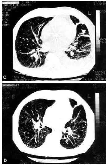
Cont. Fig. 1 - Radiological examinations in case 1 - C) chest computed tomography showing the presence of bilateral pleural effusion, subpleural infiltration (arrow), nodular images, and septal thickenings; D) chest computed tomography after regular use of diuretics showing regression of the pleural effusion and of the pulmonary infiltrations.
The physical examination was almost normal. His heart rate was 70 bpm and his blood pressure was 130/60 mmHg. The results of the ventilatory function test perfor-med in the preceding year showed a forced expiratory volu-me in 1 second to a forced vital capacity ratio (FEV1/ FVC) of 37%. The first measurement of the arterial gases showed a
PO2 of 60.2 mmHg and a PCO2 of 48.3 mmHg. The chest
ra-diographies showed the presence of free pleural effusion. The patient underwent thoracocentesis with a pleural biop-sy, but again the results were unspecific. Pulmonary scinti-graphy was negative for thromboembolic disease.
In May 2000, the patient complained of intolerance to effort for the first time, spontaneous and complete regres-sion of the pleural effuregres-sion then being observed. In January 2001, the patient returned complaining of fatigue. The elec-trocardiogram showed fibrosis in the inferior wall. In the fol-lowing 6 months, dyspnea increased, nocturia and cough appeared, and rales persisted in the left pulmonary base (the patient used to lie in the left lateral decubitus position). In July 2001, the patient had edema of the lower limbs, being hospitalized then for the first time. A new evaluation of the ventilatory function confirmed the presence of severe obs-tructive syndrome. Chest computed tomography identified signs of pulmonary edema in the left inferior lobe (figs. 2B and 2C). Sequential measurements of the arterial gases sho-wed persistence of hypoxemia (fig. 3). Partial and temporary improvements were obtained with the use of diuretics, vaso-dilators, and theophylline. Five echocardiograms were per-formed throughout the evolution period, all of which sho-wed preservation of systolic function with the left ventricu-lar ejection fraction greater than 70%.
In October 2001, the patient was hospitalized again. During this new hospitalization, a negative angiotomogra-phy for thromboembolic pulmonary disease and the marked elevation in the serum levels of the cerebral natriuretic pepti-de – 931 pg/mL (fluorescent immunoassay – Biosite, BNP) – contributed to confirmation of the diagnosis of heart failure. Irreversible deep coma occurred due to cardiopulmonary arrest, and the patient died in December 2001.
Discussion
Respiratory findings conditioned by congestive heart failure have long been well known; in 1833, Hope introduced
the term cardiac asthma2. Currently, the knowledge about
the pulmonary alterations associated with chronic conges-tive heart failure has been the object of extensive reviews2-4,
but a report on the pulmonary manifestations attributed ex-clusively to diastolic heart failure is still lacking.
The respiratory function assessed at rest in patients with chronic congestive heart failure has long been known to be impaired. The alterations are as follows: irregularity between ventilation and perfusion; a reduction in pulmona-ry distensibility; obstruction of the airways; bronchial hy-perreactivity; a reduction in the capacity of pulmonary dif-fusion; and a reduction in the strength and resistance of the respiratory muscles 2,3.
Obstructive ventilatory alterations in patients with congestive heart failure have been reported by several
au-thors according to reviews. Light and George 3 reported
evi-dence of obstructive ventilatory disorder in 53% of their nonsmoking patients with the disease. However, only one of them had the FEV1/FVC ratio below 60% in the best test
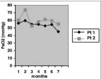
Fig. 3 - Sequential values of PO2 corresponding to the best result obtained during the months the examination was performed, with patients spontaneously breathing room air.
tained during the period observed, indicating that, in that group, most patients had mild obstructive ventilatory
disor-der. Petermann et al 4 also reported a mild obstructive
disor-der in 60 patients with decompensated congestive heart fai-lure, with no significant impairment in gas exchanges. Other authors reported a significant bronchodilating response to the use of salbutamol and ipratropium bromide, indicating that chronic congestive heart failure may result in a mild obstructive ventilatory disorder. Experimental evidence as-sociating peribronchial vascular congestion and increased
airway resistance supports this finding 5.
Caruana et al 6 ruled out the diagnosis of diastolic heart
failure in some of their patients because they had FEV1 results < 70% of the foretold value, which, according to the above re-ported, is unacceptable. These same authors did not try to correlate this finding with antecedents of smoking and other clinical and radiological findings characteristic of primary obstructive pulmonary disease, which should have been considered to correctly establish the diagnosis of chronic obstructive pulmonary disease. Despite the flaws in their methods, these authors were recently cited in 2 studies 7,8, as
providing examples of patients with respiratory disorders in-correctly diagnosed as having diastolic heart failure. In fact, we believe that, at least in the population older than 75 years, the inverse has been more frequent, ie, patients with diastolic heart failure and associated obstructive ventilatory disorder are erroneously labeled as having chronic obstructive pul-monary disease.
Spirography of our 2 patients revealed the presence of severe obstructive ventilatory disorder, although both res-ponded to the use of salbutamol. In their cases, the diagno-sis of chronic obstructive pulmonary disease was ruled out due to the lack of chronic cough, significant smoking, radio-logical signs of emphysema, and historadio-logical alterations compatible with disease in lung biopsy, among others. The unequivocal signs of fluid accumulation in the lungs and pleural space contributed to rule out the diagnosis of the
disease (figs. 1 and 2), because these are not part of the na-tural history of that illness. These data left us with no option other than attributing this severe obstructive disorder to diastolic heart failure corroborating the findings of Light
and George 3, However, in contrast with the mild
abnormali-ties described by these authors, our patients showed seve-re obstruction (FEV1/FVC<50%). This diffeseve-rence can be, in part, attributed to patient's age, since these authors pa-tients aged 75 years or less.
Severe and persistent alterations in gas exchanges have also been observed in our patients. The insaturation of arterial oxygen, which has been referred to as uncommon in chronic congestive heart failure, even when measured du-ring exercise, was present in our patients throughout seve-ral months of follow-up (fig. 3). In the first patient, a PO2 of 53.7 mmHg measured with oxygen supplementation at 5 L/min through a nasal catheter prior to the introduction of diuretics evolved to 56.3 mmHg with room air in 5 days of re-gular use of that medication. However, his best result did not exceed 59.8 mmHg with room air during ambulatory follow-up.
PCO2 was elevated, characterizing the presence of
se-vere hypoventilation during the exacerbations of the disea-se. The substantial improvement observed in the first pa-tient 5 days after the regular introduction of diuretics trans-lated the improvement in the ventilatory function obtained with the removal of the fluid accumulated in the lungs and in the pleural cavity (fig. 1D), with a consequent reduction in the respiratory work overload.
These functional alterations maintained even after confirming the regression of the radiological signs of fluid accumulation in the lungs and in the pleural cavities were attributed to structural alterations induced in the lungs by
chronic hydrostatic pulmonary edema 2. They consisted of
intense proliferation of fibroblasts and histiocytes in the al-veolar septa, resulting in dense deposits of collagen fibers
that Bachofen et al 5 denominated congestive pulmonary
fi-brosis, which may lead to compression of the small adjacent airways. The pulmonary blood vessels of all dimensions undergo important changes, among which are the deposi-tion of dense connective tissue in the intima and adventitia
layers 2,5. An intense proliferation of type II pneumocytes is
still observed, translating into the presence of a reparative activity, as are alveolar macrophages phagocytizing pro-teins and red blood cells. Fragmentation of the capillary basal membrane, when present, correlates with the duration and severity of the disease.
These morphological and functional alterations asso-ciated with chronic hydrostatic pulmonary edema have been intensely investigated aiming at clarifying their possi-ble participation in the pathophysiology of intolerance to
exercise, characterizing chronic congestive heart failure 2,9.
References
1. Sharpe N, Doughty R. Epidemiology of heart failure and ventricular dysfunc-tion. Lancet 1998; 352(suppl I): 3-7.
2. Mancini DM. Pulmonary factors limiting exercise capacity in patients with heart failure. Prog Cardiovasc Dis 1995; 37: 347-70.
3. Light RW, Geoge RB. Serial pulmonary function in patients with heart failure. Arch Intern Med 1983; 143: 429-33.
4. Petermann W, Barth J, Entzian P. Heart failure and airway obstruction. Int J Car-diol 1987; 17: 207-09.
5. Bachofen H, Bachofen M, Weibel ER. Ultrastructural aspects of pulmonary ede-ma. J Thorac Imag 1988; 3: 1-7.
6. Caruana L, Petrie MC, Davie AP, McMurray JJV. Do patients with suspected heart failure and preserved left ventricular systolic function suffer from “diastolic heart failure” or from misdiagnosis? A prospective descriptive study. Br Med J 2000; 321: 215-18.
7. Vasan RS, Benjamin EJ. Diastolic heart failure – No time to relax. N Eng J Med 2001; 344: 56-9.
8. Senni M, Redfield MM. Heart failure with preserved systolic function. A different natural history? J Am Coll Cardiol 2001; 38: 1277-82.
9. Myers J, Salleh A, Buchanan N, et al. Ventilatory mechanisms of exercise intole-rance in chronic heart failure. Am Heart J 1992; 124: 710-19.
The pulmonary manifestations induced by chronic congestive heart failure tend toward irreversibility. The des-cription of extensive fibrosis in the alveolar septa and
pul-monary vessels points towards this direction 5. Other
indi-cations of irreversibility are the persistence of the reduced capacity of pulmonary diffusion after cardiac transplanta-tion and absence of enhancement in the alteratransplanta-tions of pul-monary function after improvement in left ventricular func-tion obtained with the use of carvedilol. These irreversible alterations or alterations of complex reversibility seem to in-fluence the prognosis of the disease, worsening it.
In conclusion, our patient cases suggest that, with an insidious course in elderly individuals, diastolic heart
fai-lure may causepulmonary impairment, whose severity may
apparently overlap that of the hemodynamic disorder cau-sing it. Its slow start, fortunately documented in one of our
patients, seems to be marked by mild clinical and radiolo-gical manifestations, which are spontaneously reversible. Over time, structural and functional alterations end up resul-ting in severe respiratory findings, marked by the presence of obstructive ventilatory syndrome and changes in arterial gases. In this stage, the use of diuretics, although able to revert the characteristic images of hydrostatic pulmonary edema, is not able to revert the functional changes.