www.bjorl.org
Brazilian
Journal
of
OTORHINOLARYNGOLOGY
ORIGINAL
ARTICLE
The
potential
role
of
amlodipine
on
experimentally
induced
bacterial
rhinosinusitis
夽
Arzu
Tatar
a,∗,
Mukadder
Korkmaz
b,
Muhammed
Yayla
c,
Elif
Polat
d,
Hakan
Uslu
e,
Zekai
Halici
f,
Secil
N.
Parlak
daAtaturkUniversity,MedicalFaculty,DepartmentofOtorhinolaryngology,HeadandNeckSurgery,Erzurum,Turkey bOrduUniversity,MedicalFaculty,DepartmentofOtorhinolaryngology,HeadandNeckSurgery,Ordu,Turkey cKafkasUniversity,MedicalFaculty,DepartmentofPharmacology,Kars,Turkey
dAtaturkUniversity,MedicalFaculty,DepartmentofEmbryologyandHistology,Erzurum,Turkey eAtaturkUniversity,MedicalFaculty,DepartmentofMedicalMicrobiology,Erzurum,Turkey fAtaturkUniversity,MedicalFaculty,DepartmentofPharmacology,Erzurum,Turkey
Received16January2016;accepted9August2016 Availableonline28September2016
KEYWORDS
Rhinosinusitis; Non-antibiotic; Amlodipine; Antioxidants; Guineapig
Abstract
Introduction:Antibioticsarefrequentlyusedforthetreatmentofrhinosinusitis.Concernshave beenraised regardingtheadverseeffectsofantibioticsandgrowingresistance. Thelackof developmentofnewantibioticcompoundshasincreasedthenecessityforexplorationof non-antibioticcompoundsthathaveantibacterialactivity.Amlodipineisanon-antibioticcompound withanti-inflammatoryactivity.
Objective: Inthisstudyweaimedtoinvestigatethepotentialroleofamlodipineinthe treat-mentofrhinosinusitisbyevaluatingitseffectsontissueoxidativestatus,mucosalhistologyand inflammation.
Methods:Fifteen adult albino guinea pigs were inoculatedwith Staphylococcus aureus and treatedwithsaline,cefazolinsodium,oramlodipinefor7days.Thecontrolgroupwascomposed by five healthy guinea pigs. Animals were sacrificed after the treatment. Histopathologi-calchanges wereidentifiedusingHematoxylin-Eosin staining.Inflammationwas assessedby PolymorphonuclearLeukocyteinfiltrationdensity.Tissuelevelsofantioxidants(superoxide dis-mutase,glutathione)andanoxidativeproduct(malondialdehyde)weredetermined.
Results:In rhinosinusitis induced animals, amlodipine reduced loss ofcilia, lamina propria edema and collagendeposition compared to placebo (saline) andalthough notsuperior to cefazolin,amlodipinedecreasedpolymorphonuclearleukocyteinfiltration.Thesuperoxide dis-mutaseactivityandglutathionelevelswerereduced,whereasthemalondialdehydelevelswere
夽 Pleasecitethisarticleas:TatarA,KorkmazM,YaylaM,PolatE,UsluH,HaliciZ,etal.Thepotentialroleofamlodipineonexperimentally inducedbacterialrhinosinusitis.BrazJOtorhinolaryngol.2017;83:619---26.
∗Correspondingauthor.
E-mail:[email protected](A.Tatar).
PeerReviewundertheresponsibilityofAssociac¸ãoBrasileiradeOtorrinolaringologiaeCirurgiaCérvico-Facial.
http://dx.doi.org/10.1016/j.bjorl.2016.08.006
increasedsignificantlyinallthree-treatmentgroupscomparedtothecontrolgroup.Amlodipine treatedgroupshowedsignificantlyincreasedsuperoxidedismutaseandglutathionelevelsand decreasedmalondialdehydelevelscomparedtoalltreatmentgroups.
Conclusion:Thenon-antibioticcompoundamlodipinemayhavearole inacuterhinosinusitis treatmentthroughtissueprotective,antioxidantandanti-inflammatorymechanisms.
© 2016 Associac¸˜ao Brasileira de Otorrinolaringologia e Cirurgia C´ervico-Facial. Published by Elsevier Editora Ltda. This is an open access article under the CC BY license (http:// creativecommons.org/licenses/by/4.0/).
PALAVRAS-CHAVE
Rinossinusite; Nãoantibiótico; Amlodipina; Antioxidantes; Cobaia
Opapelpotencialdaamlodipinanarinossinusitebacterianainduzida experimentalmente
Resumo
Introduc¸ão:Antibióticos sãofrequentemente utilizadospara otratamento de rinossinusite. Questõestêmsidolevantadassobreosefeitosadversosdosantibióticosearesistência cres-cente.Afaltadedesenvolvimentodenovoscompostosantibióticosaumentouanecessidadeda explorac¸ãodecompostosnão-antibióticosquepossuematividadeantibacteriana.Aamlodipina éumcompostonão-antibióticocomatividadeanti-inflamatória.
Objetivo:Oobjetivodesseestudofoiinvestigaropapelpotencialdaamlodipinanotratamento darinossinusite,avaliandoseusefeitossobreoestadooxidativodotecido,histologiadamucosa einflamac¸ão.
Método: QuinzecobaiasalbinasadultasforaminoculadascomStaphylococcusaureusetratadas comsoluc¸ãosalina,cefazolina,ouamlodipinadurante7dias.Ogrupocontroleincluiucinco cobaiassaudáveis.Osanimaisforamsacrificadosapósotratamento.Alterac¸õeshistopatológicas foramidentificadasutilizando-seacolorac¸ãodeHematoxilina-Eosina.Ainflamac¸ãofoi avali-ada peladensidade deinfiltrac¸ão deleucócitospolimorfonucleares.Foramdeterminadosos níveisteciduaisdeantioxidantes(superóxidodismutase,glutationa)eumprodutodeoxidac¸ão (malondialdeído).
Resultados: Emanimaiscomrinossinusiteinduzida,aamlodipinareduziu aperdadoscílios, edemadalâminaprópriaedeposic¸ãodecolágenoemcomparac¸ãocomogrupoplacebo(soluc¸ão salina)eemboranãosejasuperioràcefazolina,aamlodipinadiminuiuainfiltrac¸ãodeleucócitos polimorfonucleares.Osníveisdeatividadedasuperóxidodismutaseeglutationaforam reduzi-dos,enquantoosníveisdemalondialdeídoaumentaramsignificativamentenostrêsgruposde tratamentoemcomparac¸ãoao grupocontrole. Ogrupo tratadocomamlodipinaapresentou aumentosignificantedosníveisdesuperóxidodismutaseeglutationaediminuic¸ãodosníveis demalondialdeídoemcomparac¸ãocomtodososgruposdetratamento.
Conclusão:Ocompostonãoantibióticoamlodipinapodeterumpapelnotratamentoda rinoss-inusiteagudaatravésdemecanismosprotetoresdetecido,antioxidanteseanti-inflamatórios. © 2016 Associac¸˜ao Brasileira de Otorrinolaringologia e Cirurgia C´ervico-Facial. Publicado por Elsevier Editora Ltda. Este ´e um artigo Open Access sob uma licenc¸a CC BY (http:// creativecommons.org/licenses/by/4.0/).
Introduction
Rhinosinusitis is characterized by inflammation of the sinus and nasal mucosa. It is one of the most common healthproblems and accountsfor moreoutpatient antibi-otic prescriptions than any other diagnosis.1 Both host and environmental factors play a role in the develop-mentof rhinosinusitis. The pathophysiologycausing acute rhinosinusitis involves a series of changes that lead to obstruction of the sinus ostia, swelling and inflammation ofthemucosa,mucousstasis, impairedmucociliary clear-ance, and microbial infection. The goals of treatment areto reduce inflammation, eradicate infection,improve drainageandaerationofnasalandsinusmucosa,andrestore
mucociliaryfunction.2Commonmedicaltherapiesforacute rhinosinusitis include antibiotics, nasal saline irrigation, decongestants, antihistamines, mucolytics, topicalor sys-temic corticosteroids, and anti-inflammatorydrugs. Little evidence supports the use of decongestants and antihis-tamines,althoughtheymayhelptoreducerhinorrhoeaand nasalcongestion.Nasalirrigationwithsalineisusuallyused inacuterhinosinusitisandmayimprovemucociliary clear-ancebutevidenceislimitedtosupportitsuse.3,4
trialsshowedthatuse ofantibiotics for acute rhinosinusi-tisoffersasmalltherapeutic benefitover placebo,witha correspondingriseintheriskforadverseevents.5
ReactiveOxygenSpecies(ROS)areproducedunder phys-iological conditions and their production and antioxidant activityarebalanced.ExcessiveaccumulationofROScauses lipid and protein peroxidation and can lead to cell dam-age and death. Cells overcome this oxidative stress via
anti-oxidantdefence mechanisms thatinclude Superoxide Dismutase (SOD), Glutathione Peroxidase (GPx), Catalase (CAT),Glutathione(GSH),andperoxiredoxins. Malondialde-hyde(MDA),aproductoflipidperoxidation,isthereforea goodindicator ofcellulardamage.Recent studies showed thatROSactsassecond messengersfor inflammation acti-vationandplayaroleininflammation.6,7
Amlodipine (AML) is a dihydropyridine L-type cal-ciumchannelblockerwithsignificantantibacterialactivity against several gram-positive and gram-negative strains. It is reportedly the most powerful of the cardiovascu-lar drugs with antibacterialactivity.8,9 AML alsohas been reportedtohaveanti-inflammatoryactivity.AMLhasbeen showntodecreaseischemia-reperfusioninjurybyimproving theoxidativestatusinileumischemia-reperfusioninduced rabbits.10
The growthofantibioticresistanceandthelack of dis-covery of new antibiotic compounds have increased the necessityfor explorationofnon-antibioticcompounds like AML that have antibacterial activity. In both acute and chronicsinusitis,inflammationandedemaleadsto obstruc-tionofsinusostiathatfurtherexacerbatesthedisease.We supposethatanti-inflammatoryandantibacterialeffectsof AMLcanbebeneficialinthetreatmentofrhinosinusitis.
Theaimofthepresentstudywastoinvestigatepossible effectsof AML onoxidative status,inflammation, and tis-sueintegrityinananimalmodelofexperimentallyinduced acuterhinosinusitis.
Methods
Animals
Twentyadultalbinoguineapigswithnoevidenceofupper respiratorytractinfectionswereused.Eachanimalweighed 330---370g, and all were obtained from Ataturk Univer-sity’sExperimentalAnimalLaboratoryattheMedicinaland ExperimentalApplicationandResearchCentre.Theanimal experimentsandprocedureswereperformedinaccordance withnationalguidelinesfortheuseandcareoflaboratory animals, and the study was approved by Ataturk Univer-sity’slocalanimalcarecommittee(approvaln◦2014-1/12). The guineapigswere housedin standard plasticcages on sawdust bedding in an air-conditioned room at 22◦±1◦C and a 12:12h dark:light cycle. Standard guinea pig food andtapwaterweregivenadlibitum.Theadaptationtime beforetheexperimentwas2weeks.Theanimalswere ran-domly divided into four groups (five animals per group): one group served asa negative control (Group C; no rhi-nosinusitis induced, only treatedwith Intraperitoneal [IP] salineinjections),onegrouphadrhinosinusitisbutwasonly treated with IP saline injections (Group S; positive con-trol),onegrouphadrhinosinusitisandwastreatedwithoral
amlodipine(GroupSA),andonegrouphadrhinosinusitiswas treatedwithcefazolin(GroupSC).Animalsweresacrificed at7days.
Developmentoftheanimalmodel
Merocel(MedtronicXomed,Jacksonville,FL,USA)wascut withmicroforceps. Microscissors were used to shape and insertaspongeintothenostrils.Allfiveanimalsin theS, SC,andSAgroups,butnottheCgroup,wereadministered ketamine(i.m.,50mg/kg;Ketalar Pfizer,Istanbul,Turkey) and diazepam (i.m., 2mg/kg; Diazem, Deva, Istanbul, Turkey). The nasal dorsum was sterilized with povidone-iodine and gelatin sponges were inserted into the right nasal cavities. The nostrils were then inoculated with
Staphylococcusaureus(0.5mL)usingahypodermicsyringe
(Staphylococcus aureus strain ATCC25923 wassuspended
ata concentration of 900×106CFU [Colony-forming units] permL).At24hafterthebacterialinoculation,theMerocel wasremovedfromthenasalcavities.Purulentnasal secret-ionswereobservedinthenasalcavitiesofanimals.
Guineapigsweretreated twicedailyfor a7 dayswith injection of saline IP in Group S and Group C and cefa-zolinsodium (i.m., 50mg/kg/day) in group SC. Group SA was treated with per oral amlodipine 5mg/kg/day (dis-solvedinsaline,twicedaily)for7days.The animalswere thensacrificedwithalethaldoseofthiopentalsodiumand decapitated.Theexternalnasaldorsumwassterilizedwith povidone-iodineswab,andafterskinelevation,thelateral nasalwalls andmaxillary sinuses were removed fromthe right nasalcavity. The mucosae of thelateral nasal walls werestoredat−80◦Cforbiochemicalanalysis.
Histopathologicalanalysis
Thespecimens of allguineapig wererapidlyfixed in 10% bufferedformalinfor24hforhistologicalexamination.After thefixation, tissue sampleswere routinely processedand embedded in paraffin wax from that 5m-thick sections werecut.Then,sixsectionswereobtainedforeachguinea pigtissueandplacedontopositivelychargedslides.The sec-tionswerestainedwithMayer’sHematoxylin&Eosinafter the deparaffinization and rehydration of them. Later, all sections were examined and photographed under a light photomicroscope(NikonEclipseE600,Japan)for histopatho-logicalexamination.
Semi-quantitativeanalysis
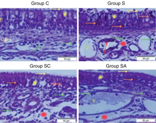
Group C
Group S
Group SC
Group SA
V
V
E
L.P
Figure1 Micrographoftissuesforallgroups.E,Epithelium;LP,LaminaPropria;V,Vasodilatationsinveins,yellowstar;goblet cell,redstar;laminapropriaedema,greenarrow;neutrophiles,redarrow;macrophages,orangearrow;degenerativeepithelium cell,yellowarrow;cilias,H&Estaining.
Stereologicalanalysis
Stereologicalanalysesweremadeusingastereology work-stationconsistingofstereologysoftware(StereoInvestigator version9.0,Microbrightfield,Colchester,VT,USA),a mod-ified light microscope (Leica DM4000 B, Germany) and a motorizedsystemwhichcanmovethesectioninbothxandy
direction.Theopticfractionatorframemethodwasusedto countPolymorphonuclearLeukocytes(PMNLs)inthelamina propriaofsixtissuesectionsforeachguineapig.The sec-tionswereexaminedundera40×LeicaPlanApoobjective (NA=1.40).ThedensityofPMNLswasestimatedaccording todefinedbyKaraandcolleagues.13
Biochemicalanalysis
Lateral nasal wall mucosa samples sized 0.5cm2 were usedforbiochemicalanalysis.SuperoxideDismutase(SOD) activity,14 Glutathione(GSH)15 levels,andMalondialdehyde (MDA)16 levelsweredeterminedinduplicateforeach sam-plesupernatantand standardsat roomtemperature using a modified method and an ELISA reader. The average absorbanceofeach sampleandstandard werecalculated. A standard curve was plotted and the linear relationship equation was obtained from the absorbance of the stan-dards. Linear SOD, GSH, and MDA concentrations were calculatedaccordingtothisequation.TheresultsforSOD, GSH,andMDAinthetissueswereexpressedasU/mgprotein, mmoL/mLprotein,andnmoL/mgprotein,respectively.All dataarepresentedasmean±standarddeviation(SD)based onpermgprotein.Proteinconcentrationsweredetermined bythe Lowry methodusing commercial proteinstandards
(Totalproteinkit-TP0300-1KT;SigmaChemicalCo.,Munich, Germany).
Statisticalanalysis
All data are expressed as group mean±SD and analyzed using SPSS (IBM SPSS Statistics 20.0, IBM Corporation, Somers, NY, USA). Kolmogorov---Smirnov test was applied for analysis of data distribution. The stereological and semi-quantitativehistopathologicaldatawereanalyzed by the nonparametric Kruskal---Wallis test followed by the Mann---Whitney U-test. The parametric test of analysis of variance (ANOVA) and LSDtest for mean separation were usedtoanalyzethebiochemicaldata. Acriticalvaluefor significanceofp<0.05wasusedthroughoutthestudy.
Results
Histopathologicalresults
Group C(Control) ---epithelial structurewasseen normal. Nocilialosswasobserved.Littleinflammatorycell infiltra-tionwasevidentin thelamina propriaandnoedemawas observed(Fig.1;Table1).
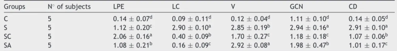
Table1 Thestatisticalresultsofsemi-quantitativeassessmentofhistopathologicalchangesofallgroups.
Groups N◦ofsubjects LPE LC V GCN CD
C 5 0.14±0.07d 0.09±0.11d 0.12±0.04d 1.11±0.10d 0.14±0.05d
S 5 1.12±0.20c 2.90±0.10a 2.85±0.19b 2.94±0.16a 2.91±0.10a
SC 5 2.06±0.16a 0.40±0.09b 1.70±0.27c 1.18±0.18c 1.07±0.06b
SA 5 1.08±0.21b 0.16±0.09c 2.92±0.08a 1.98±0.47b 1.01±0.17c
C,controlgroup;S,rhinosinusitis+salinegroup;SC,rhinosinusitis+cefazolingroup;SA,rhinosinusitis+amlodipinegroup;LPE,lamina propriaedema;LC,lossofcilia;V,vasodilatation;GCN,gobletcellnumber;CD,collagendeposition.
Thedifferentsuperscriptletters(a,b,candd)showthatthevaluesinthesamecolonare statisticallydifferentfromeachother (p<0.05).Valuesgiveninthetablearemean±standarddeviation.UsedtheKruskal---Wallisrangetestoptionandwereconsideredto besignificantatp<0.05(allgroups).
Table2 Thestatisticalresultsofthenumericaldensities ofPolymorphonuclearLeukocyte(PMNL) infiltrationinthe treatmentandcontrolgroups.
Groups PMNL(mean±SD)
C 1.405±0.034d
S 4.105±0.062a
SC 1.796±0.008c
SA 2.514±0.064b
Thedifferentsuperscriptletters(a,b,candd)showthatthe valuesarestatisticallydifferentfromeachother(p<0.05). Val-uesgiveninthetablearemean±standarddeviation.Usedthe Kruskal---Wallisrangetestoptionandwereconsideredtobe sig-nificantatp<0.05(allgroups).
C, control group; S, rhinosinusitis group; SC, rhinosinusi-tis+cefazolingroup;SA,rhinosinusitis+amlodipinegroup.
lamina propria weredistinguished. Differently fromother groups macrophage infiltrations were seen and collagen depositionwasincreasedinthisgroup(Fig.1;Table1).
Group SC(Rhinosinusitis+cefazolinsodium)---nociliary loss wasdetected. The number of goblet cells and colla-gendepositionwerelessthanGroupS.Dilationsofvessels weresimilartoGroupC.Themostlaminapropriaedemawas seeninthisgroup.Degenerativeepitheliumcellswereless thanGroupS. Differentlyfromtheothergroups increased connectivetissuecellswereprominent(Fig.1;Table1).
Group SA (Rhinosinusitis+amlodipine)--- this groupwas similar to Group C than the other experimental groups. Nociliary loss wasdetected. The numbers of gobletcells werelessthanGroupS,collagendepositionandlamina pro-pria edema were similar to Group C. Vasodilatations was increased than theother experimental groups. Degenera-tiveepitheliumcellswerelessthantheotherexperimental groups(Fig.1;Table1).
Stereologicalresults
Statisticalanalysisrevealedthatthereweresignificant dif-ferences between all groups (p<0.05). When the mean numerical densities of PMNLs for all groups compared,it wasseenGroupShadthehighestvalue.TheGroupSAvalue followedGroupS,while theGroup SChadsimilarvalueto GroupC(Table2).
60
50
40
30
20
10
0 C d
a
b
c
S SC
Groups
SOD activity U/ml
SA
Figure2 Superoxidedismutase(SOD)activitiesofthestudy groups.All groups showed a statistical difference fromeach other. Group S presented the lowest SOD activity. C, con-trol group; S, sinusitis group; SC, sinusitis+cefazolin group; SA,sinusitis+amlodipinegroup.Used theanalysisofvariance (ANOVA)andLSDtestsandwereconsideredtobesignificantat
p<0.05(allgroups).Thebarsinthedifferentserialshownby thedifferentletters(a,b,c,andd)arestatisticallydifferent fromeachother.
Biochemicalresults
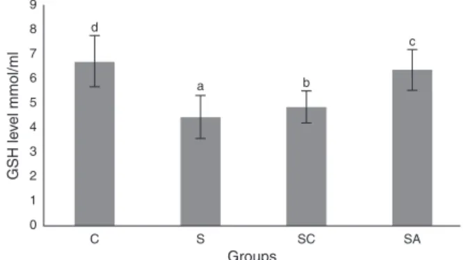
Theactivity of SODandthe levelof GSH, aswell aslipid peroxidation(MDA) levels,were evaluatedin allanimals. The resultsareshown in Figs. 2---4,respectively. The SOD (p<0.004)activityandGSH(p<0.003)levelswerereduced, whereastheMDA (p<0.003)levelswere increased signifi-cantlyinallthree-treatmentgroupscomparedtothecontrol group.Withinthetreatmentgroups,SOD(p<0.004)andGSH (p<0.003)levelswerehighestintheSAgroup,followedby theSCgroup,andwerelowestintheSgroup.MDA(p<0.003) levelswerelowestintheSAgroup,followedbytheSCgroup, andwerehighestintheSgroup.
Discussion
Themostcommonlyreportedpathogensinacutebacterial rhinosinusitis are streptococcus pneumonia, Haemophilus
influenzae,Moraxellacatarrhalis,Streptococcuspyogenes,
and Staphylococcus aureus. The Infectious Diseases
9 8 7 6 5 4 3 2 1 0 C d a b c
S SC SA
Groups
GSH le
vel mmol/ml
Figure3 Glutathione (GSH) levels ofthe study groups.All groupsshowedastatisticaldifferencefromeachother.Group SpresentedthelowestGSHlevel.C,controlgroup;S,sinusitis group;SC,sinusitis+cefazolingroup;SA,sinusitis+amlodipine group. Used the Used the analysis of variance (ANOVA) and LSDtestsandwereconsideredtobesignificantatp<0.05(all groups).The barsinthedifferentserial shownby the differ-entletters(a,b,c,andd)arestatisticallydifferentfromeach other. 3.00 2.50 2.00 1.50 1.00 0.50 0.00
C S SC
Groups MD A le vel nmol/ml a d c b SA
Figure4 Malondialdehyde(MDA)levelsofthestudygroups. All groups showed a statistical difference from each other. Group S presented the highest MDA level. C, controlgroup; S, sinusitis group; SC, sinusitis+cefazolin group; SA, sinus-itis+amlodipinegroup.Usedtheanalysisofvariance(ANOVA) andLSDtestsandwereconsideredtobesignificantatp<0.05 (allgroups).Thebarsinthedifferentserialshownbythe differ-entletters(a,b,c,andd)arestatisticallydifferentfromeach other.
antibiotictherapiesisaworldwideclinicalthreat.Itis essen-tial todevelop novel antibacterialsto modulate thedrug stressresponseofbacteriaandhost defence mechanisms. Althoughourstudyisrepresentativeofacuterhinosinusitis, tissueprotectiveandanti-inflammatoryeffectsofAMLmay havearoleinpreventingtheprolongationofthediseaseand progressiontochronicstate.
Medicinal compounds used for the therapy of non-infectiouspathologyandthathaveantimicrobialproperties are called non-antibiotics. These compounds are divided into two classes: those with direct antibacterial activ-ity are called ‘‘antimicrobial non-antibiotics’’, while the second group consists of ‘‘helper compounds’’ and ‘‘macrophage modulators’’.18---20 The most well-known non-antibiotic compounds are phenothiazines,21 chlor-promazine, thioridazine,22,23 the anti-inflammatory drug diclofenac,24 antihistamines such as promethazine and
diphenhydramine,andcardiovasculardrugssuchas amlodip-ine,dobutamine,lacidipine,nifedipine,andoxyfedrine.25---27 Theexactmechanismofnon-antibioticactionhasnotbeen determinedbuttheyhavebeenproposedtomodifycell per-meabilityandaffectpotassiumandcalciumeffluxpumpsin susceptiblebacteria.Drugresistancealsohasbeenreversed by additionof non-antibiotics suchasphenothiazines and chlorpromazine toan antibiotictowhichthe bacteriaare initiallyresistant.19
Amlodipine has been reported to have the most powerful antibacterialactivity amongcardiovascular non-antibiotics.11 In vitro studies alsosuggested that dihydro-pyridinecalciumantagonistsactasantioxidantsandreduce leukocyte-induced oxidationof low density lipoproteins.28 AMLalsohasbeensuggestedtoimproveendothelial dysfunc-tionindiabetesthroughantioxidantandanti-inflammatory mechanisms.29Ithasbeenshowntoactsynergisticallywith antibiotics andto have in vivoand in vitro antimicrobial activityagainstseveralbacteria.25
Inourstudy,histopathologicalexaminationshowedthat AML improvedlamina propriaedema, collagendeposition, decreasedciliarylosscomparedtosalinetreatedand cefa-zolin treated groups. Amlodipine group showed reduced PMNLinfiltrationandgobletcellnumbercomparedtosaline treated group. However in cefazolin treated group, gob-let cell number and PMNL infiltration was significantly lowerthanothertreatmentgroups.Amlodipinealsocaused increased vasodilatationcomparedtoall groups that may be attributed to the direct vasodilatatory effect of the compound. SOD and GSH are major components of the antioxidant mechanisms in tissue for combating ROS. In thepresentstudy,SODandGSHlevelswerehighestinthe healthycontrolgroupfollowedbytheSA,SC,andSgroups. LevelsofMDA,whichindicatetissuedamagebylipid perox-idation,werelowest inthecontrolgroupandfollowed by theSA, SC,andS groups,in increasingorder.In ourstudy AML better improved the oxidative status than cefazolin sodium. These findingssuggest thatAML improved antiox-idantdefencemechanismsandreducedMDAlevels,thereby preventingthetissuedamagecausedbyROS.
Although host defence mechanisms are basic ways to clearpathogenicmicroorganisms,theseverityofinfectious disease needs to be furthercontrolled by additional pro-tectivemechanismsthatlimittheextentoftissuedamage. Thisisreferredtoasdiseasetolerance,whichisabiologic phenomenonmainlybasedonprotectionofself-tissuesfrom immuneattack.Tissuedamagecontrolisaveryimportant componentofhostdefencemechanismsagainstinfectionas thisenforcesthebarrierfunctionofepithelialcellsto pre-ventpathogenaccesstohosttissuesandlimitsthedisease severitywithoutinterferingwithpathogenload.30
severecasescomparedtocontrolsubjects,withagreater decreaseinseverecases.AstudyconductedbyWesterveld etal.reportedasignificantreductioninreducedglutathione levelsinmucosalsamplestakenfrompatientswithchronic sinusitis compared to healthy controls, but no informa-tionwasprovidedregardingtheseverityofdiseaseintheir study.32Improvementinoxidativestatusofsinonasalmucosa anddecreaseininflammationandtissuedamagemay poten-tially help to treat acute rhinosinusitis and may prevent furtherprogressionofthediseasetochronicstate.
The exact mechanism of antimicrobial action of AML remains to be fully established. However, a reduction in theminimuminhibitoryconcentrationsofantibioticsbyits usecouldmakeitabeneficialauxiliarycompoundfor treat-ment of severe or recalcitrantbacterial infections of the upperrespiratorytract.AMLcanimproveantioxidantstatus andtissueintegrityand decreaseinflammation associated withbacterialrhinosinusitis.Thisdrugmaythereforehave apotential rolein thetreatment of rhinosinusitisby con-trollingtissuedamageandlimitingtheseverityofdisease.
The results of the present study indicated that AML improved SOD and GSH activities while decreasing MDA level,therebyenhancingtheoxidativestatusanddecreasing lipidperoxidationinthesinonasalmucosa.
Conclusion
This study evaluated the effect of AML on inflammation andoxidativestatusofsinonasalmucosaasasingleagent. ThecombinationofAMLandantibioticmayproduceamore dramaticdecreaseinoxidativestressandinflammationby acting synergistically withthe antibiotic.We believe that future studies with different microorganisms and studies thatinvestigatetheeffectofAMLco-administeredwithan antibiotic both in acuteand chronic rhinosinusitis models wouldbebeneficial.
Ethical
approval
Theanimalexperimentsandprocedureswereperformedin accordancewithnationalguidelinesfortheuseandcareof laboratoryanimals,andthestudywasapprovedbyAtaturk University’slocalanimalcarecommittee(approvalnumber: 2014-1/12).
Conflicts
of
interest
Theauthorsdeclarenoconflictsofinterest.
Acknowledgement
WethankProf.HasanTurkez,fromErzurumTechnical Uni-versityforstatisticalanalyses.
References
1.SmithSS,EvansCT,Tan BK,ChandraRK,Smith SB,KernRC. National burden of antibiotic use for adult rhinosinusitis. J AllergyClinImmunol.2013;132:1230---2.
2.Taw MB,Nguyen CT, Wang MB. Complementaryand integra-tive treatments: rhinosinusitis. Otolaryngol Clin North Am. 2013;46:345---66.
3.KasselJC,KingD,SpurlingGKP.Salinenasalirrigationforacute upper respiratory tract infections. Cochrane Database Syst Rev. 2010;3:CD006821, http://dx.doi.org/10.1002/14651858. CD006821.pub2.
4.Peters AT, SpectorS, HsuJ, Hamilos DL,Baroody FM, Chan-dra RK, et al. Diagnosis and management of rhinosinusitis: a practice parameter update. Ann Allergy Asthma Immunol. 2014;113:347---85.
5.FalagasME,GiannopoulouKP,VardakasKZ,DimopoulosG, Kara-georgopoulos DE.Comparison ofantibiotics withplacebo for treatment ofacute sinusitis: a meta-analysis ofrandomized controlledtrials.LancetInfectDis.2008;8:543---52.
6.Harijith A, Ebenezer DL, Natarajan V. Reactive oxygen species at the crossroads of inflammasomes and inflamma-tion. Front Physiol. 2014;5:352, http://dx.doi.org/10.3389/ fphys.2014.00352.
7.Heid ME, Keyel PA, Kamga C, Shiva S, Watkins SC, Salter RD. Mitochondrial reactive oxygen species induces NLRP3-dependentlysosomaldamageandinflammasomeactivation.J Immunol.2013;191:5230---8.
8.Mazumdar K, Asok KumarK, DuttaNK. Potentialrole ofthe cardiovascularnon-antibiotic(helpercompound)amlodipinein thetreatmentofmicrobialinfections:scopeandhopeforthe future.IntJAntimicrobAgents.2010;36:295---302.
9.Kumar KA, Ganguly K, Mazumdar K, Dutta NK, Dastidar SG, ChakrabartyAN.Amlodipine:acardiovasculardrugwith power-fulantimicrobialproperty.ActaMicrobiolPol.2003;52:285---92. 10.CoskunAK,GunalA,HaliciZ,OralA,SeyrekM,BayirY,etal.The effectsofamlodipineonthebiochemicalandhistopathological changesintherabbitileumsubjectedtoischemia-reperfusion. EurasianJMed.2011;43:33---8.
11.SimsekN,BayraktarogluAG,AltunayH.Localizationofinsulin immunpositive cells and histochemical structureof the pan-creasinfalcons(FalcoAnaumanni).AnkaraUnivVetFakDerg. 2009;56:241---7.
12.Mont MA,Elmallah RK, CherianJJ, BanerjeeS, Kapadia BH. Histopathologicalevaluationoftheanteriorcruciateligament inpatientsundergoingprimarytotalkneearthroplasty.J Arthro-plasty.2016;31:284---9.
13.KaraA,AkmanS,DemirciT,ArabacıT.Influenceofalphalipoic acidonepithelialapoptosisinexperimentalperiodontitis.Turk JMedSci.2013;43:747---55.
14.SunY,OberleyLW,LiY.Asimplemethodfor clinicalassayof superoxidedismutase.ClinChem.1998;34:497---500.
15.SedlakJ,LindsayRH.Estimationoftotal,protein-bound,and nonproteinsulfhydrylsgroupsintissuewithEllman’sreagent. AnalBiochem.1968;25:192---205.
16.Ohkawa H,OhishiH, Yagi K. Assayfor lipidperoxidein ani-mal tissues by thiobarbutiric acid reaction. Anal Biochem. 1979;95:351---8.
17.ChowAW,BenningerMS,BrookI,BrozekJL,GoldsteinEJ,Hicks LA,etal.InfectiousDiseasesSocietyofAmerica.IDSAclinical practiceguidelineforacutebacterialrhinosinusitisinchildren andadults.ClinInfectDis.2012;54:e72---112.
18.KristiansenJE,ThomsenVF,MartinsA,ViveirosM,AmaralL. Non-antibioticsreverseresistanceofbacteriatoantibiotics.In Vivo.2010;24:751---4.
19.KristiansenJE,AmaralL.Thepotentialmanagementof resis-tantinfectionswithnon-antibiotics.JAntimicrobChemother. 1997;40:319---27.
21.Chan YY, OngYM, Chua KL. Synergistic interaction between phenothiazinesand antimicrobialagentsagainstBurkholderia pseudomallei.AntimicrobAgentsChemother.2007;51:623---30. 22.DasguptaA,MukherjeeS,ChakiS,DastidarSG, HendricksO, ChristensenJB,etal.Thioridazineprotectsthemousefroma virulentinfectionbySalmonella entericserovarTyphimurium 74.IntJAntimicrobAgents.2010;35:174---6.
23.Amaral L, Molnar J. Mechanisms by which thioridazine in combination withantibiotics cures extensivelydrug-resistant infectionsofpulmonarytuberculosis.InVivo.2014;28:267---71. 24.Dutta NK, Annadurai S, Mazumdar K, Dastidar SG, Kris-tiansenJE,MolnarJ,etal.Potentialmanagementofresistant microbial infections with a novel non-antibiotic: the anti-inflammatorydrugdiclofenacsodium.IntJAntimicrobAgents. 2007;30:242---9.
25.DuttaNK,MazumdarK,DasGuptaA,DastidarSG.Invitroand invivoefficaciesofamlodipineagainstListeriamonocytogenes. EurJClinMicrobiolInfectDis.2009;28:849---53.
26.MazumdarK,GangulyK,KumarKA,DuttaNK,ChakrabartyAN, DastidarSG.Antimicrobialpotentialityofanewnon-antibiotic: the cardiovascular drug oxyfedrine hydrochloride. Microbiol Res.2003;158:259---64.
27.Dasgupta A, Jeyaseeli L, Dutta NK, Mazumdar K, Karak P, Dastidar SG, et al. Studies on the antimicrobial poten-tial of the cardiovascular drug lacidipine. In Vivo. 2007;21: 847---50.
28.Suchanova B, Kostiainen R, Ketola RA. Characterization of the in vitro metabolic profile of amlodipine in rat using liquid chromatography-mass spectrometry. Eur J Pharm Sci. 2008;33:91---9.
29.Toma L, Stancu CS, Sanda GM, Sima AV. Anti-oxidant and anti-inflammatory mechanisms of amlodipine action to improve endothelial cell dysfunction induced byirreversibly glycated LDL. Biochem Biophys Res Commun. 2011;411: 202---7.
30.SoaresMP,GozzelinoR,WeisS.Tissuedamagecontrolindisease tolerance.TrendsImmunol.2014;35:483---94.
31.KassimSK,ElbeigermyM,NasrGF,KhalilR,NassarM.Therole ofinterleukin-12,andtissueantioxidantsinchronicsinusitis. ClinBiochem.2002;35:369---75.