Accuracy of intraoperative consultation in thyroid
nodules: analysis of 2,040 cases performed at Instituto
Nacional de Câncer in 12 years
Acurácia da consulta intraoperatória nos nódulos da tireoide: análise de 2.040 casos
realizados no Instituto Nacional de Câncer em 12 anos
Theresinha C. Fonseca; Ana Lúcia A. Eisenberg; Mário Lúcio C. Araújo Jr.
Instituto Nacional de Câncer José de Alencar Gomes da Silva (Inca).
First submission on 25/06/15; last submission on 11/07/15; accepted for publication on 17/07/15; published on 20/10/15
ABSTRACT
Introduction: The thyroid nodule is the most common clinical manifestation of thyroid diseases, and, among the procedures for its diagnosis, cytological examination is the most used. Due to the high accuracy of cytological examination, the value of intraoperative consultation (IOC) has become a matter of controversy. IOC is one of the most important and dificult procedures that a pathologist adopts, enabling the patient to be diagnosed, treated and staged in the same intervention. Objective: To evaluate the accuracy of IOC in thyroid at Instituto Nacional de Câncer (Inca). Methods: This study analyzed IOC in thyroid held in Inca from January 2001 to December 2012. The IOC diagnosis was compared with the histopathologic diagnosis, considered the gold standard, and was classiied as concordant, discordant and indeterminate. From these data we calculated sensitivity, speciicity and accuracy. Results: Among the 2,040 held IOCs, diagnoses were concordant in 1,770 cases (87%), discordant in 45 (2%) and indeterminate in 225 (11%). Among the 1,770 concordant cases, 1,252 (61%) were true negative, and 518 (25%), true positive. Among the 45 discordant cases, 38 (2%) were false negative, and seven (0,3%), false positive. The test sensitivity was 0.94%, speciicity of 0.99% and accuracy of 0.97%. Conclusion: Our results indicate that IOC in thyroid is a highly accurate test, contributing to the surgical conduct.
Key words: thyroid neoplasms; dimensional measurement accuracy; frozen sections; pathology.
INTRODUCTION
Thyroid nodules are a common finding in the population. Most of these lesions are benign, while 5%-20%
are malignant(1, 2). Cytological examination of material
obtained by fine-needle aspiration (FNA) biopsy is widely employed as a diagnostic method, changing the diagnostic and the therapeutical approaches of thyroid nodules. The main objective of cytological examination is to distinguish cases positive for malignancy (in which surgical resection is indicated) from negative cases(3). Cytological examination
also provides pathologists with valuable information during intraoperative consultation (IOC)(4, 5). Due to the high
accuracy of cytological examination in the thyroid, IOC value became a matter of controversy(6-8). This happens because of
the high diagnostic accuracy of cytological examination in papillary carcinomas and the low diagnostic sensitivity of IOC in follicular lesions(6-9). However, IOC can be very useful, and
pathologists are frequently asked to perform it(4, 10-12).
The objective of IOC is to offer rapid diagnoses that enable surgeons to take immediate decisions during a surgical procedure(13, 14). It may assist in cases of thyroid nodules with
undeterminate cytopathological diagnosis and when IOC is malignant, once it permits an immediate thyroidectomy, avoiding a second surgical procedure(3).
IOC in the thyroid gland is indicated for: 1) conirmation of cytological diagnosis; 2) diagnosis of suspicious nodules cytologically diagnosed as indeterminate; 3) identiication of masses found during surgery; 4) examination of regional lymph nodes to verify the presence of metastasis(3, 4).
In follicular lesions there are diagnostic limitations as to both cytological examination and IOC, but the combination of both analyses increases the diagnostic accuracy of IOC(3, 8, 10, 15, 16).
The objective of this study is to assess the diagnostic accuracy of IOC of thyroid nodules carried out at Instituto Nacional de Câncer (Inca).
METHODS
This is a retrospective descriptive study of IOC in lesions of the thyroid gland performed at Inca between January 2001 and December 2012. After approval by the research ethics committee of Inca (CAE: 12435613.0.0000.5274), a survey was conducted in the computerized system of the Division of Pathology (Dipat); 7,888 thyroid cases were reported in this period. IOCs were performed in 2,040 (25.86%) individuals.
The cases were deined as:
1) IOC diagnosis (IOCD) – diagnosis established during IOC; 2) histopathological diagnosis (HPD) – inal HPD;
3) gold standard – standard exam used for comparison with the exam to be tested, aiming at assessing its exactness. In this study HPD was the gold standard;
4) positive concordant cases – positive HPD and IOCD (true positive [TP]);
5) negative concordant cases – negative HPD and IOCD (true negative [TN]);
6) positive discordant cases – positive HPD and negative IOCD (false negative [FN]);
7) negative discordant cases – negative HPD and positive IOCD (false positive [FP]);
8) indeterminate cases (IC) – cases in which IOCD was not conclusive;
9) sensitivity – capacity of the exam to offer correct positive (TP)
diagnoses;
10) speciicity – capacity of the exam to offer correct negative (TN) diagnoses;
11) positive predictive value (PPV) – probability of the positive cases to be actually positive;
12) negative predictive value (NPV) – probability of the negative cases to be actually negative;
13) accuracy – ability of an exam to obtain results equal to those
obtained by the gold standard.
The histopathological classiication of the World Health Organization (WHO) is used as a routine for thyroid diagnoses at Dipat/Inca(2). The cases with descriptive HPD, thyroid tissue free
from neoplasia and cysts were included in the group “absence of malignancy”.
Statistical analyses were described through absolute frequencies and percentages for qualitative variables, and mean (standard deviations) and median for quantitative variables. Data sets were organized by univariate, bivariate and contingency tables. For proportional comparison between sexes, the ratio male:female was used. Sensitivity, speciicity, PPV, NPV and accuracy were used to compare diagnostic methods, that is, IOCD with HPD (gold standard). Statistical decisions were taken at the signiicance level a = 0.05 (5%). In order to calculate sensitivity, speciicity, PPV, NPV, and accuracy, a double-entry table was used for the results of IOCD and HPD (gold standard) tests, considering: A) positive concordant cases (TP); D) negative concordant cases (TN); B) positive discordant cases (FP); C) negative discordant cases (FN); AC) total of HPD positive cases; BD) total of HPD negative cases; AB) total of IOC positive cases; CD) total of IOC negative cases; ABCD) total of cases. Based on these pieces of information, sensitivity [= A/(A + C) × 100]; speciicity [= D/(B + D) × 100]; PPV [= A/(A + B) × 100]; NPV [= D/(C + D) × 100]; and accuracy [= (A + D)/(A + B + C + D) × 100] of IOCD were calculated.
A search in the literature was conducted in the US National Library of Medicine/National Institutes of Health (PubMed), Cochrane database, Medline, and Scientiic Electronic Library Online (SciELO). The key words used for the research were: intraoperative consultation, accuracy of intraoperative consultation, sensitivity of intraoperative consultation, speciicity of intraoperative consultation, thyroid tumors.
RESULTS
Among the 2,040 IOCs, diagnoses were concordant in 1,769 cases (87%), discordant in 46 (2%), and indeterminate in 225 (11%). Among the 1,769 concordant cases, 1,251 were TN and 518 were TP. Among the 46 discordant cases, 38 were FN and eight were FP (Tables 1 and 2).
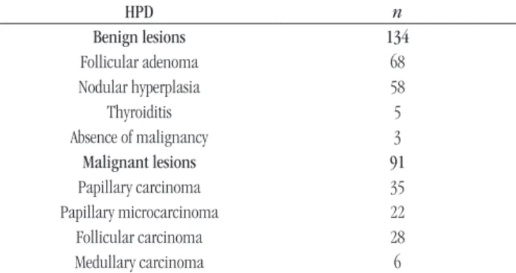
Among the 225 cases with indeterminate IOCD, 134 (60%) had HPD of benign lesion, and 91 (40%), of malignant lesion
(Table 3).
TABLE 5 – Thyroid neoplasms at Inca between January 2001 and December 2012. HPD (n = 2,040)
HPD Total (%)
Benign 1,393 (68)
Nodular hyperplasia 965 (43.3) Absence of malignancy 180 (8.8)
Thyroiditis 138 (6.2)
Follicular adenoma 111 (5.4)
Malignant 647 (32)
Papillary carcinoma 569 (27.89) Follicular carcinoma 51 (2.5) Medullary carcinoma 20 (0.98) Poorly differentiated carcinoma 4 (0.19)
Anaplastic carcinoma 3 (0.14)
Total 2,040 (100)
Inca: Instituto Nacional de Câncer; HPD: histopathological diagnosis.
TABLE 1 – IOC of thyroid lesions at Inca from January 2001 to December 2012. FP (n = 8)
HPD n IOCD n
Nodular hyperplasia 5 Papillary carcinoma 5 Thyroiditis 3 Papillary carcinoma 3 IOC: intraoperative consultation; Inca: Instituto Nacional de Câncer; FP: false positive; HPD: histopathological diagnosis; IOCD: diagnosis established during IOC.
TABLE 2 – IOC of thyroid lesions at Inca from January 2001 to December 2012. FN (n = 38)
HPD n IOCD n
Papillary carcinoma 21
Nodular hyperplasia 16 Absence of malignancy 4
Follicular adenoma 1
Minimally invasive follicular
carcinoma 11
Follicular adenoma 8 Nodular hyperplasia 2 Absence of malignancy 1 Invasive follicular carcinoma 1 Follicular adenoma 1
Medullary carcinoma 4 Nodular hyperplasia 3 Absence of malignancy 1 Anaplastic carcinoma 1 Absence of malignancy 1 IOC: intraoperative consultation; Inca: Instituto Nacional de Câncer; FN: false negative; HPD: histopathological diagnosis; IOCD: diagnosis established during IOC.
TABLE 3 – IOC of thyroid lesions at Inca from January 2001 to December 2012. Indeterminate cases with HPD of malignant neoplasm (n = 225)
HPD n
Benign lesions 134
Follicular adenoma 68
Nodular hyperplasia 58
Thyroiditis 5
Absence of malignancy 3
Malignant lesions 91
Papillary carcinoma 35
Papillary microcarcinoma 22
Follicular carcinoma 28
Medullary carcinoma 6
IOC: intraoperative consultation; Inca: Instituto Nacional de Câncer; HPD: histopathological diagnosis.
TABLE 4 – Relationship between IOCD and HPD in cases of IOC performed in thyroid lesions at Inca from January 2001 to December 2012 (n = 1,815)
HPD
Positive Negative Total
IOCD
Positive A = 518 B = 8 A + B = 526 Negative C = 38 D = 1,251 C + D = 1,289
Total A + C = 556 B + D = 1,259 A + B + C + D = 1,815 IOC: intraoperative consultation; IOCD: diagnosis established during IOC; HPD: histopathological diagnosis; Inca: Instituto Nacional de Câncer; A: true positive cases; D: true negative cases; B: false positive cases; C: false negative cases; A + C: total of HPD positive cases; B + D: total of HPD negative cases; A + B: total of IOC positive cases; C + D: total of IOC negative cases; A + B + C + D: total of cases.
Among the 2,040 IOCs performed in the thyroid gland, HPD were benign in 1,393 (68%) cases, and malignant in 647 (32%)
(Table 5).
DISCUSSION
Most thyroid nodules are benign lesions, and those considered potentially malignant both clinically and by imaging exams must be assessed so that a precise diagnosis is established. Clinical history, physical examination, and ultrasonography are important to determine whether the patient presents high risk to develop a
malignant lesion(1, 4).
Cytological examination of the material obtained by FNA is the irst procedure for the diagnosis of thyroid gland lesions(4-8, 11).
In the cases with diagnosis reported as positive for malignancy, the surgical procedure can be planned previously(4-8, 11).
Although the cytological examination has caused a decrease in the number of IOCs, it has limitations. IOC can be useful in the cases cytologically diagnosed as unsatisfactory, suggestive of malignancy and indeterminate. Another indication of IOC is the analysis of nodules found during the surgery, and the evaluation of surgical margins(3, 4, 10, 11).
False positive cases
The eight FP cases had IOCD of papillary carcinoma, of which ive had HPD of nodular hyperplasia, and three, of thyroiditis. Both nodular hyperplasia and papillary carcinoma may present papillary formations, what may cause a wrong interpretation of IOC. The papillary formations of nodular hyperplasia are associated with dilated follicles, and nuclear alterations typical of papillary carcinoma are not observed(8, 17-19). In lymphocytic thyroiditis
and Hashimoto’s thyroiditis, epithelial cells may present nuclear alterations, which mimic those found in papillary carcinomas. The presence of lymphocytic iniltration and the absence of nuclear alterations typical of papillary carcinoma suggest the diagnosis of
thyroiditis(3, 19). In our work, FP diagnoses amounted to 0.3% of all
cases, similarly to data of the consulted literature(8, 17, 18).
False negative cases
Among the 38 FN cases, HPD was papillary carcinoma in 21, minimally invasive follicular carcinoma in 11, medullary carcinoma in four, anaplastic carcinoma in one, and invasive follicular carcinoma in one.
The differential diagnosis between papillary carcinoma, nodular hyperplasia and follicular adenoma may be dificult at IOC. The presence of nuclear alterations typical of papillary carcinoma is fundamental for differential diagnosis(2). In our
study, among the 21 papillary carcinomas that had IOCD of FN, 16 had IOCD of nodular hyperplasia, four of absence of malignancy, and one of follicular adenoma.
IOCD of minimally invasive follicular carcinoma is very dificult due to the dificulty of inding capsular or vascular invasion in the frozen sections; in these cases, the lesion may be interpreted as adenoma or nodular hyperplasia. In our study, this problem occurred in 11 cases(3).
Invasive follicular carcinoma may be identiied grossly and microscopically at IOC, but in our work one case was diagnosed as adenoma at IOC(3).
The medullary carcinoma is generally recognized as a carcinoma or a malignant neoplasm at IOC, due to its unusual cytological and histological aspects. However, well-differentiated medullary carcinomas may be mistaken for follicular neoplasms or nodular hyperplasia(3). In our study, three cases of medullary
carcinoma were diagnosed as nodular hyperplasia, and one, as absence of malignancy.
The anaplastic carcinoma is a high-grade neoplasm that can be identiied at IOC. In our study, one case of anaplastic carcinoma had IOCD of absence of neoplasia due to sampling problems.
The FN rate in the consulted literature ranged from 0.4% to 9%, and the rate found in this work was 2%(8, 17, 18).
Indeterminate cases
In our analysis, among the 225 cases with undeterminate IOCD, HPD was benign lesion in 134 cases (60%), and malignant lesion in 91 (40%). In the cases with indeterminate IOCD and HPD of benign lesion, in general there is no harm to the patient. However, when HPD is malignant lesion, an additional treatment may be necessary(3).
In our study, among the cases with HPD of benign lesion, 68 were diagnosed as follicular adenoma; 58, as nodular hyperplasia; ive, as thyroiditis; one, as absence of malignancy. The distinction between follicular adenoma and nodular hyperplasia in a single node may be a problem at IOC. Grossly, nodular hyperplasia is in general nodular, partially encapsulated and with degenerative alterations. Microscopically, nodular hyperplasia exhibits scant follicular cells, oncocytes and abundant colloid.
Follicular adenomas are typically single and microscopically present high cellularity with numerous microfollicles and little colloid. As previously described, for the diagnosis of follicular carcinoma, demonstration of capsular and/or vascular invasion is necessary, but they are rarely found during IOC(20, 21). These
dificulties support those who argue against IOC in lesions with pre-operative cytological diagnosis of follicular neoplasia. Surgeons that request IOC in follicular lesions must know that these lesions have chances to be given indeterminate IOCD(3).
In the group of cases with indeterminate IOCD and whose HPD was malignant, 35 had HPD of papillary carcinoma; 22, of papillary microcarcinoma; 28, of follicular carcinoma; six, of medullary carcinoma. In the consulted literature, indeterminate IOCD ranged from 0% to 30%. In this research, the rate was 5%(8, 17, 18).
We found 93.4% sensitivity, 99.5% speciicity, and 98% accuracy, data consistent with the literature(7, 9, 10, 17, 22-27). In our
study, among the 2,040 IOCs performed in thyroid, HPD was benign in 1,393 cases (68%) and malignant in 647 (32%). Among the benign lesions, nodular hyperplasia was the most common; among malignant lesions, papillary carcinoma, data compatible with the literature(3).
CONCLUSION
REFERENCES
1. Dean DS, Gharib H. Epidemiology of thyroid nodules. Best Pract Clin Endocrinol Metab. 2008; 22(6): 901-11.
2. DeLellis R, Lloyd R, Heitz P, Eng C, editors. World Health Organization classiication of tumors. Pathology and genetics of tumours of endocrine organs. Lyon: IARCPress; 2010.
3. Taxy JB, Husain AN, Montag AG. Biopsy interpretation series. Biopsy interpretation: the frozen section. 2 ed. LWW; 2014. 426 p.
4. Anton RC, Wheeler TM. Frozen section of thyroid and parathyroid specimens. Arch Pathol Lab Med. 2005; 129: 1575-84.
5. Muratli A, Erdogan N, Sevim S, Unal I, Akyuz S. Diagnostic eficacy and importance of ine-needle aspiration cytology of thyroid nodules. J Cytol. 2014; 31(2): 73-8.
6. Cooper DS, Doherty GM, Haugen BR, et al. Revised thyroid association management guidelines for patients with thyroid nodules and differentiated thyroid cancer. Thyroid. 2009; 19(11): 1167-214. 7. Cetin B, Aslan S, Hatiboglu C, et al. Frozen section in thyroid surgery. Is it a necessity? Cancer J Surg. 2004; 47(1): 29-33.
8. Huber GF, Dziegielewski P, Matthews W, et al. Intraoperative frozen-section analysis for thyroid nodules. A step toward clarity or confusion? Arch Otolaryngol Head Neck Surg. 2007; 133(9): 874-81.
9. Miller MC, Rubin CJ, Cunnane M, et al. Intraoperative pathologic examination: cost effectiveness and clinical value in patients with cytologic diagnosis of cellular follicular thyroid lesion. Thyroid. 2007; 17(6): 557-65.
10. Almeida JPA, Couto Netto SD, Rocha RP, Pfuetzenreitter EG, Dedivitis, RA. The role of intraoperative frozen section for thyroid nodules. Braz J Otorhinolaryngol. 2009; 75(2): 256-60.
RESUMO
Introdução: O nódulo tireoidiano é a manifestação clínica mais comum de doenças da tireoide, e entre os procedimentos para o seu diagnóstico, o exame citológico é o mais usado devido à alta acurácia. Como consequência, o valor da consulta intraoperatória (CIO) tornou-se motivo de controvérsia. A CIO é um dos mais importantes e difíceis procedimentos que o patologista utiliza. Ela possibilita que o paciente seja diagnosticado, tratado e estadiado em uma mesma intervenção. Objetivo: O objetivo deste trabalho é avaliar a acurácia da CIO em tireoide no Instituto Nacional de Câncer (Inca). Métodos: Este estudo analisou as CIOs em tireoide realizadas no Inca no período de janeiro de 2001 a dezembro de 2012. O diagnóstico delas foi comparado com o diagnóstico histopatológico, considerado o padrão ouro, e foi classificado em: concordante, discordante e indeterminado. A partir desses dados, foram calculadas a sensibilidade, a especificidade e a acurácia. Resultados e conclusão: Das 2.040 CIOs realizadas, os diagnósticos foram concordantes em 1.770 casos (87%), discordantes em 45 (2%) e indeterminados em 225 (11%). Dos 1.770 casos concordantes, 1.252 (61%) foram verdadeiros negativos e 518 (25%), verdadeiros positivos. Dos 45 casos discordantes, 38 (2%) foram falsos negativos e sete, falsos positivos (0,3%). A sensibilidade do exame foi de 0,94%; a especificidade, de 0,99%; e a acurácia, de 0,97%. Nossos resultados indicam que a CIO em tireoide é um exame de alta acurácia, o que contribui para a conduta cirúrgica.
Unitermos: doenças da glândula tireoide; congelamento; patologia; precisão da medição dimensional.
11. Basolo F, Ugolini C, Proietti A, Iacconi P, Berti P, Miccoli P. Role of frozen section associated with intraoperative cytology in comparison to FNA and FS alone in the management of thyroid nodules. Eur J Surg Oncol. 2007; 33(6): 769-75.
12. Nixon IJ, Shaha AR, Patel SG. Surgical diagnosis: frozen section and the extent of surgery. Otolaryngol Clin North Am. 2014; 47(4): 519-28. 13. Novis DA, Gephardt GN, Zarbo RJ; College of American Pathologists. Interinstitutional comparison of frozen section consultation in small hospitals: a College of American Pathologists Q-Probes study of 18,532 frozen section consultation diagnoses in 233 small hospitals. Arch Pathol Lab Med. 1996; 120(12): 1087-93.
14. Gephardt GN, Zarbo RJ. Interinstitutional comparison of frozen section consultations. A College of American Pathologists Q-probes study of 90,538 cases in 461 institutions. Arch Pathol Lab Med. 1996; 120(9): 804-9.
15. Liu FH, Liou MJ, Hsueh C, Chao TC, Lin JD. Thyroid follicular neoplasm: analysis by ine needle aspiration cytology, frozen section, and histopathology. Diag Cytopathol. 2009; 38(11): 801-5.
16. Winter C, Graem N. Accuracy of frozen section diagnosis: a retrospective analysis of 4785 cases. APMIS. 2010; 119: 259-62.
17. Farah-Klibi F, Blel A, Neji O, Ferjaoui M, Ben Jilani S, Zermani R. The value of intraoperative frozen section in surgical management of thyroid nodules: report of 409 cases. Ann Pathol. 2009; 29(2): 80-5.
18. Osamura RY, Hunt JL. Current practices in performing frozen sections for thyroid and parathyroid pathology. Virchows Arch. 2008; 453: 433-40. 19. Rosai J, Kuhn E, Carcangiu ML. Pitfalls in thyroid tumour pathology. Histopathology. 2006; 49: 107-20.
21. Leteurtre E, Leroy X, Pattou F, et al. Why do frozen sections have limited value in encapsulated or minimally invasive follicular carcinoma of the thyroid? Am J Clin Pathol. 2001; 115(3): 370-4.
22. Chang HY, Lin JD, Chen JF, et al. Correlation of ine needle aspiration cytology and frozen section biopsies in the diagnosis of thyroid nodules. J Clin Pathol. 1997; 50: 1005-9.
23. Chao TC, Lin JD, Chen MF. Surgical treatment of Hurthle cell tumors of the thyroid. World J Surg. 2005; 29(2): 164-8.
24. Giuliani D, Willemsen P, Verhelst J, Kockx M, Vanderveken M. Frozen section in thyroid surgery. Acta Chir Belg. 2006; 106(2): 199-201.
25. Lee TI, Yang HJ, Lin SY, et al. The accuracy of ine-needle aspiration biopsy and frozen section in patients with thyroid cancer. Thyroid. 2002; 12(7): 619-26.
26. Lumachi F, Borsato S, Tregnaghi A, et al. Accuracy of fine-needle aspiration cytology and frozen-section examination in patients with thyroid cancer. Biomed Pharmacother. 2004; 58(1): 56-60.
27. Rios ZA, Rodriguez GJM, Sola PJ, Soria CT, Galindo FPJ, Parrilla PP. Utility of frozen-section examination for diagnosis of malignancy associated with multinodular goiter. Thyroid. 2004; 14(8): 600-4.
MAILING ADDRESS
Theresinha C. Fonseca