On the Role of Water in the Protein Activity
L. Degr`eve, G.H. Brancaleoni, C.A. Fuzo, M.R. Lourenzoni,
F.M. Mazz´e, A.M. Namba, and D.S. Vieira
Grupo de Simulac¸˜ao Molecular, Departamento de Qu´ımica,
Faculdade de Filosofia, Ciˆencias e Letras de Ribeir˜ao Preto, Universidade de S˜ao Paulo
14040-350 Ribeir˜ao Preto (S.P.), Brazil
Received on 05 October, 2003.
The role of the supporting medium of water molecules in some protein activities is examined under different aspects as in the cases of a monomeric peptide, thebasic fibroblast growth factor, of a dimeric peptide, the human neutrophil peptide 3, of a peptide that acts in non-aqueous environment, thegramicidin Adimer, of a water molecule present in the binding of a co-factor in aphospholipasepeptide, and under the general point of view of the hydrophilic/hydrophobic properties described by a hydropathy scale. These examples illustrate the importance of water in the hydrogen bond formation that is, of main importance in keeping the peptide structures that cannot be defined without the water contributions. The conclusions confirm that living systems are like they are because water is an outstanding and abundant molecule present everywhere in living matter.
1
Introduction
Almost all mechanisms occuring in the cells depend on proteins [1]-[6] that constitute most of biological macro-molecules providing large variety of functions[2]-[4]. Their importance can be emphazised by noting that the ge-netic information is fundamentally expressed as protein molecules[5]-[7]. Specific DNA segments contain the ge-netic information on the peptide amino acids sequence. Thousands of different proteins in the cells, precisely cod-ified by the genes, realize specific functions. The individ-uality of the proteins is directly related with their three-dimensional structure that provides the ideal conditions for realizing correctly their functions[8]. However, in spite of the importance of the proteins in the celular life, the role of the supporting medium, in particular of the solvent molecules,i.e.the water molecules, is equally important be-cause they constitute the most part of the cell, about 70%, and because the contribution of the water molecules is essen-tial in the peptide activities[9]. The water molecules[10, 11], beyond the small mass, present high multipolar moments that contribute to the formation of hydrogen bonds, HB. Consequently, the water molecules perform an essential structural role in the organization and activity of the bio-logical medium. Many of the protein activities depend on the protein stability, on associations with other proteins or ligands while the catalytic activity depend on the structure, on thermodynamic and dynamic properties, properties that are deeply influenced by the solvent.
All the most important biological molecules like
pep-tides, saccharides, nucleic acids have the common feature that they contain hydrogen-bonding functional groups[8]. The hydrogen bonds have some interesting features like:(i)
low enthalpy formation, about 20% of the chemical bond enthalpy;(ii)total unspecificity;(iii)the association, or dis-sociation, of molecules through HB are fast enough to per-mit to check their formation and to correct misformations. The large number of HB, that are generally present between water and biological materials, result in the high specificity of the HB networks which include intra and intermolecular bonds.
2
Material
2.1
The
basic fibroblast growth factor
The function of the growth factors peptides is to induce their specific target cells to grow and/or to make a differentia-tion. The first step in their activity is to bind to heparin or to the heparan sulfate chains of proteoglycans located on the cell-surface receptor[2]-[4]. Thebasic fibroblast growth factor(bFGForFGF-2) is a mitogenic, neurotrophic, and angiogenic polypeptide that is a member of a protein fam-ily containing until the present nine known different species (constituting theFGFs family) that interact with three, re-lated but distinct, receptors[13, 14]. Several members of the
FGFs family are oncogenes: the angiogenic properties of
bFGFsuggest an involvement in tumor growth and cancer. The basic fibroblast growth factoralso has wound healing properties that made it an attractive candidate as a thera-peutic drug. Even though the three-dimensional structure of
bFGFhas been recently elucidated relatively little is known about its structure-function relationship. Two regions in the primary structure of the 155 amino acid bFGFhave been proposed to be involved in the receptor-binding and mito-genic activity of this factor. These regions correspond to residues 33-77 and 115-124[6, 12, 16].
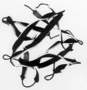
The secundary structure ofbFGFcomprises twelve an-tiparallelβ strands arranged in a pattern with approximate threefold internal symmetry[6, 12, 16]. The strands are numbered sequentially from the amino terminus. The β strands 1, 4, 5, 8, 9 and 12 form a six stranded antiparal-lel β barrel that is closed at one end by β sheet interac-tions involving strands 2, 3, 6, 7, 10 and 11. The three-dimensional structure of the basic fibroblast growth factor
is displayed in Fig. 1.
Figure 1. The tertiary structure of thebasic fibroblast growth fac-tor.
2.2
The
defensin HNP-3
Antibacterial cationic peptides are important components of the innate defenses of all species of life[17]. Most of them interact with the bacterial membranes by disrupting the or-der of the phospholipid bilayer, causing loss of membrane integrity [18]-[20]. Two main mechanisms have been sug-gested for peptide permeation of the bacterial membrane: (i) the barrel-stave mechanism, where bundles of peptides form transmembrane pores through the bacterial membrane[21] and (ii) the carpet-like mechanism, where membrane de-struction/solubilization occurs via parallel binding of the peptides to the bacterial membrane, covering the membrane in a carpet like manner[22].
The human neutrophil peptide 3,HNP-3, is a member of theα-defensinfamily, one of the most common classes of cationic antimicrobial peptides[21, 23]-[26]. The primary structure ofHNP-3is displayed in the Fig. 2. The common structural feature of the α-defensin family is a hydrogen-bonded pair of antiparallelβ−strands (strands 2 and 3) con-nected by a short turn to form aβ−hairpin. The crystal structure ofHNP-3[26] shows a dimeric structure formed by two monomers connected by theirβ− hairpin regions through HB. The resulting quaternary structure is a six-strandedβ−sheet (Fig. 3) stabilized by HB, six disulfide bridges and hydrophobic contacts. Moreover, the crystallo-graphic analysis shows ordered internal water molecules sta-bilizing the peptide structure[26]. The tertiary structures of the two monomers, monomer 1 and monomer 2, are slightly different.
D C Y C R I P A C I A G E R R Y G T C I Y Q G R L W A F C C
β1 β2 β3
5 10 15 20 25 30
Figure 2. Primary structure of theHNP-3. The disulfide bridges are represented by the solid lines.
2.3
The
gramicidin A
The dimergramicidinA (GA) is a 30 amino acid residues peptide with antibiotic properties that is produced by
Bacil-lus brevis. GA is active against Gram-positive bacterial
membranes[27, 28] by forming one of the best-characterized ion channels. The GA channel increases the membrane per-meability to water molecules and to monovalent ions[2]-[4]. It has been used as a model for ion channels in numerous experimental and theoretical studies. It has been accepted that GA is a N-terminal to N-terminal right handedβ-helix dimer during the channel formation[4, 29, 30]. GA is con-stituted by an amino acid residues sequence where D and L configurations are alternated. The N-terminus is formylated and the C-terminus is bound to an ethanolamine group. The secundary structure is depicted in the Fig. 4. The GA bio-logical activity is intimately related to its structure so that it may be influenced by different factors such as the nature of the solvent[8, 31]-[34]. Consequently the importance of the water molecules in the maintenance of the structure of the GA in the active form is a main object of study.
Figure 4. The secundary structure ofgramicidin A.
2.4
Phospholipase
s A2
Phospholipases A2 (PLA2 EC 3.1.1.4) catalyze the hydroly-sis of the sn-2 acyl bonds of sn-3 phospholipids. Snake ven-oms constitute a rich source of PLA2 with several structural and functional diversity[35]. The classification of venom PLA2s into two classes is based on the basis of primary structure[36]. Classes I/II snake venom PLA2s display sev-eral pharmacological properties[37]. The structural basis of the catalytic function involves the highly conserved active site residues His48, Asp49, Tyr52 and Asp99, in which the essential Ca2+co-factor is bound to Asp49 and to carbonyl
main chain oxygen atoms of the aptly named calcium bind-ing loop. A schematic diagram of the interaction between
phospholipase A2 and a phospholipid is observed in Fig.
5[38]. The mechanism of action ofphospholipase A2 in-volves a His48 and Asp99 catalytic diad that activates one water molecule (into the dashed circle in Fig. 5) for nucle-ophilic attack on the ester while Ca2+stabilizes the
oxyan-ion transitoxyan-ion state[38]. A sub-family of catalytically inac-tive PLA2s has been characterized with Asp49 substituted
by Lys49. The presence of the NH+3 group of Lys49 makes impossible the binding of the Ca2+ co-factor resulting in
the lack of catalytic activity. However, despite the lack of catalytic activity, these Lys49-PLA2 homologues retain a Ca2+ independent membrane damaging activity. In spite
of the lack of catalytic activity be related in many works, some papers refute this fact and report a residual catalytic activity[39, 40]. Furthermore, as such as in the case of other snake venoms, PLA2s isolated from snake venom, Lys49-PLA2s, also processes combined myotoxic and cytolytic pharmacological activities.
Figure 5. Schematic diagram of a productive interaction between phospholipase A2and a phosphatidylethanolamine.
Figure 5
Figure 6. Ribbon representation of the Lys49-PLA2, in which are indicated the different secondary structures and the catalytic site (I).
sterically hinders the binding of the essential co-factor Ca2+
[35]. The C-terminal region and a single double-strandedβ -sheet are linked by disulphide bridges. Several studies have identified amino acid clusters located in the active site and lipid substrate binding regions[35] however the mechanisms of myotoxic and cytolytic activities of Lys49PLA2s are still unknown.MyotoxinII, (BaspMT-II), isolated fromBothrops Asper, is a basic dimeric Lys49-PLA2s. Molecular simula-tion was employed to study the hydrasimula-tion structure and the structural changes in monomer of the BaspMT-II.
2.5
An amphipathy scale
Considering the fundamental importance in the knowledge of the structure of proteins to understand their functions, notable efforts have been dedicated to predict and to eluci-date the three-dimensional shape of amino acids sequences. Some works were directed to the analysis of the pro-tein’s hydrophobicity using amphipathy scales that show the relative hydrophobicity of amino acids. In these re-searches, different pathways were pursued based on: the transfer free energies[41]-[43], the accessible amino acid surfaces in proteins[44], statistical techniques using soluble proteins[45], the interactions between amino acids[46], the modification of existent experience based data scales[47], statistical methods using membrane proteins[48], etc. In some of the above cited cases, different procedures were used: for example, the determination of the free energy of transfer was determined using water and different solvents. The transfer free energy was obtained from the equilibrium between water solutions and: ethanol or dioxane[41], N -methylacetamine[42] and n-octanol or palmitoyloleoylphos-phocholine solutions[43].
It can be noted from the analysis of the amphipathy scales that the hydrophobicities of the amino acids are lack-ing of uniformity because the residue hydrophobicity is too much correlated with the different kind of methods used in its determination. In the light of the general observation that the interior of soluble proteins is predominantly com-posed by hydrophobic amino acids, while the hydrophilic side chains are preferentially located on the external surface of the proteins where they are free to interact with the sol-vent molecules, the present example shows how it is possi-ble to elaborate a new amphipathy scale that determinesin situthe hydrophobicity of amino acids in water by molec-ular simulation without using any arbitrary equilibrium. It is highly probable that molecular simulation is to be consid-ered as the good method to weigh correctly the hydropho-bicity of the amino acids because this technique is able “to reproduce” the environment protein-water as so as charac-teristics that depend on the physical and chemical properties of water[49, 50, 51, 52].
3
Method
All the results were obtained from molecular simulation us-ing the molecular dynamics method[53]-[55]. The simu-lated systems consist of the solute molecule(s) immersed in water molecules or in a mixed medium water-carbon tetra-chloride in the GA case. The solvent concentrations were adjusted to match, in the pure solvent regions, the experi-mental solvent density. In the aqueous phases, ions were introduced to neutralize electrically the system. The peri-odic boundary conditions and minimum image convention were applied[53]-[55]. The Gromos96 force field was used to model all the molecules and interactions[56] including the spc/e model for water molecules. The bond lengths were controlled using the SHAKE constraint algorithm[55] as so as to maintain the rigidity of the solvent molecules. Exper-imental X-ray or NMR data were used as initial guess for the peptides coordinates excluding in the amphipathy calcu-lations that start with random configurations. The molecular dynamic simulations were conducted at 298 K during 2.0 to 4.0 ns after relaxing the systems. The equations of mo-tion were integrated using the Verlet algorithm with a 2.0 fs time step. A cut-off was applied at 1.0 nm and a generalized Poisson-Boltzmann cavity field method was used to take into account the long-range interactions corrections[57].
4
Results and discussion
4.1
The
basic fibroblast growth factor
The initial structure used in the simulation is the structure obtained by X-ray crystallographic difraction (pdb code: 1BFF[60]). As a first analysis, the structural stability of
bFGFwas investigated from thermsd data that are ploted in Fig. 7. Thermsdcalculated using the X-ray structure as the reference. These data show that the simulated structures are very similar to the experimental one confirming that the simulation data are reliable for an investigation of thebFGF
structure.
Figure 7. The root mean square deviations for thebasic fibroblast growth factor.
The backbone hydration was analysed by considering the carbonyl CO and amide NH atoms that are hydrogen bonded to water molecules. Similar conclusions are given by the analysis of the rdfs and P(E) distributions in the case of intermolecular HB formation.The gCO,H(r) are character-ized by a first peak with a maximum located at 1.80-1.90 ˚A, while the gN H,O(r) feature first peaks with maxima at 1.75-2.00 ˚A. The CO P(E) distributions present a peak in the in-termolecular attractive region with maxima located around -6 kcal/mol. However, in some cases, the CO-water pair energies give rise to distributions with a shoulder in the in-termolecular attractive region instead of a peak. The results for nHB, shown in Figs. 8 and 9, indicate that 52% of the CO and and 30% of the NH atoms of thebFGFforms inter-molecular HB.
Figure 8. The HB of thebasic fibroblast growth factor CO atoms.
Figure 9. The HB of thebasic fibroblast growth factorNH atoms.
The degree of exposition to the solvent of thebFGFside chains is appropriately described focusing on the interaction of polar or charged groups with water molecules. As such as it was found in the backbone hydration, each functional group exhibits a solvation pattern characterized by rdfs with similar profiles and peak positions. However, these rdf dif-fer in intensity. The hydratation of the lateral chains was analyzed considering only the polar lateral chains, charged or not, since no intermolecular HB were found in the apolar side chains.
The gHZ,O(r) for the lysine residues HZ atoms exhibit similar profiles featured by peaks with maxima at 1.95 - 2.00
˚
A. The gHE,O(r) and gHH,O(r) functions HE and HH argi-nine atoms present distinct profiles indicating that the hy-dration structures around the NH and NH2 groups are dif-ferent. In the cases of the arginine residues, the HE-Ow rdfs present similar first peaks located at 2.05 ˚A. In the HH-OW rdfs peaks were observed with maxima at 1.90 ˚A. The pair energy distributions for the interactions between water molecules and the HZ, and HE/HH atoms of the lysines and arginines, respectively, reveal that these interaction energies span from -6.5 to 8.75 kcal/mol. The OE1-Hw, OE2-Hw, OD1-Hw, OD2-Hw rdfs for the aspartic and glutamic acid carboxylate oxygen atoms present a first peak at 2.00 ˚A and 2.05 ˚A, respectively. The corresponding interaction energies give rise to distributions that span from -10 to -9 kcal/mol, characterized by peaks in the region of attractive interac-tions. The carboxyl groups of the aspartic and glutamic acids are similarly solvated as such as the solvations of their amino groups are similar.
The hydration structures of the hydroxyl groups of the serine, tyrosine, threonine residues exhibit similar patterns. Analysis of the rdfs OH-Hw presents peaks at 1.75 ˚A. The HHOw rdfs present a striking sharp first peak at 1.85 -2.05 ˚A indicating a strong hydrogen bond. The pair energy distributions for the OH atoms reveal that their hydrogen bonds with water molecules at energies in the range from -9.5 kcal/mol. The pair energy distributions for the HH atoms occur through energies in the range from -8.3 to -13.5 kcal/mol.
and intermolecular hydrogens bonds. In these cases, the hy-dration numbers are smaller than the hyhy-dration numbers of the atoms that are only hydrogen bonded to water, Figs. 8 and 9.
The reasons of the structural stabilization ofbFGFare clear from the detailed analysis of the set of intermolecu-lar hydrogen binding. The structural stabilization promoted by the HB intramolecular were complemented by the inter-actions with solvent. The nucleus of bFGF is practically inaccessible to the solvent while the external region of this globular protein is in immediate contact with the solvent since 76.7% of the residues of bFGFare solvated. These residues can be divided in a group of 36.6% that makes only intermolecular HB while the other 40,1% are involved in both intra and intermolecular HB. The external surface of the protein is rich in hydrophilic and charged amino acids, in particular lysine, arginine, glutamic acid and aspartic acid residues. Consequently the intense hydration of the residues promotes the existence of a peptide/solvent interface where the intermolecular HB insert the surface structure of the protein in the water HB network resulting in a good stabi-lization of the protein/water contacts. The amide and car-boxyl terminal group are ionized at physiological pH and strongly solvated. Hydroxyl groups are present in many sol-vated side chains contributing by means of intermolecular HB formed to the more attractive intermolecular energies. These energies are so attractive that they surpass the ener-gies of the hydrogen bonds between water molecules in pure water phase[10, 11]. The hydration of the hydroxyl groups is therefore a decisive factor in the stabilization ofbFGFin aqueous solution.
4.2
The
defensin HNP-3
The structural organization due to the water near the peptide interface together with intramolecular HB were found to stabilize the crystal structure. The intramolec-ular HB are: OD1(Asp2)-NH(Cys3), OE1(Glu14)-HE(Arg6), OE2(Glu14)-HH2(Arg6), OE1(Glu14)-NH(Ile11), OH(Tyr17)-HH2(Arg15), OH(Tyr17)-HE(Arg15), CO(Gly18)-HE(Arg16), OG1(Thr19)-HH2(Arg16) and O(Cys31)-NH(Arg15) .
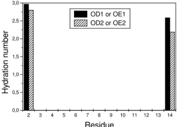
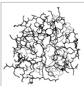
The polar amino acids side chains are directed to the po-lar solvent in aqueous solution differently of the intramolec-ular HB found in the crystal structure (Fig. 10, crystal struc-ture, and Fig. 11, aqueous solution structure). In solution, the donor and acceptor atoms are involved in intermolecular HB with water molecules disrupting the HB found in solid state. The nHBvalues of the polar side chain groups in the monomer 1, Figs. 12-15, indicate that each peptide atom ex-hibits its own hydration shell. Similar results are observed with the monomer 2. The HNP-3 aqueous solution structure shows that the six Arg residues (Arg6, Arg15 and Arg16 of the both monomers) form an equatorial ring around the dimer, while the apolar side chains of Tyr17, Tyr22, Trp27, Phe29, Cys5 and Cys20 are interacting with each other, repeling water molecules, resulting in a hydrophobic core (Fig. 11). The hydrophobic mini-channel (Fig. 16), that
completely crosses the dimer, does not contain conserved or structural water molecules. However, the presence of the hydration water molecules at the beginning (Asp2 and Tyr4) and at the end (Tyr17 and Tyr22) of the mini-channel preserves the dimer stability. The existence of a hydropho-bic mini-channel stabilized by hydration molecules at both ends, in its turn, stabilizes the overall quaternary structure of thedefensin. Such quaternary structure probably can be easily destroyed when the exposed polar groups begin to in-teract with membrane surfaces damaging the bilayers struc-tures.
Figure 10. TheHNP-3crystal structure. The polar amino acids are displayed.
Figure 11. The structure ofHNP-3in aqueous solution with the display of the polar amino acids. The polar side chains, involved in intramolecular HB in the crystal structure, are directed to the polar solvent in the aqueous medium, where they form an equatorial ring around the dimer.
2 3 4 5 6 7 8 9 10 11 12 13 14 0,0
0,5 1,0 1,5 2,0 2,5 3,0
Hy
dr
at
ion nu
mber
Residue OD1 or OE1 OD2 or OE2
6 7 8 9 10 11 12 13 14 15 16 17 18 19 20 21 22 23 24 25 0,0
0,5 1,0 1,5 2,0 2,5 3,0
Hy
drati
on numb
e
r
Residue
HE HH1 HH2
Figure 13. The HB of the arginine side chain HE, HH1 and HH2 hydrogen atoms in theHNP-3 monomer 1.
15 16 17 18 19 20 21 22 23
0,0 0,5 1,0 1,5 2,0 2,5 3,0
H
ydr
ati
o
n nu
mb
er
Residue OE1 HE2
Figure 14. The HB of the OE1 and HE2 glutamine residue atoms in theHNP-3monomer 1.
4 5 6 7 8 9 10 11 12 13 14 15 16 17 18 19 20 21 22 0,0
0,5 1,0 1,5 2,0 2,5 3,0
Hy
dr
at
ion numb
e
r
Residue
OH or OG1 HH or HG1
Figure 15. The HB of the OH, HH Tyr and OG1, HG1 Thr residues atoms in theHNP-3 monomer 1.
The selected gA,O(r) and gB,H(r) and corresponding P(E) profiles ( not shown ) indicate that the polar side chains of many amino acids present well defined hydration shells. The spatial arrangement of the cationic residues on the pep-tide surface enhances the action of the hydrophobic forces in the dimer inner region. Hydrophobic interactions are usually thought as resulting from a partial reversal of the solvation process that can be seen as due to the weak exposure or the lack of exposure of apolar groups to the water phase with
mini-channel
Figure 16. The hydrophobic mini-channel that completely crosses theHNP-3dimer.
the consequently unfavorable entropy contribution to the free energy of solvation, is minimized. The HNP-3 am-phiphilic character is probably the key of the affinity of this peptide, and other of the same class, to negatively charged bacterial membranes.
4.3
The
gramicidin A
The initial structure of the GA used in the simulations was obtained by NMR in dimyristoilphosphatidilcoline bilayer (DMPC)[29], pdb code: 1MAG[60]. The simulated sys-tem was constituted by a GA dimer and a mixture of po-lar (water) and apopo-lar (carbon tetrachloride) solvents. This system intends to mimic internal apolar region of the bio-logical membranes and their polar neighborhoods. The sim-ulation box was divided in three slices containing the apo-lar phase in the central slice and the poapo-lar phases in the two other slices. The central part of the simulation box embodies the GA channel structure excepting the ethanolamine polar groups (−N H−CH2CH2OH) that must remain in
con-tact with the aqueous phase.
Figure 17. The last structure of thegramicidin Aobtained by sim-ulation.
The stability of the structure generated during the sim-ulation can be evaluated by monitoring of the intramolec-ular HB involving the oxigen and hydrogen atoms of the polar groups in all residues. It can be concluded that the GA channel structure is maintained in its active form by the stabilizing effects due to the intermolecular HB with water molecules and by direct interaction between the monomers. The datas relative to the intermonomer HB are presented in Table 1 where it can be seen that the intermonomer HB were detected during more than 70%of the time indicating that the dimer structure exhibits a high stability. These HB stabi-lize both the internal region of the channel as so as its con-tacts with the aqueous phase at both ends. The maintenance of the intramolecular HB network is enough to character-ize the stability of the channel structure in the active form. The structural stability is a consequence, among other fac-tors, of the intermolecular interactions in which the water molecules carry out the important function of stabilizing the peptide main chain.
Table 1. The HB between the two monomersgramicidin A
in the GA channel.
CO NH xm´ax( ˚A) Fr
Val(1) Ala(20) 1.95 0.96 Ala(5) Val(16) 1.95 0.75 Ala(3) Ala(18) 1.95 0.98 Ala(20) Val(1) 1.95 0.86 Val(16) Ala(5) 1.95 0.95 Ala(18) Ala(3) 1.95 0.99
Figure 18. The water molecules that occupy the center of the chan-nel in the last structure of thegramicidin Aobtained by simulation.
4.4
The
myotoxin II
The trajectory of BaspMT-II reveals that its structure re-mains highly stable after 0.5 ns because thermsdoscillates near 3.0nm, Fig. 19, showing also that the secondary struc-tures of the helix-I, II and III are well defined and stable. This stabilization occurs by means of intra and intermolec-ular HBs. The intramolecintermolec-ular HBs that were found are 112 HBs between H and O backbone atoms, 111 HBs between backbone and side chains atoms and 45 HBs between atoms of side chains. Intermolecular HBs with the backbone atoms were observed with the larger frequency inβ-wing and C-terminal regions. The helices are stabilized mainly by in-tramolecular HB because the number of inin-tramolecular HB is larger than the number of intermolecular HB, Fig. 20.
0,0 0,5 1,0 1,5 2,0 2,5
0 1 2 3
t / (ns)
rm
sd
/
(Å) AspMT-II
Figure 19. Thermsdof theBaspMT-II. The reference structure is
the initial structure of theBaspMT-II used in the simulations.
a new short helix formed by four residues (115-118) with charged side chains located in the C-terminal region. The β-wing and C-terminal loop are the most hydrated regions on account of the presence of charged side chains exposed to the solvent, Fig. 20. The Lys49 charged group N H3+
is directed to the calcium binding loop with the three hy-drogen atoms hyhy-drogen bonded with three water molecules. In the final structure of the simulation, one of these water molecules is hydrogen bonded with the ND1 atom of the His48 residue. An HB network is observed around these water molecules. These results are supported by the nHB of the hydrogen atoms of theN H3+group in Lys49 and of
the ND1 atom of His48. The water molecule that is hydro-gen bonded with the ND1 atom of the His48 residue is an hydration water molecule (W-His48).
0 200 400 600 800 1000 1200
0,0 0,5 1,0 1,5 2,0 2,5 3,0 Calcium binding loop
Number of the protein atoms
0 200 400 600 800 1000 1200
0,0 0,5 1,0 1,5 2,0 2,5
3,0 Calciumbinding Helix-2 B - wing Helix-3 C-terminalloop
loop Short helix Helix 1
O or H
Nunb er of H ydr ogen bond
Figure 20. The intermolecular HB between oxygen (O) and hy-drogen (H) atoms ofBaspMT-II of protein and water molecules. The plot shows HB in the each of the secondary structure of the BaspMT-IImonomer. The data of the backbone atoms are plotted in the first part of the figure and the side chain data in the second part.
The HB network, located into hydrophobic channel, has an essential role in the stabilization of the local structure. The water molecule, W-His48, detected in the catalytic site, is the molecule shown into the circle in the Fig. 5. The lo-calization of W-His48 is in agreement with the experimental results[38] and with the mechanism proposed to explain the catalytic activity[35, 38]. Other water molecules can also be identified in the channel forming a HB network with W-His48.
The simulation showed that the W-His48 is present in the Lys49 PLA2s, as is Asp49 PLA2s. Consequently, the pres-ence of W-His48 cannot guarantee the ability to catalyze. The lack of the catalytic activity in the Lys49 PLA2s is ex-plained by the existence of aN H3+ group, in place of the
co-factor Ca2+in Asp49 PLA2s, coordinated with calcium
binding loop carbonyl oxygen atoms. TheN H3+ group of
the Lys49 PLA2s residue plays a role in the stability of the local structural but cannot stabilize the oxyanion transition state in the same way as the co-factor Ca2+can do it. The
access of the Ca2+co-factor is completely hindered because
theN H3+group of the Lys122 residue, that is located on the
other side of the loop, interacts strongly with oxygen car-bonyl atoms of the loop forming intramolecular HBs.
The structure of themyotoxinII is stabilized in great part by intramolecular HBs. Parts of the secondary structure are stabilized by intermolecular HBs excepting the helix-2 and helix-3. The local structure of the hydrophobic channel is stabilized by a HB network. The maintenance of the W-His48 plays an important role in the structure and function of both PLA2s, Lys49 PLA2 and Asp49 PLA2. The Ca2+
co-factor is essential to realize the catalyze, but it is not es-sential to stabilize the structure of the Lys49 PLA2s.
4.5
An amphipathy scale
In order to determine a hydrophobicity scale, molecular sim-ulations of small peptides in spc/e water were performed with small peptides likeaa(1aa),Gly−aa−Gly(3aa) andGly−Gly−aa−Gly−Gly (5aa), whereaa cor-responds to the amino acids commonly found in the pro-tein primary structures. The amino acid glycine was used as reference because its side chain is constituted by only one hydrogen atom that is unable to interact strongly with other atoms. The mean configurational energies peptide-water molecules were focused for all the peptides. They can be splitted in two parts: the backbone average config-urational energy, Ebb(iaa)withi≡1, 3 or 5, and side chains average configurational energy, Esc(iaa).
The values of Ebb(1aa), Ebb(3aa)and Ebb(5aa)listed in Table 2 indicate that the backbone configurational en-ergy are somewhat constant since it does not depend on the nature of the centralaaresidue. The Ebb(1aa)average is equal to (-113,2±6,8) kcal/mol, (-224,8±6,1) kcal/mol for Ebb(3aa)and (-231,9±14,3) kcal/mol in the Ebb(5aa)case. Of course, deviations of Ebb(iaa)from the average are ob-served being the larger deviation obob-served with the proline amino acid because its side chain is a cycle and the hy-bridization of the N atom of its main chain issp2notsp3as
for the other amino acids since theN-terminal of proline is an imino not an amino group so that proline would be more correctly classified as an imino acid. In this way, the lack of one hydrogen atom bonded to the nitrogen atom in the proline amino acid results in less atractive Ebb(iPro).
Table 2. The backbone configurational energies of1aa,3aaand5aa.
Amino acid Ebb(1aa)(kcal.mol−1) Ebb(3aa)(kcal.mol−1) Ebb(5aa)(kcal.mol−1)
Ala -119.02 -227.58 -216.69
Arg -115.17 -226.07 -215.66
Asn -115.18 -230.49 -221.62
Asp -102.51 -216.38 -228.68
Cys -115.91 -222.30 -222.90
Gln -112.70 -230.20 -240.02
Glu -108.05 -217.43 -219.01
Gly -118.91 -229.32 -240.96
His -112.50 -226.82 -245.44
Ile -113.54 -221.58 -254.78
Leu -115.69 -228.73 -249.10
Lys -122.71 -229.61 -245.68
Met -112.19 -231.04 -245.68
Phe -114.00 -222.51 -228.53
Pro -90.89 -207.99 -204.59
Ser -114.05 -229.03 -216.08
Thr -112.75 -217.42 -239.62
Trp -117.14 -228.67 -249.32
Tyr -118.54 -223.77 -222.51
Val -112.30 -229.51 -229.99
Table 3. The side chain configurational energies of1aa,3aaand5aa.
Amino acid Esc(1aa)(kcal.mol−1) Esc(3aa)(kcal.mol−1) Esc(5aa)(kcal.mol−1)
Ala -1.81 -1.70 -1.59
Arg -56.50 -63.21 -57.37
Asn -16.26 -18.67 -18.40
Asp -90.59 -110.89 -108.19
Cys -4.48 -4.58 -4.34
Gln -19.14 -19.45 -18.60
Glu -112.42 -115.84 -115.94
Gly 0 0 0
His -20.33 -24.01 -23.55
Ile -5.91 -5.54 -5.41
Leu -5.96 -5.84 -5.62
Lys -76.74 -79.50 -81.48
Met -6.90 -6.80 -6.67
Phe -11.74 -11.65 -11.31
Pro -2.99 -3.95 -3.92
Ser –9.06 -13.58 -13.68
Thr -9.68 -13.79 -14.32
Trp -17.22 -17.26 -17.42
Tyr -24.19 -24.51 -24.57
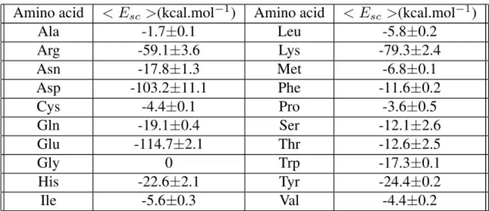
Table 4. The side chain average configurational energies.
Amino acid < Esc>(kcal.mol−1) Amino acid < Esc>(kcal.mol−1)
Ala -1.7±0.1 Leu -5.8±0.2
Arg -59.1±3.6 Lys -79.3±2.4
Asn -17.8±1.3 Met -6.8±0.1
Asp -103.2±11.1 Phe -11.6±0.2
Cys -4.4±0.1 Pro -3.6±0.5
Gln -19.1±0.4 Ser -12.1±2.6
Glu -114.7±2.1 Thr -12.6±2.5
Gly 0 Trp -17.3±0.1
His -22.6±2.1 Tyr -24.4±0.2
Ile -5.6±0.3 Val -4.4±0.2
The values of Esc(1aa), Esc(3aa) and Esc(5aa) are listed in Table 3. They are quite invariable in function of the number of Gly residues in the peptides1aa, 3aa and 5aa. The model1aa,3aaand5aapeptides are small so that they are unable to protect their hydrophobic side chains from direct interactions with the solvent. Table 4 lists the aver-age values,< Esc>, of Esc(1aa), Esc(3aa)and Esc(5aa). Consistently with our model,< Esc >= 0for the glycine amino acid where the side chain is a hydrogen atom not ex-plicitly identified in the Gromos96 force field[56]. From a single inspection of Table 4 is can be seen that the amino acids can be grouped in 3 sets: one where the amino acids with energies< Esc >between0and -7 kcal/mol are put together constituting the group of the apolar amino acids. The second group aggregates the polar amino acid with
< Esc >found in the range −12 to -25 kcal/mol. The last group is the group of charged polar amino acid: their < Esc >energies are in the -60 to -115 kcal/mol range. It is interesting to note that the Gromos96 force field[56] is consistent with the fact that the phenilalanine and trypto-phan amino acids are weakly polar so that a charge less than 0.2eis attributed to the aromatic hydrogen atoms. Moreover, the three-dimensional structure of the proteins depends not solely on the hydrophobicity of the side chains but depends also on the steric effects due to the different extensions of the space hindered by the different secondary structures in the interior of proteins[61, 62].
As a final classification, the results listed in table 4 can provide the yearned hydrophobicity sequence of the 20 amino acids:
⌋
largest hydrophobicity→Gly→Ala→Pro→(Cys, Val)→(Ile, Leu)→Met→Phe→(Ser, Thr)
→Trp→Asn→Gln→His→Tyr→Arg→Lys→(Asp, Glu)→largest hydrophilicity
⌈
The< Esc >of the amino acids put together are very close. The < Esc > side chain energies configurational present some advantages when compared with other am-phipathy scales:
1. they consider the amino acids when they are bonded with other residues forming peptide chains;
2. they analyse the complete interation between the sol-vent and the amino acids (backbone and side chain); 3. they are able to reproduce exactly the environment
amino acid-water;
4. they do not depend on unclear differences of standard free energies;
5. they do not depend on hypothesis frequently included in many amphipathy scale determination.
Nevertheless, the quality of the present results has to be confirmed by analyzing the interaction energies between the
solvent and the side chains of proteins in aqueous phases and comparing these results with present< Esc >scale. For this analysis, the next proteins used were: basic fibroblast growth factor[63], defensin[26], gramicidin[64] and two
re-gions present less attractive energies, they are specifically the regions 36-40, 61-65, 69-73, 108-114, 119-126 and 144-152. It is interesting to observe that these regions are hy-drophobic regions localized in the interior of basic fibrob-last growth factor. The regions shown in Fig. 23 are hy-drophilic regions situated on the external part of the basic fibroblast growth factor. It is, consequently, clear that parts of secundary structures can be predicted from the< Esc > scale.
-80 -60 -40 -20 0
-120 -100 -80 -60 -40 -20 0
<
Esc
>
(kcal
.mo
l
-1)
< Eproteins > (kcal.m ol -1)
Figure 21. The plot of< Eproteins>vs.< Esc>. The correla-tion coefficient is equal to 0.97.
40 60 80 100 120 140
0 -20 -40 -60 -80 -100 -120
E1B
F
F
(
kcal
.mo
l
-1)
Residue
Figure 22. The sequence of the< Esc >in thefibroblast growth factorprimary structure. The gray regions are the hydrophobic re-gions.
5
Conclusion
In this paper, important features of the role of water in the protein activity were presented and discussed. The fun-damental function of the intermolecular HB in the tertiary and quaternary protein structures was described focusing the maintenance of local structures as so as the insertion of the peptides into the larger, and very stable, HB network formed by the interactions between the solvent molecules. The ex-amples proposed to enlight the water importance are clear
Figure 23. The position of hydrophobic regions detected in Fig. 22 in thefibroblast growth factorcrystal structure.
in the aspects that HB are of main importance considering that:
1. the basic fibroblast growth factor is unable to de-fine its tertiary structure without the solvent contri-bution since onlyβ-strands, and no disulfide brigde, are present in its structure;
2. the importance of the water molecules is enhanced in considering the dimerdefensinwhere the hydropho-bic channel is stabilized by water molecules at both ends. The immediate consequence is that highly re-active polar and charged side chain groups are firmly exposed to external interactions;
3. the same stabilizing influence of the water molecules are observed in the case of the gramicidin channel since both interior region and mouthes suffer the in-fluence of the water that can pass through the chan-nel as it must occur as well whengramicidinforms a channel in bacterial membranes resulting in the loss of protoplasmic materials and in the dead of the bacteria; 4. the double function of a water molecule in the main-tenance of the catalytic center structure and in the hy-drolysis of the sn-2 acyl bonds of sn-3 phospholipids is outstanding in thephospholipasestudy;
Acknowledgments
This work was supported in part by the Conselho Na-cional de Desenvolvimento Cient´ıfico e Tecnol´ogico and by the Fundac¸˜ao de Amparo `a Pesquisa do Estado de S˜ao Paulo.
References
[1] C. Branden, J. Tooze,Introduction to Protein Structure, Gar-land Publishing, New York (1999).
[2] A. L. Lehninger, D. L. Nelson, and M. M. Cox,Princ´ıpios de Bioqu´ımica, Sarvier, S˜ao Paulo (1995).
[3] L. Stryer,Biochemistry, W. H. Freeman and Company, New York (1995).
[4] D. Voet, J. Voet,Biochemistry, John Wiley and Sons, New York (1995).
[5] J. Darnell, H. Lodish, and D. Baltimore,Molecular Cell Bi-ology, Scientific American Books, New York (1986).
[6] B. Alberts, A. Johnson, J. Lewis, M. Raff, K. Roberts, and P. Walter,Molecular Biology of The Cell, Garland Science, New York (2002).
[7] J. ´Etienne, Bioqu´ımica Gen´etica e Biologia Molecular, Livraria Santos Editora, S˜ao Paulo (2003).
[8] G. A. Jeffrey, W. Saenger,Hydrogen Bonding in Biological Structures, Springer-Verlag (1994).
[9] R. B. Gregory ed., Protein-solvent interactions, Marcel Dekker, New-York (1995)
[10] L. Blum, L. Degr`eve, Mol. Phys.88, 585 (1996).
[11] L. Degr`eve, L. Blum, Physica A224, 550 (1996).
[12] J. Zhang, L. S. Cousens, P. J. Barr, S. R. Sprang, Proc. Natl. Acad. Sci. USA,88, 3446 (1991).
[13] G. G. Gallego, G. Conn, V. B. Hatcher, K. A. Thomas, Biochem. and Boophysical Res. Comm.135, 541 (1986).
[14] H. Ago, Y. Kiagawa, A. Fujishima, Y. Matsuura, and Y. Kat-sube, J. Biochem.110, 360 (1991).
[15] B. Alberts, D. Bray, J. Lewis, M. Raff, K. Roberts, and J. D. Watson, Biologia Molecular da C´elula, Artes M´edicas, Porto Alegre, 1997.
[16] F. J. Moy, A. P. Seddon, P. Bohlen, and R. Powers, Biochem-istry,35, 13552 (1996).
[17] R. E. W. Hancock, R. Lehrer, Trends Biotechnol. 16, 82 (1998).
[18] R. M. Epand, H. J. Vogel, Biochim. Biophys. Acta1462, 11 (1999).
[19] J. Patterson-Delafield, D. Szklarek, R. J. Martinez, and R. I. Lehrer, Infect. Immun.31, 723 (1981).
[20] R. I. Lehrer, A. Barton, K. A. Daher, S. S. L. Harwig, T. Ganz, and M. E. Selsted, J. Clin. Invest.84, 553 (1989).
[21] S. H. White, W. C. Wimley, and M. E. Selsted, Curr. Opin. Struct. Biol.5, 521 (1995).
[22] Y. Shai, Trends Biochem. Sci.20, 460 (1995).
[23] T. Ganz, M. E. Selsted, D. Szklarek, S. S. L. Harwig, K. Da-her, D. F. Bainton, and R. I. Lehrer, J. Clin. Invest.76, 1427 (1985).
[24] T. Ganz, R. I. Lehrer, Curr. Opin. Immun.6, 584 (1994).
[25] T. Ganz, R. I. Lehrer, Pharmac. Ther.66, 191 (1995).
[26] C. P. Hill, J. Yee, M.E. Selsted, and D. Eisenberg, Science,
251, 1481 (1991).
[27] P. Yeagle, The Structure of Biological Membranes, CRC Press, (1992).
[28] R. B. Gennis, Biomembranes - Molecular Structure and Function, Springer-Verlag, New York, (1989).
[29] R. R. Ketchem, W. Hu, T. A. Cross, Science,261(10), 1457 (1993)
[30] B. A. Wallace, Annu. Rev. Biophys. Biophys. Chem.19, 127 (1990).
[31] W. R. Veatch, E. T. Fossel, and E. R. Blout, Biochemistry,13, 5249 (1974).
[32] W. R. Veatch, E. R. Blout, Bichemistry,13, 5257 (1974).
[33] B. E. Isbell, C. Rice-Evans, and G. H. Beaven, FEBS LET-TERS,25, 192 (1972).
[34] E. T. Fossel, W. R. Veatch, Y. A. Ovchinnikov, and E. R. Blout, Bichemistry,13, 5264 (1974).
[35] R. K. Arni, R. J. Ward, Toxicon34, 827 (1996).
[36] E. A. Dennis, J. Biol. Chem.269, 13.057 (1994).
[37] P. Rosenberg, in: W. T. Shier, D. Mebs (Eds.),Handbook of Toxicology, Marcel Dekker, New York, (1990).
[38] D. L. Scott, S. P. White, Z. Otwinowski, W. Yuan, M. H. Gelb, and P. B. Sigler, Science250, 1545 (1990).
[39] C. J. Van den Bergh, A. J. Slotboom, H. M. Verheij, and G. H. De Haas, J. Cell. Biochem.39, 379 (1989).
[40] Y. Li, B. Yu, H. Zhu, M. Jain, and M. Tsai, Biochemistry33, 14714 (1994).
[41] Y. Nozaki, C. Tanford, J. Biol. Chem.246, 2211 (1971).
[42] S. Damodaran, K. B. Song, J. Biol. Chem.261, 7220 (1986).
[43] S. H. White, W. C. Wimley, Annu. Rev. Biophys. Biomol. Struct.28, 319 (1999).
[44] C. Chothia, J. Mol. Biol.105, 1 (1976).
[45] P. Manavalan, P. K. Ponnuswamy, Nature,275, 673 (1978).
[46] S. Fraga, Can. J. Chem.60, 2606 (1982).
[47] R. M. Sweet, D. Eisenberg, J. Mol. Biol.171, 479 (1983).
[48] L. A. Kuhn, J. S. Leigh, Biochim. Biophys. Acta,828, 351 (1985).
[49] H. Tanaka, J. Chem. Phys.86(3), 1512 (1987).
[50] G. Hummer, S. Garde, A. E. Garcia, M. E. Paulaitis, and L. R. Pratt, J. Phys. Chem. B102(51), 10469 (1998).
[51] W. Blokzijl, J. B. F. N. Engberts, Angew. Chem. Int. Ed. Engl.
32, 1545 (1993).
[52] C. Chothia, Nature,248, 338 (1974).
[53] B. J. Alder, T. E. Wainwright, J. Chem. Phys. 31, 459 (1959).
[54] G. Cicotti, D. Frenkel, and I. R. McDonald,Simulation of Liquids and Solids,North-Holland, Amsterdam, (1990).
[56] W. F. van Gunsteren, S. R. Billeter, A. A. Eising, P. H. H¨unenberger, P. Kr¨uger, A. E. Mark, W. R. P. Scott, and I. G. Tironi,Biomolecular Simulation: The GROMOS 96 Man-ual and User Guide, Biomos, Groeningen, (1996).
[57] I. G. Tironi, R. Sperb., P. E. Smith, and W. F. van Gunsteren, J. Chem. Phys. 102,5451 (1995).
[58] F. H. Stillinger, A. Rahman, J. Chem. Phys.60, 1545 (1974).
[59] L. B. Silva, L. Degr`eve, Mol. Phys.100, 3111 (2002).
[60] http://www.rcsb.org/pdb
[61] S. Lifson, C. Sander, Nature,282, 109 (1979).
[62] J. Janin, C. Chothia, J. Mol. Biol.143, 95 (1980).
[63] J. S. Kastrup, E. S. Eriksson, H. Dalboge, and H. Flodgaard, Acta Crystallogr. D Biol. Crystallogr.53, 160 (1997).
[64] R. R. Ketchem, K. C. Lee, S. Huo, and T. A. Cross, J. Biomol. NMR,8, 1 (1996).
[65] S. Farr-Jones, G. P. Miljanich, L. Nadasdi, J. Ramachandran, and V.J. Basus, J. Mol. Biol.248, 106 (1995).