53 Radiol Bras. 2012 Jan/Fev;45(1):53–58
Imaging findings of abdominal gossypibomas
*
Avaliação por imagem dos gossipibomas abdominais
Francisco Abaeté das Chagas Neto1, Paulo Moraes Agnollitto2, Fernando Marum Mauad3, André Rodrigues Façanha Barreto4, Valdair Francisco Muglia5, Jorge Elias Junior5
Gossypiboma is a term utilized to describe a mass developed from a matrix of cotton fibers surrounded by inflammatory granulomatous reaction. Its incidence is estimated at 0.15% to 0.2% of laparotomies. A foreign body within the abdominal cavity may provide a niche for proliferation of microorganisms, acting as primary focus for development of an abscess and peritonitis. Several studies have demonstrated the relevance of clinical correlation with the findings of different imaging methods (conventional radiography, ultrasonography, computed tomography and magnetic resonance imaging) in the diagnosis of gossypibomas. The present pictorial essay is aimed at demonstrating a series of typical cases of abdominal gossypibomas and illustrating the several presentations of such mass, with emphasis on the findings at different imaging methods in order to familiarize radiologists with this entity and with the main differential diagnosis.
Keywords: Gossypiboma; Abdominal; Imaging diagnosis.
O termo gossipiboma é usado para descrever uma massa formada a partir de uma matriz de algodão cercada por uma reação inflamatória/granulomatosa. Sua incidência é estimada em 0,15% a 0,2%. O corpo estranho na cavidade abdominal pode servir de nicho para a proliferação de microrganismos e agir como foco primário para formação de abscessos e de peritonite. Vários estudos têm demonstrado a importância da correlação clínica com os diversos métodos de imagem (radiografia convencional, ultrassonografia, tomografia computadorizada e ressonância magnética) no diag-nóstico dos gossipibomas. Este ensaio tem por objetivo demonstrar uma série de casos típicos de gossipibomas abdo-minais e ilustrar suas diversas formas de apresentação, com ênfase nos achados dos diferentes métodos de imagem, visando a familiarizar os radiologistas com esta enfermidade e seus principais diagnósticos diferenciais.
Unitermos: Gossipiboma; Abdominal; Diagnóstico por imagem.
Abstract
Resumo
* Study developed at Hospital das Clínicas da Faculdade de Medicina de Ribeirão Preto da Universidade de São Paulo (HC-FMRPUSP), Ribeirão Preto, SP, Brazil.
1. MD, Radiologist, Fellow MSc degree, Program of Health Sciences applied to the Locomotor System, Faculdade de Me-dicina de Ribeirão Preto da Universidade de São Paulo (FMRP-USP), Ribeirão Preto, SP, Brazil.
2. MD, Resident, Division of Radiology, Department of Med-ical Practice, Faculdade de Medicina de Ribeirão Preto da Uni-versidade de São Paulo (FMRPUSP), Ribeirão Preto, SP, Brazil.
3. MD, Physician Assistant, Division of Radiology, Department of Medical Practice, Faculdade de Medicina de Ribeirão Preto da Universidade de São Paulo (FMRPUSP), Ribeirão Preto, SP, Brazil.
4. MD, Radiologist, Hospital das Clínicas da Faculdade de Medicina de Ribeirão Preto DA- Universidade de São Paulo (HC-FMRPUSP), Ribeirão Preto, SP, Brazil.
5. Professor, PhDs, Division of Radiology, Department of Med-ical Practice, Faculdade de Medicina de Ribeirão Preto da Uni-versidade de São Paulo (FMRPUSP), Ribeirão Preto, SP, Brazil.
Chagas Neto FA, Agnollitto PM, Mauad FM, Barreto ARF, Muglia VF, Elias Jr J. Imaging findings ao abdominal gossypibomas. Radiol Bras. 2012 Jan/Fev;45(1):53–58.
which originate gossypibomas. Absorbable materials most frequently utilized include gelatin foam (Gelfoam®), oxidized cellu-lose (Surgicel®, Oxycel®) and microfibril-lar collagen (Avitene®), which are left in the manipulated site to avoid repeated hem-orrhage. It is important to differentiate the cases that the identified foreign body is an intentional component of the surgical pro-cedure (absorbable materials), from those involving unintentional components (non-absorbable materials)(6).
Non-absorbable hemostatic materials induce two types of inflammatory reac-tions. The first type is an aseptic inflamma-tory reaction to the presence of a foreign body, with fibroblastic reaction and com-plete encapsulation of the process. There-fore, such type of reaction is generally as-ymptomatic, sometimes being identified as a palpable mass(6–8). The other is the
exu-dative type, and frequently determines the development of an abscess, in association or not with a secondary bacterial infec-Its incidence is estimated to be 0.15%
to 0.2%, with one case for every 500 to 1,000 laparotomies, sometimes determin-ing important complications, with a mor-tality rate of up to 10% to 18%(3–5).
The term gossypiboma is utilized to de-scribe a mass which develops from a cot-ton matrix surrounded by an inflammatory reaction. The word comes from the Latin gossypium (cotton) and from the Kiswahili boma (place of concealment, or hiding)(6).
There are basically two types of hemo-static materials utilized in surgeries: those constituted by absorbable materials and those that are non-absorbable. Examples of non-absorbable materials are swabs, cotton and synthetic rayon synthetic gauzes,
INTRODUCTION
The first case report about intra-ab-dominal foreign body after laparotomy dates back to 1884(1,2). Despite all
precau-tions taken by surgeons and by other spe-cialists, besides other preventive measures, postoperative intracavitary retention of for-eign bodies is a relatively frequent event.
Mailing Address: Dr. Francisco Abaeté das Chagas Neto. Se-cretaria do Setor de Radiologia (CCIFM) – Hospital das Clínicas da Faculdade de Medicina de Ribeirão Preto da Universidade de São Paulo. Avenida Bandeirantes, 3900, Campus Universitário, Monte Alegre. Ribeirão Preto, SP, Brazil, 14048-900. E-mail: [email protected]
tion(6–8). In such a type of reaction, more
exuberant symptoms, such as abdominal pain and fever, are observed. Upper or lower intestinal obstruction, enterocuta-neous fistulas, erosion or perforation of intestinal loops, adhesions and even granu-lomatous peritonitis may be associated.
On the other hand, absorbable agents trigger a more controlled inflammatory re-action along the material absorption period. In most cases such a process is asymptom-atic(6).
Thus, gossypibomas represent a feared problem in medicine as, besides the com-plications to the patient, their impact on the physician-patient relationship and the medical-legal implications(7) are in both
cases, very challenging.
The diagnosis of gossypibomas may be extremely challenging for the radiologist because of their variable presentation, usu-ally resulting from the investigation of complaints such as abdominal pain, pal-pable mass, intestinal obstruction, fistulas, or as part of routine postoperative imaging studies. In other occasions, the diagnosis results from an incidental finding, years after an abdominal surgery. Eventually, a suspicion may be raised by the clinical team that indicates imaging studies to cor-roborate the diagnosis of gossypiboma.
Main factors involved in the occurrence of abdominal gossypibomas are the fol-lowing: emergency surgeries, unexpected changes during the surgical act, two or more different teams involved in the surgical act,
changes in the nursing team during the course of surgery, patient’s obesity and occurrence of intraoperative hemorrhage. The early identification of such a condition is of most importance, as it results in reduc-tion of morbidity, and may prevent poten-tially fatal complications, besides mitigat-ing medical-legal complications(4,9).
The present essay is aimed at demon-strating a series of typical cases of
abdomi-Figure 1A. Female, 35-year-old patient submitted to renal transplantation, complaining of bulging and pain in the right iliac fossa in the late postopera-tive period. Digital radiography (scanogram) show-ing bulgshow-ing on the right lower quadrant associated with hyperdense linear serpiginous image on a pro-jection of the right iliac fossa (arrow).
nal gossypibomas and illustrating their dif-ferent presentations, emphasizing the find-ings at different imaging methods, by means of conventional radiology, ultra-sonography (US), computed tomography (CT) and magnetic resonance imaging (MRI), with the purpose of familiarizing radiologists with this disorder and its main differential diagnoses.
IMAGING EVALUATION
OF ABDOMINAL GOSSYPIBOMAS
Conventional radiology
Conventional radiology is the imaging method most commonly utilized in the postoperative period for the detection of retained sponges or swabs (gossypibomas), which, in most cases, can be identified by the presence of radiopaque markers incor-porated in their composition, with a sensi-tivity above 90% in such cases (Figures 1A and 2)(10).
Radiographs obtained at the surgery room, by means of bedside apparatuses, technical restrictions limiting the images quality therefore, the manipulation and post-processing of the images can be help-ful in certain dubious cases.
The appropriateness of the field of view in the study must also be considered, as well as the careful evaluation of the periph-ery of the obtained image, which may in-clude part of a gossypiboma.
The imaging finding of gossypiboma at plain radiography is variable. The most
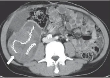
Figure 1B. Pre-contrast axial CT section of the abdomen demonstrating a mass with soft tissue density with hyperdense serpiginous structures inside (arrow).
common presentation includes radiodense irregular linear images, some of them ser-piginous, associated or not with increase in volume and density of adjacent soft tissues. In some cases, amorphous radiolucent im-ages are observed, probably caused by gas entrapment or secondary infection by gas-forming germs(10).
However, eventually small fragments of surgical material may not include radio-dense filaments, so their direct identifica-tion by X-ray emitting imaging methods is difficult. In such cases, US and MRI are extremely useful.
Ultrasonography
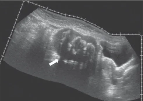
Ultrasonography allows the identifica-tion of practically all types of gossypibomas, including radiolucent gossypibomas, be-sides providing information on their ana-tomical relationships (Figure 3A).
Sonographic findings in cases of gossypiboma can be divided into three types as follows: 1) hyperechogenic image with posterior acoustic shadowing; 2) well defined mass with cystic contents and echogenic, undulated internal structures; 3) non-specific finding of complex and/or hypoechoic mass. Invariably, internal vas-cular flow is absent at Doppler study (Fig-ure 3B)(10–12).
Posterior acoustic shadowing is gener-ally present and may be related to the
at-tenuation of the acoustic beam by the for-eign body itself, as well as by the presence of gas and, occasionally, by the presence of calcified areas(10–12).
Some studies in the literature suggest that US may be considered the imaging method of choice for the diagnosis of gossypibomas, since it does not rely on ionizing radiation, presents high sensitiv-ity (between 95% and 98%) and can be useful in the differential diagnosis with
other types of postoperative complica-tions(10–12).
However, US presents some limitations, such as being operator-dependent, besides the possibility of not identifying foreign bodies at greater depths or located posteri-orly to gas containing hollow viscus. Also, it may present false-positive results in cases where scars and calcifications of other eti-ologies are present.
Figure 2. Male, one-year-old patient submitted to surgical correction of imperforate anus, progress-ing with pain and bulgprogress-ing in the right iliac fossa in the late postoperative period. Presence of sponta-neously hyperdense linear image, projected into the
right iliac fossa (arrow). Figure 3A. Female, 25-year-old patient complaining of increased abdominal volume, and bulging in the hypogastric region. History of gynecologic surgery (tubal desobstruction) three years ago. Panoramic US, sagittal scan, demonstrating a complex mass in the hypogastric region, adjacent to the urinary bladder and the uterus (arrow).
Computed tomography
At CT, gossypibomas are generally identified as a mass with well defined con-tours, with soft tissues density, high or even mixed densities, sometimes containing air bubbles and high density capsule that may presents enhancement in the post-contrast phase (Figures 1B, 1C, 3C and 4)(10,12–14).
Sterile gas bubbles may be identified next to the gossypiboma up to six months after surgery(10). However, the presence of
gas densities within the gossypiboma should alert the radiologist to the possibil-ity of associated infection (Figure 3C).
The CT scout image or scanogram must always be evaluated as many times it holds the key for a correct diagnosis, particularly in those cases where beam hardening arti-facts impair the identification of the radio-paque marker on tomographic sections.
Gossypibomas should not be confused with a fluid collection, in spite of the pos-sibility of association with the development of abscess.
Multidetector CT (MDCT) allows ref-ormation of images acquired in different planes, including oblique ones, besides three-dimensional reconstruction,
provid-Figure 3E. Paramedian sagittal abdominal MRI, T2-weighted section demonstrating the relationship between complex abdominal cystic lesion and the bladder (arrow).
Figure 3F. Paramedian sagittal, contrast-enhanced abdominal MRI, T1-weighted section demonstrat-ing the capsular reinforcement of the lesion (arrow).
ing the method with extremely high sensi-tivity and accuracy. However, the utiliza-tion of ionizing radiautiliza-tion and iodinated contrast media constitute limiting factors in this method that should be taken in consid-eration.
The tomographic differentiation be-tween hemostatic absorbable and
non-ab-sorbable materials lies in the fact that the latter present a radiopaque marker that is not present in the several absorbable ma-terials. However, the clinical team mem-bers interaction and the knowledge on the utilized surgical techniques are determi-nants of an appropriate interpretation of postoperative images.
Figure 3C. Coronal reformatation of contrast-en-hanced abdominal CT showing predominantly hypodense mass in the hypogastric region, with lin-ear hyperdense images inside and adjacent to the urinary bladder, with some small hypoattenuating foci (gas) inside and subtle capsular enhancement (arrow).
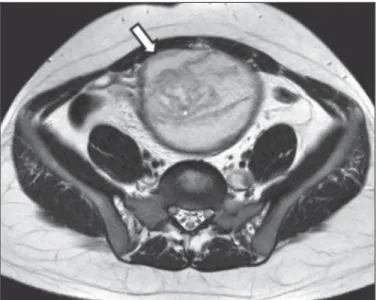
Figure 5A. Female, 53-year-old patient presenting a palpable mass in the mesogastric region. History of previous abdominal surgery for colon adenocar-cinoma. Coronal MRI, T2-weighted section demon-strating ovoid, circumscribed lesion with heteroge-neous signal intensity, located in the mesogastric region (arrow).
Figure 5B. Coronal MRI T1-weighted section dem-onstrating the same lesion identified on Figure 5A, with predominant intermediate signal intensity (ar-row).
Figure 5C. Coronal MRI T1-weighted section ac-quired after intravenous gadolinium administration, demonstrating a low signal capsule, with no sign of contrast uptake (arrow).
Magnetic resonance imaging
Few experimental studies on the utili-zation of MRI for the detection of gossy-pibomas are found in the literature. How-ever, in some studies, such method seems to present a sensitivity close to 100%(6,10).
Also, there are reports describing cases of patients with retained gossypibomas, sub-mitted to other diagnostic modalities such as plain radiography and CT, in whom only MRI was capable of identifying them, or
was utilized as a complementary diagnosis method(10,11,15,16).
Therefore, MRI should be utilized only in those cases where other more easily available imaging methods have not been capable of identifying direct or indirect signs suggestive of gossypibomas, or in those cases where those methods were in-conclusive.
Gossypibomas appearance at MRI is varied, being most commonly identified as
a heterogeneous mass, sometimes present-ing a solid-cystic component, with well de-fined contours, surrounded by a well delim-ited capsule. Hyposignal and hypersignal predominate respectively on T1-weighted and T2-weighted images, including inter-nal serpiginous and irregular images with intermediate signal on both T1- and T2-weighted imaging (Figures 3D, 3E, 3F, 5A and 5B)(6,10,11,15,16).
The capsule usually presents with low signal intensity on all pre-contrast se-quences, and may or may not present en-hancement on the sequences after admin-istration of paramagnetic contrast agent (Figures 3F and 5C)(6,10,11,15,16).
DIFFERENTIAL DIAGNOSIS
The main differential diagnoses of gossypibomas include intra-abdominal expansile masses/lesions such as hemato-mas, abscesses/collections, neoplastic le-sions and fecalomas(17).
Hematomas are seen in the early post-operative period and in general present pro-gressive resorption at follow-up studies.
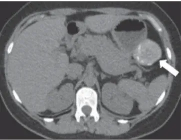
Abscesses are visualized as fluid den-sity masses with well defined walls with capsular reinforcement. In some cases, gas may be identified within the lesion, produc-Figure 4. Asymptomatic, female, 39-year-old patient submitted to partial gastrectomy for
ing air-fluid level. However, the develop-ment of an abscess may occur as a compli-cation related to the presence of a gossy-piboma.
Neoplastic lesions simulating gossypi-bomas are in general identified as an ab-dominal palpable mass in asymptomatic patients or in patients with non specific abdominal complaints associated with a previous history of abdominal surgery. In cases of oncologic surgery, the differentia-tion between residual lesion, tumor recur-rence and gossypiboma may represent a diagnostic challenge to radiologists.
Fecalomas may present irregular con-tours and poorly defined limits at CT, but are located inside colon loops and do not present a well defined and thick capsule.
Other conditions, such as postoperative adhesions, intestinal invagination, mesen-teric panniculitis and retained absorbable hemostatic materials should be remem-bered and considered among diagnostic possibilities(18).
CONCLUSION
Imaging findings of gossypibomas have variable presentations; however their diag-nosis may be suggested in cases where there is a correlation with a history of pre-vious abdominal surgery, and should be considered among the differential diagno-sis of abdominal masses/collections.
In the suspicion of gossypiboma, imag-ing findimag-ings should be impartially and ob-jectively reported by the radiologist. How-ever, the knowledge on the utilized surgi-cal techniques is necessary in order to al-low the radiologist differentiating between intentionally implanted materials/absorb-able hemostatic materials and gossypi-bomas.
Thus, the radiologist must have the knowledge on the different usual presenta-tions of gossypibomas and be prepared to interact with the assisting medical team in order to propose strategies that can better support the specific diagnostic and thera-peutic approaches for the patient.
REFERENCES
1. Iglesias AC, Salomão RM. Gossipiboma intra-abdominal – análise de 15 casos. Rev Col Bras Cir. 2007;34:105–13.
2. Mafalda LG, Caitano MJC, Magioni FM, et al. Textiloma simulando tumor de cólon e mesenté-rio, assintomático durante 40 anos. ABCD Arq Bras Cir Dig. 2009;22:186–7.
3. Rappaport W, Haynes K. The retained surgical sponge following intra-abdominal surgery. A con-tinuing problem. Arch Surg. 1990;125:405–7. 4. Gawande AA, Studdert DM, Orav EJ, et al. Risk
factors for retained instruments and sponges af-ter surgery. N Engl J Med. 2003;348:229–35. 5. Gonzales-Ojeda A, Rodríguez-Alcantar DA,
Are-nas-Marquez H, et al. Retained foreign bodies following intra-abdominal surgery. Hepatogastro-enterology. 1999;46:808–12.
6. Martins MCB, Amaral RPG, Andrade CS, et al. Características de imagem na ressonância mag-nética de gossipiboma intracraniano: relato de
caso e revisão da literatura. Radiol Bras. 2009; 42:407–9.
7. Schanaider A, Manso JEF. Corpos estranhos pro-venientes de acessos cirúrgicos à cavidade abdo-minal: aspectos fisiopatológicos e implicações médico-legais. Rev Col Bras Cir. 2006;33:250–5. 8. Olnick HM, Weens HS, Rogers JV Jr. Radiologi-cal diagnosis of retained surgiRadiologi-cal sponges. JAMA. 1955;159:1525–7.
9. Kaiser CW, Friedman S, Spurling KP, et al. The retained surgical sponge. Ann Surg. 1996;224: 79–84.
10. O’Connor AR, Coakley FV, Meng MV, et al. Im-aging of retained surgical sponges in the abdomen and pelvis. AJR Am J Roentgenol. 2003;180: 481–9.
11. Zbar AP, Agrawal A, Saeed IT, et al. Gossypiboma revisited: a case report and review of the litera-ture. J R Coll Surg Edinb. 1998;43:417–8. 12. Malik A, Jagmohan P. Gossypiboma: US and CT
appearance. Indian J Radiol Imaging. 2002;12: 503–4.
13. Ariz C, Horton KM, Fishman EK. 3D CT evalu-ation of retained foreign bodies. Emerg Radiol. 2004;11:95–9.
14. Kalovidouris A, Kehagias D, Moulopoulos L, et al. Abdominal retained surgical sponges: CT ap-pearance. Eur Radiol. 1999;9:1407–10. 15. Sugimura H, Tamura S, Kakitsubata Y, et al.
Mag-netic resonance imaging of retained surgical sponges. Case report. Clin Imaging. 1992;16:259– 62.
16. Matsuki M, Matsuo M, Okada N. Case report: MR findings of a retained surgical sponge. Radiat Med. 1998;16:65–7.
17. Pelandré GL, Djahjah MC, Nobre LF, et al. As-pectos tomográficos do tumor estromal gastrin-testinal de origem gástrica: estudo de 14 casos. Radiol Bras. 2008;41:297–303.