V
Radiol Bras 2006;39(5):V–VII
WHICH IS YOUR DIAGNOSIS?
Marcelo Souto Nacif1
, Amarino Carvalho de Oliveira Júnior2
, Denise Madeira Moreira3
, Mônica Nagano4 , José Hugo Mendes Luz4
, Paulo Roberto Dutra da Silva5
, Antônio Sergio Rocha5
, Carlos Eduardo Rochitte6
Study developed at Hospital Pró-Cardíaco, Rio de Janeiro, RJ. 1. Professor at Faculdade de Medicina de Teresópolis (FESO) and for Post-Gradu-ation in Radiology at IPGMCC-VOT Imagem, Master of Radiology by Universidade Federal do Rio de Janeiro, MD Radiologist at Hospital Pró-Cardíaco, Hospital de Clínicas de Niterói and Hospital Universitário Antônio Pedro (UFF). 2. Coordinator for the Service of Radiology and Diagnostic Imaging at Hospital Pró-Cardíaco. 3. Doctor Professor for Department of Radiology at UFRJ, Sub-Coordinator for Service of Radiology and Diagnostic Imaging at Hospital Pró-Cardíaco. 4. MD, Radiologists at Hospital Pró-Cardíaco. 5. MD, Cardiologists at Hospital Pró-Cardíaco. 6. MD, Consultant for Hospital Pró-Cardíaco, Professor, Private Docent by Universidade de São Paulo (USP). Mailing address: Prof. Dr. Marcelo Souto Nacif. Rua Álvares de Azevedo, 130, ap. 704/A, Icaraí. Niterói, RJ, Brazil, 24220-042. E-mail: [email protected]
A 42-year-old female patient, weighting 58 kg, 1.60 m in height, 74 bpm, blood pressure 140/80 mmHg, presenting a long history of abnormal findings on electrocardiograms and a fa-milial history of heart disease has been referred to the Service of Radiology and Diagnostic Imaging at Hospital Pró-Cardíaco for undergoing a heart magnetic resonance imaging (MRI).
At physical examination, extrasystoles or arrhythmias have not been detected. An electrocardiogram (ECG) has dem-onstrated sinusal bradycardia, left ventricular hypertrophy and negative T wave. Echocardiogram (ECHO) has showed that the left ventricular (LV) global systolic function was normal (Simpson ejection fraction = 78%).
Figure 1. Images acquisition with ECG-gating, in cine Fiesta sequence (SSFP), T1FGRET and T1FGRIR, outflow tract plane diastole (A) and systole (B); short-axis (C); delayed enhancement – outflow tract (D) and two LV chambers (E).
A B C
VI Radiol Bras 2006;39(5):V–VII Images description
Figure 1 – Acquisition with ECG-gat-ing, in cine Fiesta sequence (SSFP), T1FGRET and T1FGRIR, outflow tract plane diastole (A) and systole (B); short-axis (C); delayed enhancement – outflow tract (D) and two LV chambers (E). Ob-serve a mild concentric hypertrophy, with an increase of the myocardial thick-ness predominating in the apical and anteroseptal segments, associated with a moderate decrease the LV end relaxation and accentuated endomyocardial trabe-culation.
Diagnosis: Apical hypertrophic car-diomyopathy (AHCM).
COMMENTS
The AHCM is characterized by myo-cardial hypertrophy, predominantly from the apex to the left ventricle, and was first described in Japan, by Sakamoto et al.(1),
in 1976, and universally disseminated by Yamaguchi et al.(2), in 1979.
The AHCM is a rare form of hyper-trophic cardiomyopathy (HCM), with higher predominance amongst male Asians (5:1), with a lower ratio amongst Caucasians (2.5:1)(3). The incidence ranges
between 2% and 10% of patients in the general population, achieving 25% in the Japanese population. In Rio de Janeiro, a study performed by Albanesi Fº et al.(4)
has found an incidence of 8.34%(3,4).
Despite the good prognosis, it is known that, in some cases, after long term follow-up periods, there is a possibility of evolution with severe arrhythmias, such as fibrilation or atrial flutter and ven-tricular tachycardia, myocardial infarc-tion (with or without atherosclerosis), with formation of apical aneurysms or as-sociated with severe mitral and/or tricus-pid regurgitation and sudden death(4–6).
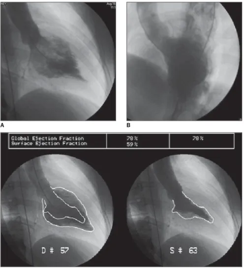
Typical findings of this disease are: a) large inverted T waves (> 10 mm) at ECG; b) the finding of “suit of spades” at left ventriculography (Figure 2).
From the histological point of view, an extensive disarrangement of myocardial fibers and myofribrillar and myocyte al-terations are observed, more restricted to the ventricular end, indistinguishable from other forms of HCM, reinforcing the
concept that the apical type is a HCM variant (7,8).
The echocardiogram has been the first-line method for evaluating patients with HCM, but its limitation in the study of the apex, mainly in the hands of inex-perienced professionals, complicates the diagnosis of AHCM. The MRI of the heart should be performed in the suspect of AHCM, since this study minimizes these problems because it is not opera-tor-dependent as much as ECHO, does not present any limitation to the window size, has multiplanar capacity and pre-sents an excellent contrast between soft parts(4,7–9).
In athletes, it seems that exercises would increase the left ventricular (LV) wall stress and work, factors which could affect the development of the localized LV hypertrophy. Little reference is made
to this hypothesis in the study of the AHCM, and up to this moment, no rela-tion has been established between the sports practice and the onset of the dis-ease(3,5,8).
The MRI of the heart has been per-formed for better studying the myocar-dium thickness, LV function and perfu-sion and, mainly, the myocardial en-hancement, ruling out the likelihood of an associated myocardial fibrosis and, through a non-invasive method, exclud-ing the possibility of endomyocardiofi-brosis or amyloidosis.
With the huge development of the non-invasive cardiac study, mainly by means of new methods like MRI and, most recently, CT, we must be careful with these diagnoses, particularly in cases of asymptomatic patients presenting a fa-milial history of heart disease.
A B
C
VII
Radiol Bras 2006;39(5):V–VII
REFERENCES
1. Sakamoto T, Tei C, Murayama M, Ichiyasu H, Hada Y. Giant T wave inversion as a manifesta-tion of asymmetrical apical hypertrophy (AAH) of the left ventricle. Echocardiographic and ultra-sonocardiotomographic study. Jpn Heart J 1976; 17:611–629.
2. Yamaguchi H, Ishimura T, Nishiyama S, et al. Hypertrophic nonobstructive cardiomyopathy with giant negative T waves (apical hypertrophy): ven-triculographic and echocardiographic features in 30 patients. Am J Cardiol 1979;44:401–412. 3. Codd MB, Sugrue DD, Gersh BJ, Melton LJ 3rd.
Epidemiology of idiopathic dilated and
hyper-trophic cardiomyopathy. A population-based study in Olmsted County, Minnesota, 1975-1984. Cir-culation 1989;80:564–572.
4. Albanesi Fº FM, Castier MB, Lopes AS, Ginefra P. Is the apical hypertrophic cardiomyopathy seen in one population in Rio de Janeiro city similar to the found in the East? Arq Bras Cardiol 1997;69: 117–123.
5. Webb JG, Sasson Z, Rakowski H, Liu P, Wigle ED. Apical hypertrophic cardiomyopathy: clini-cal follow-up and diagnostic correlates. J Am Coll Cardiol 1990;15:83–90.
6. Suzuki J, Shimamoto R, Nishikawa J, et al. Mor-phological onset and early diagnosis in apical hypertrophic cardiomyopathy: a long term
analy-sis with nuclear magnetic resonance imaging. J Am Coll Cardiol 1999;33:146–151. Erratum in: J Am Coll Cardiol 1999;33:1750.
7. Gavaliatsis IP, Kouvousis NM, Rallidis LS, et al. Recurrent atrial flutter in apical hypertrophic cardiomyopathy. Jpn Heart J 1992;33:499–504. 8. Eriksson MJ, Sonnenberg B, Woo A, et al.
Long-term outcome in patients with apical hypertrophic cardiomyopathy. J Am Coll Cardiol 2002;39:638– 645.