Maria Adelina Jerónimo
Dissertation presented to obtain the Ph.D. degree in Evolutionary Biology
Instituto de Tecnologia Química e Biológica António Xavier | Universidade Nova de Lisboa
Oeiras, November, 2016
novelty: eyespots and immunity
Esta dissertação é o resultado do meu próprio trabalho desenvolvido entre Maio de 2011 e Maio de 2015 no laboratório da Dra. Patrícia Beldade, Instituto Gulbenkian de Ciência em Oeiras, Portugal, no âmbito do Programa da FCT de Doutoramento (2010). Com base no trabalho desenvolvido, a minha orientadora e eu esperamos submeter dois artigos científicos para publicação.
This dissertation is the result of my own research, carried out between May 2011 and May 2015 in the laboratory of Dr. Patrícia Beldade, Instituto Gulbenkian de Ciência in Oeiras, Portugal, under the FCT Doctoral Programme (2010). From this work, my supervisor and I expect to submit two papers for publication.
Apoio Financeiro/Financial Support
Apoio financeiro da FCT e do FSE no âmbito do Quadro
Comunitário de Apoio, bolsa de doutoramento #
SFRH/BD/73658/2010, dos projectos atribuidos a P. Beldade (PTDC/BIA-BEC/ 099808/2008 e PTDC/BIA-EVF/2170/2012) e da Fundação Calouste Gulbenkian.
Financial support for this thesis was provided by the FCT and FSE thourgh the Quadro Comunitário de Apoio, doctoral fellowship # SFRH/BD/73658/2010, research grants to P. Beldade (PTDC/BIA-BEC/ 099808/2008 and PTDC/BIA-EVF/2170/2012) and Fundação Calouste Gulbenkian.
Acknowledgements
I would like to thank the IGC for giving me the opportunity to do my Ph.D..
To all elements of my thesis committee: in the very beginning Victor Barbosa, António Jacinto, and more recently Élio Sucena and Jorge Carneiro.
To all the present and past members of Variation: Development & Selection Lab, Evolution and Development Lab and Development, Evolution and the Environment lab. Thank you for all your help, patience and fruitful discussions that contributed a lot for this work.
For all my friends and family outside IGC that encouraged me throughout this journey. Thank you for your patience and your curiosity about my work.
Finally, I would like to thank to Patrícia Beldade, the supervisor that gave me the opportunity, the guidance and the freedom to dive into the wonderful word of evolution and development.
Summary
The infinitude of forms across living beings always fascinated scientists and laymen alike. Phenotypic variation is the raw material for natural selection and is transversal in biological systems. Evolutionary novelties are lineage specific traits with adaptive value. How such traits arise in organisms is still an open question. To understand this process we used the butterfly eyespots as a model, which are evolutionary novelty. Our testing hypothesis was that butterflies had rewired ancient genetic mechanisms used for the wound-response to develop eyespots, co-option. The major fact supporting this hypothesis is that wounding an early pupal wing can develop an eyespot-like pattern around wound site. This pinpoints the existence of genetic and cellular commonalities between eyespot development and wound response.
First, we characterized the genetic wound response on the wings of our model system, Bicyclus anynana, comparing the genetic profiles of wounded and non-wounded wing pairs (Chapter 2). Wounded wings were enriched for immunity-related genes. These genes are expressed in circular patterns around wound sites. Additionally, we observed faint expression patterns at the presumptive eyespot center, especially for the antimicrobial peptide gene Gloverin 2. Antennapedia, a transcription factor involved in eyespot development was also observed at wound sites. At presumptive eyespot centers and wound sites there are higher cell density due to migration of hemocyte-like cells.
Secondly, we explored the role of immunity into the development of wound-induced patterns (Chapter 3). We manipulated the immune system activation during pupal development and quantified the dorsal
adult wing patterns. Wounds with high immunity activation developed larger wound-induced eyespots. This enlargement is mediated through local immunity. With this experiments we noticed that not only dorsal wound-induced patterns had changed, but also the ventral wing patterns were different.
Thirdly, we characterized the immunity-mediated developmental plasticity, one genotype producing different phenotypes according to the environmental conditions, on ventral wing patterns (Chapter 4). As in low developmental temperature individuals, high immunity activation induced long developmental time, smaller eyespots, lower wing color contrast, and lower ecdyson hormone levels. This effect was independent of the organ where the immunity treatment was applied. To our knowledge, this is the first time that immunity is mediating developmental plasticity in butterfly wing patterns.
Lastly, we explored the immunity effects on wing size and shape (Chapter 5). Contrary to low developmental temperature, wings of high immunity individuals were smaller. However, this effect only occurred when immune treatment was applied in one of the developing wings, which reduced the proliferation rate. High immunity and low developmental temperature induce similar shape changes, pulling the wing shape in the same direction. This indicates that different stresses activate a common developmental program determining wing shape.
In summary, this work reinforces the hypothesis of wound genetic circuitry co-option for the evolutionary origin of butterfly eyespots. Moreover, it uncovered the crucial role of immunity in the butterfly wing patterns development. It also open up new perspectives to understand the origin and diversification of novelties, developmental plasticity and the ecological meaning of phenotypic variation on butterfly wing patterns.
Sumário
A infinitude de formas dos seres vivos sempre fascinou os cientistas e demais pessoas. A variação fenotípica é a base da seleção natural e é transversal aos sistemas biológicos. Novidades evolutivas são características exclusivas de uma linhagem e têm valor adaptativo. Como surgem num organismo ainda é uma questão em aberto. Para compreender este processo utilizámos os ocelos da borboleta que são uma novidade evolutiva. A hipótese que testámos foi que as borboletas reutilizaram mecanismos genéticos preexistentes necessários para a resposta às feridas, para formarem ocelos, co-opção. O facto mais relevante que suporta esta hipótese é que uma ferida na asa da pupa pode desenvolver um padrão de cores semelhante a um ocelo à volta da ferida. Isto indica que existem semelhanças genéticas e celulares entre o desenvolvimento dos ocelos e a resposta às feridas.
Primeiro, nós caracterizámos geneticamente a resposta à ferida nas asas do nosso modelo biológico, Bicyclus anynana, comparando os perfis genéticos de pares de asas feridas e não feridas (Capítulo 2). As asas feridas expressam níveis mais elevados de genes relacionados com a imunidade. Estes genes são expressos em padrões circulares à volta das feridas. Também observámos ténues padrões de expressão no presumível centro dos ocelos, especialmente para o péptido antimicrobiano Gloverin 2. Antennapedia, um factor de transcrição envolvido no desenvolvimento dos ocelos também foi detectado nas feridas. Nos presumíveis centros dos ocelos e nas feridas há maior densidade cellular devido à migração de células semelhantes a hemócitos.
Em segundo lugar, nós investigámos o papel da imunidade no desenvolvimento de padrões induzidos pela ferida (Capítulo 3). Nós manipulámos a ativação do sistema imune durante o desenvolvimento
maiores. Este efeito é mediado pela imunidade local. Observámos também que não só os padrões dorsais induzidos pela ferida foram alterados, como também os padrões ventrais das asas eram diferentes.
Em terceiro lugar, caracterizámos a plasticidade no desenvolvimento (quando um genótipo produz diferentes fenótipos de acordo com as condições ambientais) mediada pela imunidade nos padrões das asas ventrais (Capítulo 4). Tal como a baixa temperatura, a elevada ativação da imunidade induziu um desenvolvimento mais longo, ocelos menores, menor contraste de cor nas asas, e níveis mais baixos da hormona ecdisona. Este efeito foi independente do órgão afectado. Do que pesquisámos, esta é a primeira vez que a imunidade está a mediar plasticidade no desenvolvimento dos padrões das asas das borboletas.
Por fim, nós explorámos os efeitos de imunidade no tamanho e forma das asas (Capítulo 5). Contrariamente ao efeitos da baixa temperatura, as asas dos indivíduos com maior activação da imunidade foram menores. No entanto, este efeito só ocorreu quando o tratamento imunológico foi aplicado em uma das asas, o qual induz a redução da taxa de proliferação cellular. Uma activação elevada da imunidade e baixas temperaturas induziram diferenças de forma similares, na mesma direção. Isto indica que diferentes stresses activam o mesmo programa de desenvolvimento determinando a forma da asa adulta.
Em resumo, este trabalho reforça a hipótese de co-opção do circuito genético usado para responder às feridas na origem evolutiva dos ocelos das borboletas. Além disso, demonstrámos que a imunidade tem um papel crucial no desenvolvimento dos padrões das asas das borboletas. O trabalho aqui apresentado abre também novas perspectivas para entender a origem e diversificação das novidades evolutivas, a plasticidade no desenvolvimento e o significado ecológico das variações fenotípicas dos padrões das asas das borboletas.
Table of contents
Declaração/Declaration ... I
Apoio Financeiro/Financial Support ... I
Acknowledgements ... I
Summary ... II
Sumário ... IV
Table of contents ... VI
Chapter 1 – General Introduction ... 1
1.1. Evolutionary novelties ... 1
1.1.1. Novel traits on butterfly wing patterns ... 2
1.1.2. Developmental plasticity on butterfly wing patterns ... 3
1.1.3. Eyespot development ... 5
1.1.4. The evolutionary origin of butterfly eyespots ... 8
1.2. Wound response and immunity ... 10
1.2.1. Wound response includes tissue repair and immune mechanisms ... 11
1.2.2. Regulation of insect immunity ... 12
3. Aims and thesis scope ... 14
1.4. Acknowledgements ... 17
Chapter 2 – Molecular and cellular commonalities between
wound response and pigmentation patterning ... 18
2.1. Summary ... 18
2.2. Introduction ... 19
2.3. Materials and Methods ... 22
2.3.1 Animals ... 22
2.3.2 Wing wounding and dissections ... 23
2.3.3 RNA isolation and cDNA preparation for microarray ... 24
2.3.4 Microarrays, hybridization and slide scanning ... 25
2.3.5 Data normalization and levels of gene expression ... 25
2.3.6 Gene annotation and GO enrichment analysis ... 26
2.3.7 RNA isolation and cDNA preparation for qPCR ... 27
2.3.8 qPCR reference genes ... 28
2.3.9 qPCR reaction and gene expression quantification ... 29
2.3.10 In situ hybridization (ISH) and fluorescent in situ hybridization (FISH) ... 31
2.3.11 Immunohistochemistry (IHC) ... 32
2.4.1 Wound response induces the expression of AMPs and
melanogenesis enzymes in B. anynana early pupal wings ... 35
2.4.3 Antp, an “eyespot gene”, is expressed at wound site ... 48
2.4.4 The wound inhibits cell proliferation ... 52
2.4.5 There are cellular similarities between wound sites and presumptive eyespot centers ... 54
2.5. Conclusions ... 56
2.6. Acknowledgements ... 56
Chapter 3 – Immunity regulates the size and scale
composition of wound-‐induced pigmentation patterns ... 57
3.1. Summary ... 57
3.2. Introduction ... 58
3.3. Materials and Methods ... 60
3.3.1 Biological Material ... 60
3.3.2 Wounding and Immune system activation ... 61
3.3.3 Melanotic Spots ... 62
3.3.4 Survival ... 63
3.3.5 Quantitative PCR ... 63
3.3.6 Phenotypic measurements ... 66
3.3.7 Local and Systemic effect of immune system activation ... 67
3.4. Results and Discussion ... 68
3.4.1 The number of melanotic spots and the mortality increase upon treatment with heat-‐killed bacteria ... 69
3.4.2 Heat-‐killed bacteria treatments increase local and systemically the expression levels of immune-‐related genes ... 72
3.4.3 Immune stimulation at wound site does not increase the probability of WIE development ... 74
3.4.4 Activation of local immune system increases the morphogen-‐ like signaling affecting the cell fate decision of cover scale ... 77
3.4.5 Severe wounding and activation of local immune system inhibit the development of cover scale ... 79
3.4.6 Reepithelialization and immunity related-‐genes might have distinct roles in WIE development ... 83
3.5. Conclusions ... 86
3.6. Acknowledgments ... 86
Chapter 4 – Immunity up-‐regulation in pupae phenocopies
effects of low temperature on development time, wing
patterns, and pupal ecdysone levels ... 88
4.1. Summary ... 88
4.2. Introduction ... 89
4.3. Materials and Methods ... 92
3.3.2 Immune challenge treatments ... 93
4.3.3 Measurement of adult wing eyespots ... 94
4.3.4 Quantification of adult wing overall darkness and color contrast ... 95
4.3.5 Quantification of 20E levels ... 96
4.3.6 Manipulation of 20E levels ... 98
4.4 Results and Discussion ... 100
4.4.1 High bacterial dosage treatment reduces the number of “fast-‐ living” individuals ... 100
4.4.2 Systemic immunity is a developmental plasticity trigger ... 104
4.4.3 Local and systemic immunity has an additive effect on native eyespot size regulation ... 106
4.4.4 Different sets of eyespot traits were affected after wing and thorax immune challenge ... 107
4.4.5 Traits related with natural selection are more sensitive to internal immunity state during development ... 109
4.4.6 Immune challenge level, but not type, affected eyespot size 112
4.4.7 Immunity up-‐regulation only partially phenocopies the low developmental temperature syndrome ... 115
4.4.8 Higher immune challenge reduces 20-‐hydroxyecdysone to levels closer to those characteristic of lower developmental temperatures ... 116
4.4.9 Hormonal treatment of immune-‐challenged pupae induced high mortality ... 119
4.5. Conclusion ... 122
4.6. Acknowledgements ... 124
Chapter 5 – Immune and thermic stress induce similar
changes in wing shape ... 125
5.1. Summary ... 125
5.2. Introduction ... 126
5.3. Material and Methods ... 129
5.3.1 Biological Material ... 129
5.3.2 Wounds and Immune Challenge ... 129
5.3.3 Wing Area and landmarks measurements ... 131
5.3.4 Immunohistochemistry ... 134
5.4. Results and Discussion ... 136
5.4.1 Bacterial treatment, on developing wing, reduces the total area of all adult wings ... 137
5.4.2 Bacterial treatment induces allometric and non-‐allometric shape changes on wounded wings ... 139
5.4.3 Wing damage induces non-‐allometric changes in wing shape ... 141
5.4.4 Bacterial treatment reduces mitotic density locally and
systemically ... 143
5.4.5 Thermal and immune stresses modulate the wing shape in same direction ... 146
5.5. Conclusions ... 148
5.6. Acknowledgements ... 149
Chapter 6 -‐ General Discussion and Perspectives ... 150
6.1. Wounds and pigmentation pattern formation ... 151
6.1.1. The “generalities” and the “particularities” of B. anynana wound response ... 151
6.1.2. The “commonalities” of wound response and eyespot formation ... 153
6.2. Immune-‐related genes are key regulators of eyespot size . 155
6.2.1. High immune levels induce larger ectopic eyespots ... 155
6.2.2. High immune levels induces smaller native eyespots ... 157
6.3. Immune challenge and developmental plasticity ... 158
6.4. Acknowledgements ... 161
Bibliography ... 162
Supplementary Material ... Error! Bookmark not defined.
A -‐ Chapter 2 ... Error! Bookmark not defined.B -‐ Chapter 3 ... 203
Chapter 1 – General Introduction
The endless diversity of morphological forms across living beings, the ecological niches colonized and the good match between the two has always fascinated scientists and laymen alike. Phenotypic variation is a universal property of biological systems and the raw material for natural selection. The molecular and genetic mechanisms and the environmental factors underlying the production of phenotypic variation are a key subject in ecological evolutionary developmental biology (eco-evo-devo). One of the most studied traits in this field is body pigmentation. There is great intra-specific variation and inter-species diversity in body pigmentation, and this trait is ecologically relevant and experimentally tractable. Here, we focused on a model for studies of the origin and modification of novel traits (i.e. traits for which there are no clear homologs in other lineages) and of developmentally plasticity (i.e. the environmental regulation of development that can result in the production of distinct phenotypes without genetic differences): wing pigmentation patterns in butterfly wings.
1.1. Evolutionary novelties
Among the diversity of morphological traits, novel traits such as flowers, bird feathers, mammalian placenta, insect wings, beetle horns and butterfly eyespots (e.g. Figure 1.1) are especially fascinating and their origin is especially challenging to explain. There is no single universally accepted definition of what constitutes an evolutionary novelty and of how that can be ascertained. Different authors consider different levels of biological organization and focus on different criteria (Ravosa 1991), including 1) lack of clear homology in the ancestral
species (Muller & Wagner 1991), 2) new ecological function within the lineage (Massimo Pigliucci 2008), or 3) new ecological function that permit new functions and open up new adaptive zones. Here, we consider as novel traits those that are restricted to a lineage and have a specific adaptive value.
Figure 1.1- Examples of evolutionary novelties. Flowers in plants, hair in mammals,
feathers in birds and scale-based wing patterns in butterflies.
1.1.1. Novel traits on butterfly wing patterns
The beauty and astonishing diversity of butterfly wing patterns have been inspiring scientists for centuries, and have been used to address questions in disciplines as different as systematics and evolution, development and genetics, biochemistry, and immunity. During last decades several evo-devo (Brakefield et al. 1996; Saenko et al. 2011; Hines et al. 2012) and eco-devo (Prudic et al. 2011; Prudic et al. 2015) studies have increased our understanding about development, genetics and ecological relevance of butterfly wing patterns. However, many questions remain. For example, the evolutionary origin, the genetic regulation and the role of the environment during development of the butterfly wing patterns.
Butterfly color patterns are formed by the two-dimensional arrangement of single color scales. Those patterns are evolutionary novelties and have allowed wing surfaces to perform new ecological functions in thermoregulation and visual communication (Beldade & Brakefield 2002). The diversity between species is dramatic, but there is also variation even within a population considering different wing surfaces, different sex and different seasonal forms (Nijhout 1991). Homology relationships between pattern elements are represented in the "nymphalid groundplan" (Schwanwitsch 1929; Nijhout 1991). In this model, different wing pattern elements, such as eyespots, chevrons and bands, are organized in parallel series; individual elements are repeated along the anterior-posterior wing axis within so-called wing cells that correspond to wing compartments bordered by veins. The extent to which wing pattern elements can evolve in an independent way will impact how easily they can respond to different selection pressures and it will also impact the diversification of butterfly wing patterns (Nijhout 1991; Nijhout 2001).
Several studies focusing on the genetic mechanisms underlying wing patterns development and evolution have provided important insight into homology of different wing pattern elements (Monteiro et al. 2006; Shirai et al. 2012; Saenko et al. 2011). Among the different types of wing pattern elements, eyespots that are composed of rings of contrasting colors are arguably those most studied at several levels, such as molecular, genetic, developmental, ecological and evolutionary.
1.1.2. Developmental plasticity on butterfly wing patterns Phenotypic plasticity is the ability of an organism to react to environmental input with a change in phenotype (West-Eberhard
2003). It is also defined as the ability of a single genotype to produce more than one alternative form in different environmental conditions, whether the alternative phenotypes are continuous or discontinuous (Stearns 1989). Developmental plasticity is a particular case of phenotypic plasticity where environmental input during pre-adult development induces alternative phenotypes in the adult stage (Beldade et al. 2011). Temperature-dependent sex differentiation in turtles (Pieau et al. 1999), predator-induced change in head morphology of Daphnia (Boersma et al. 1998), and nutritional regulation of horns size in scarab beetle (Moczek & Emlen 2000) are examples of developmental plasticity.
Developmentally plastic traits typically have a particular period in which organisms sensing a different environment shift their developmental programs, and environmental changes outside this sensitive period do not induce phenotypic changes (Beldade et al. 2011). The sensitive period precedes the development of the final adult phenotype by a period of time that may be as brief as a few days or as long as several months (Nijhout 1999). For example, the sensitive period for the seasonal adult wing pattern polyphenism in butterflies is during later larval instars and early pupal stage (Nijhout 1999; Kooi & Brakefield 1999).
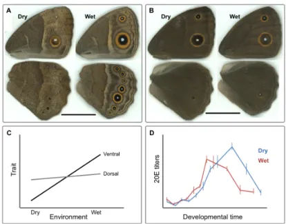
This study focuses on the eyespots of Bicyclus anynana, which are novel and developmentally plastic traits. B. anynana is an African butterfly living in tropical environments with two main seasons: the wet and the dry seasons which are very distinct in terms of temperature, humidity and food availability (Windig et al. 1994). This butterfly exhibits clear seasonal polyphenism in wing pattern and other traits (Brakefield & French 1999; Brakefield et al. 2009; Oostra et al. 2011). Wet season larvae produce adults with conspicuous ventral wing
patterns with large marginal eyespots, while dry season larvae produce adults with dull brown colors and very small eyespots (Figure 1.2A). These two alternative wing patterns are believed to correspond to seasonally different strategies to avoid predation. While the marginal large eyespots of the wet-season butterflies are thought to attract the predator’s attention to the wing margin and away from the vulnerable body, the all-brown dry-season butterflies are cryptic against a background of dry leaves (Lyytinen et al. 2003; Prudic et al. 2015; Beldade et al. 2011). Thus, this seasonal polyphenism provides an adaptive response to the alternating seasonal environments increasing the individual’s fitness.
Studies have shown that the temperature during late larval development, which predicts the season of adults, is an important environmental cue determining the development of the adult wing pattern phenotypes through changes in hormonal dynamics (Kooi & Brakefield 1999; Koch et al. 1996; Mateus et al. 2014) (Figure 2.2C and D). The eyespots on the ventral surface of B. anynana wings, the one exposed to predators when the butterfly is resting, are the ones most thermally plastic (Brakefield et al. 2009; Mateus et al. 2014) (Figure 2.2A). The dorsal eyespots, which are implicated in mate choice, are much less plastic, at least in relation to temperature (Breuker & Brakefield 2002; Westerman et al. 2014; Mateus et al. 2014) (Figure 2.2B).
1.1.3. Eyespot development
During the past decades, a number of eco-evo-devo studies focused on butterfly eyespots and provided insight into their evolutionary origin, ecology and developmental plasticity (e.g. Saenko et al. 2011; Prudic et al. 2015; Mateus et al. 2014). The experimental
tractability of developing eyespots is an advantage in evo-devo studies. For example, easy detection and access to the position of veins and developing eyespot centers on the dorsal forewing of developing pupae allows for very precise surgical manipulations under a simple stereoscope.
Figure 1.2- Seasonal polyphenism in B. anynana. In the lab, lower developmental
temperatures (e.g. 19°C) lead to the production of adults with wing patterns resembling those of the dry season, while warmer developmental temperatures (27°C) lead to the production of adult wing patterns resembling those of the wet season. Temperature during development mediates phenotypic plasticity for ventral wing patterns (A) and has a much smaller effect on dorsal patterns (B). Ventral patterns are more plastic than dorsal patterns in response to environmental temperatures (Mateus et al. 2014) D) Different 20E dynamics in the hemolymph of pupae from the different seasonal forms (cf. Oostra et al. 2011).
Wing development starts during the first larval instar when the wings greatly increase in size and the venation system increases in complexity. During this stage, the eyespot development starts with determination of eyespot centers or foci (Figure 1.3) (Brakefield et al. 1996; Saenko et al. 2011). A series of genes have been associated
with this phase of eyespot development, including Antennapedia (Antp), Distal-less (Dll), Notch (N), engrailed (en), hedgehog (hh), cubitus interruptus (ci), patched (patch) and spalt (sal) (Saenko et al. 2011; Monteiro et al. 2006; David N Keys et al. 1999). Later, in early pupal wings there is the positioning of the eyespot rings around the focus (Brakefield et al. 1996; Wittkopp & Beldade 2009). Surgical manipulations of pupal wings have led to the suggestion that focal cells produce (or degrade) a diffusible morphogen-like signal that determines the fate of surrounding cells (Nijhout 1980; Monteiro et al. 1994; French & Brakefield 1995). Depending on the concentration of the morphogen-like signal they are exposed to, surrounding cells express different (combinations of) transcription factors (during early pupal stage), which correspond to the production of different color pigments (in late pupae). In B. anynana, Dll and Sal map the black ring and en the golden ring (Brunetti et al. 2001; Beldade et al. 2002). The identity of the morphogen-like signal triggering the expression of these transcription factors is still unknown. The synthesis of pigments and their deposition is the last stage of eyespot development and takes place only during the last days of the pupal life (Figure 1.3).
Figure 1.3- B. anynana eyespot development during larval and pupal stages. Focal
pupae, the eyespot center presumably produces (or degrades) a morphogen-like signal inducing the transcription of “positioning” genes in rings around the focus (Brunetti et al. 2001). In the last days of the pupal stage, pigment synthesis takes place (Iwata & Otaki 2016). The sensitive period to induce an ectopic eyespot in this butterfly is from 6 to 18 hours post-pupation for 27°C rearing temperature individuals (Brakefield & French 1995).
1.1.4. The evolutionary origin of butterfly eyespots
The origin of evolutionary novelties is one a key question in evo-devo (Muller & Wagner 1991; Müller 2007; Moczek 2008). The factors promoting the diversification of novel traits have been studied for a long time (Stebbins 1970), but the genetic and developmental mechanisms underlying their origin have become the focus of research only in the last decades (Wagner & Lynch 2010; Saenko et al. 2008). “Co-option” has been proposed as the main mechanisms explaining the origin of evolutionary novelties (True & Carroll 2002). This mechanism refers to an evolutionary process by which evolution recycles ancestral genetic circuitries shared across lineages to produce novel traits (True & Carroll 2002; Ganfornina & Sánchez 1999). There are several studies showing that the origin of novel traits largely relies on existing genes and gene networks that are re-deployed and subsequently modified to give rise to novel morphological structures (Moczek & Nagy 2005; Saenko et al. 2011; Prum 2005). Gene recycling highlights the pleiotropy of genetic networks, which are involved in various processes during development and in different body organs. Recent whole genomic studies have raised the possibility that lineage-specific genes can also play a role (Khalturin et al. 2009; Donoghue et al. 2011; Zhou et al. 2015).
Scientists have highlighted the co-option of different genetic circuitries for the evolutionary origin of butterfly eyespots, such as leg-development, wing margin determination, anterior-posterior wing compartmentalization, embryonic development and wound response (Held 2013; Monteiro 2015; Saenko et al. 2011). In this thesis I have focused on the commonalities between eyespot development and response to epidermal wounds. The fact that some butterflies develop organized color patterns around wound sites resembling the adult eyespot patterns (Brakefield & French 1995; Monteiro et al. 2006; Otaki 2011) reveals commonalities between eyespot development and the response to wounding. While it is common to have dark pigmentation spots at wounded sites (Tang 2009; Levesque et al. 2012; Binggeli et al. 2014), the production of organized color patterns centered on wound sites is less common, but not restricted to butterflies (Ohno & Otaki 2012). In B. anynana, ectopic eyespots can be induced by wound on pupal wings from six to 18 hours post pupation, with 12 hours post-pupation corresponding to the highest probability of induction for butterflies reared at 27°C(Brakefield & French 1995).
These observation of wound-induction of ectopic eyespots led to the hypothesis of co-option of the wound response genetic circuitry for the evolutionary origin of eyespots (Monteiro et al. 2006). The genes involved in the differentiation of ectopic eyespots have not been extensively investigated and, thus, it is unclear whether native eyespots and ectopic eyespots are using the same genetic circuitry. Monteiro and co-workers investigated the gene expression of some known eyespot-related genes and saw that en, Sal and Dll were expressed in cells around wound sites several hours after wounding (Monteiro et al. 2006). In this case, eyespots are like “painted scars” on wings. This work presented here started off to investigate the
genetic and cellular basis of wound-induced eyespot formation. For this reason, the next section provides a brief overview of the mechanisms of wound response in insects.
1.2. Wound response and immunity
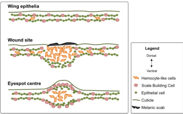
Multicellular organisms can repair their epithelia after an injury. This process is essential to maintain homeostasis (Davis & Engström 2012) and restore the integrity of the outer barrier. Wound response integrates evolutionarily conserved cellular and genetic mechanisms to form a protective scab, fight pathogens, and restore epidermis integrity (Moussian & Uv 2005; Wood et al. 2002). During the last decades, insects, and in particular Drosophila melanogaster, have become key model systems to explore the cellular and molecular mechanisms of wound healing, epithelial repair and regeneration (Wood et al. 2002; Bosch et al. 2005; Jiang et al. 2011). In insects, the wound response can be divided into five main steps: 1) early wound signaling, 2) plug formation through hemolymph coagulation and attraction of hemocytes, 3) melanin accumulation, degranulation of hemocytes and wound site stabilization, 4) phagocytosis and, 5) reepithelization (Lai et al. 2002; Galko & Krasnow 2004; Yoo et al. 2012) (Figure 1.4). Failure in any of these processes can seriously compromise survival. For example, the activation of melanogenesis is crucial and Drosophila mutants lacking hemocyte-phenoloxidases cannot properly heal compromising development and survival (Galko & Krasnow 2004; Binggeli et al. 2014).
The few existing studies of wound healing in Lepidopterans have focused on the larval stage (Madhavan & Schneiderman 1969; Rowley & Ratcliffe 1978). Wound response in Lepidopterans shared the main aspects of wound response in Drosophila and also in
mammals (Moussian & Uv 2005; Krautz et al. 2014). Despite much evolutionary conservation in wound healing mechanisms, there are also particularities to some taxa (e.g. regeneration of entire organs in adult starfish (Fan et al. 2011; Gurtner et al. 2008)) and also differences between developmental stages of the same species (e.g. embryonic wounds, unlike those of adults, usually regenerate without scar (Wood et al. 2002; Galko & Krasnow 2004)). In this thesis, I focused in a particularity of butterfly wound response, which is restricted to a very specific time window during early pupal development: induction of ectopic rings of colors.
1.2.1. Wound response includes tissue repair and immune mechanisms
After a wound, homeostasis needs to be restored rapidly and efficiently. Towards that, wounded cells send some kind of alarm signals (such as Ca2+ and H2O2) to their neighboring cells to activate
wound response (Yoo et al. 2012; Razzell et al. 2013) (Figure 1.4). In this way wounded cells behave like organizers, releasing signals to surrounding cells and to immune cells that readjust their transcriptional programs to induce melanization, coagulation, cell shape changes, the formation of functional actomyosin structures, the recruitment of immune cells, the production of AMPs and reepithelization (Niethammer et al. 2009; Cordeiro & Jacinto 2013; van der Vliet & Janssen-Heininger 2014) (Figure 1.4). During wound response, there is activation of tissue repair and immune processes. Grossly, we can distinguish various immune mechanisms: the inflammation process in which hemocytes migrate towards the wound site, melaninzation and the massive production of anti-microbial peptides (AMPs) in the fat body (the insect equivalent to the
mammalian liver). On the other hand, the formation of the plug, stabilization of the wound site, and reepithelization can be grouped as tissue repair mechanisms. Although it could be argued that wound healing is not part of the immune response, apart from in highly controlled laboratory experiments, it is nearly impossible to achieve wounding without exposure to potentially infectious organisms. Moreover, both tissue repair and immunity processes are intrinsically linked and even in sterile wounds, where in theory the immunity should not play a role, immune mechanisms are present as well as the expression of immune related genes such as AMPs (Márkus et al. 2005).
1.2.2. Regulation of insect immunity
Upon infection, insects orchestrate a humoral and a cellular immune response to control pathogens. This response integrates the production of a battery of AMPs in the fat body, which are then released into circulation (Bulet & Stocklin 2005; Aggarwal & Silverman 2008), adjustment of blood cell behavior (Williams 2007; Lavine & Strand 2002) and melanization (Nappi & Christensen 2005; Eleftherianos & Revenis 2011). These processes are tightly regulated through hormones immune signaling pathways such as Toll and immune deficiency (IMD).
The molecular events initiating the transcriptional induction of AMP genes are well characterized (De Gregorio et al. 2002; Lemaitre & Hoffmann 2007; Valanne et al. 2011; Myllymäki et al. 2014). Upon infection, pathogens are recognized through microbial patterns recognition receptors (PRRs), such as peptidoglycan recognition proteins (PGRPs) and Gram-negative binding proteins (GNBPs). Binding of pathogen-derived molecules to these receptors activates
Toll and IMD signaling cascades. Toll pathway is activated by fungal and many Gram-positive bacteria, whereas the IMD pathway responds to Gram-negative bacteria (De Gregorio et al. 2002; Tsakas & Marmaras 2010). Among insects, these pathways are more studied in Drosophila (Lemaitre & Hoffmann 2007; Valanne et al. 2011), but the conservation of the main aspects of these pathways in other insects is remarkable (Casanova-Torres & Goodrich-Blair 2013; Wang et al. 2007).
Figure 1.4- Five different stages of wound response in insects. Adapted from (Krautz
et al. 2014).
Juvenile hormone (JH) and 20-hydroxy-ecdysone (20E) are highly versatile hormones, coordinating development, growth, reproduction, and aging in insects. Changes in 20E levels during development provide key signals for initiating developmental and physiological transitions, while JH promotes or inhibits these signals in a stage-specific manner (Beckstead et al. 2005; Nijhout et al. 2007; Andersen
et al. 2013). These two hormones also control immune response in Drosophila (Flatt et al. 2008; Rus et al. 2013; Regan et al. 2013), and in other insects (Tian et al. 2010; Wang et al. 2014).
To better respond to environmental conditions, insects can adjust hormone levels and/or the sensitivity of target organs to the hormone (West-Eberhard 2003). Hormonal levels can directly affect gene expression (Herboso et al. 2015). For example, in Drosophila, higher levels of 20E increase the expression of AMPs while JH has the opposite effect (Flatt et al. 2008). In B. anynana, 20E affects eyespot development (Koch et al. 1996; Mateus et al. 2014; Monteiro et al. 2015). Target organs can change their sensitivity to hormonal levels. For example, to decrease plasticity in genitalia, Drosophila males reduce the expression of FOXO, so this organ is less sensitive to insulin (that reflect nutrition levels) (Tang et al. 2011). To increase thermal plasticity in some eyespots, B. anynana express more ecdysone receptors in those eyespots’ centers (Monteiro et al. 2015).
3. Aims and thesis scope
The origin of evolutionary novelties remains one of the most challenging topics in evo-devo. Here we investigated the involvement of an ancient genetic circuitry in the development of a novel trait through a series of different approaches, including analysis of candidate genes and less biased genetic analysis; manipulations and analysis of developing wings; characterization of various aspects of the adult wing pattern phenotype in treated and control groups.
We started investigating the 1) genetic and cellular mechanisms underlying the evolutionary origin of eyespots (Chapter 2), 2) the effects of wound-associated immune activation on wound-induced eyespots (Chapter 3), 3) the immunity-mediated developmental
plasticity in wing patterns and its hormonal regulation (Chapter 4) and 4)) the molecular basis of immune-induced regulation of organ size and shape.
Previous studies support the co-option of wound response genetic circuitry hypothesis for the evolutionary origin of butterfly eyespots. Namely 1)the fact that wounds on early pupal wings can lead to the development of an eyespot-like pattern (e.g. Brakefield & French 1995) and 2)that some “eyespot” genes are expressed around wound sites (Monteiro et al. 2006). However, if this hypothesis is correct we also expect to have expression of some typically “wound” genes in developing eyespots. In Chapter 2 we addressed this hypothesis by investigating genetic and cellular commonalities between wound response and eyespot development. We started by comparing gene expression profiles of wounded vs. non-wounded pairs of pupal wings to identify wound-induced changes in gene expression putatively associated with the formation of ectopic eyespots in B. anynana. We picked some of the genes differentially expressed after wounding to validated and extended the microarray-based analysis with a targeted study of gene expression spatial patterns and level dynamics. In this chapter, we also compare the cellular organization at the wound site and at the center of the presumptive eyespots. This study allowed us to ascertain which aspect of the wound response process (tissue repair or/and immunity) contributed to wound-induced eyespot formation and might have been involved in the evolutionary origin of eyespots.
In Chapter 3 we explored the possible involvement of immune-related mechanisms/processes in specification of cell color fate around wound sites inflicted on pupal wings. The development of a wound-induced ectopic eyespot means that wounds somehow can
change the cell fate of surrounding cells, acting much like an eyespot organizing center. Previous studies revealed that severe wounds could change the likelihood of production of an ectopic eyespot around a wound site, but not its size (Brakefield & French 1995). Our results suggest a new role of immune-related pathways into the eyespot size regulation, which does not interfere in the likelihood of production of an ectopic eyespot.
In Chapter 4, we describe the effect of an immune challenge in pupae on B. anynana adult wing patterns. This effect phenocopies the well-known effect of cooler developmental temperatures on wing pattern, as well as on ecdysone dynamics. We explored the effect of immune activation with different bacterial types and upon wounding of different body parts on developmental time, ecdysone dynamics, and adult wing patterns. These experiments open new perspectives for a role of immune challenge as a mediator of plasticity in wing color patterns.
In Chapter 5 we explored the effects of immune challenge on organ size and shape and discuss its impact on interorgan communication and readjustment of growth during development. Typically, the investment in immune defense is associated with depletion of energetic resources and, consequently, with trade-offs with various life history traits. We compared the effects of immune challenge from wound on different body parts on size and shape of adult wings. We further investigated the mechanisms behind the reduction of wing size by analyzing the effect of the immune challenge on the number of mitotic cells. We also compared differences in wing size and shape resulting from immune challenge with differences in wing size and shape between individuals reared at different temperatures. These experiments allowed us to understand how the
two factors affecting wing development (temperature and level of immune challenge) affect different axes of wing growth.
Finally, in Chapter 6, we discuss the main contributions of this work and present possible future directions of research that could further help us understanding the genetic basis of evolutionary novelties and how immune challenge can act as a cue inducing phenotypic change in butterfly wing patterns.
1.4. Acknowledgements
We would like to thank the ‘evo-devo’ community at Instituto Gulbenkian de Ciência, including the labs headed by Patrícia Beldade, Christen Mirth, and Élio Sucena, as well as Magda Atilano for fruitful discussions throughout my PhD.
Chapter 2 – Molecular and cellular
commonalities between wound
response and pigmentation
patterning
2.1. Summary
The response to epidermal wounding is an evolutionarily conserved process and is typically tightly connected to pigmentation. An interesting example occurs in some Lepidopterans that can develop organized pigmentation patterns around wound sites that resemble native pattern elements called eyespots. This suggested the existence of molecular and cellular commonalities during development of both wound-induced and native eyespots. To reveal these commonalities, we studied the wound responseon pupal developing wings of the butterfly Bicyclus anynana. Microarray analysis comparing gene expression in wounded vs. non-wounded pairs of wings reveled enrichment for antimicrobial peptides (AMPs) and melanin pathway enzymes, both related with innate immunity in insects. We validated and extended the microarray analysis through quantitative RT PCR (qPCR), and in situ hybridization (ISH). We also found that the cell organization of the epidermis was similar at wound sites and at the center of presumptive eyespots: more cells between dorsal and ventral epithelia, expressing more Actin, Antennapedia (Antp) and Gloverin 2 (Glov2) (an AMP). Higher cell density is not a consequence of higher cell proliferation around wound sites suggesting cell migration towards wound sites. The cell shape and
gene expression of those cells suggests that they are hemocytes. Our experiments brought new insight onto a specific wound response (post-growth and pre-adult stage) and also reinforce the hypothesis of the evolutionary origin of eyespots through co-option of the ancient wound response genetic mechanisms.
2.2. Introduction
Multicellular organisms can repair their epithelia after an injury. This process is essential to maintain homeostasis (Davis & Engström 2012) and restore the integrity of the outer barrier. Wound response integrates evolutionarily conserved cellular and genetic mechanisms of tissue repair and immunity (Moussian & Uv 2005; Wood et al. 2002). During the last decades, insects and in particular Drosophila melanogaster, have become a key model system to explore the mechanisms of wound healing, epithelial repair and regeneration (Wood et al. 2002; Bosch et al. 2005; Jiang et al. 2011; Moussian & Uv 2005). There are extensive similarities between insect species (Krautz et al. 2014; Eleftherianos & Revenis 2011; Kanost et al. 2004) and even between insects and mammals wound response (Moussian & Uv 2005).
After wounding, insects form a melanin clot to seal wounds and trap microorganisms, blocking their entry into the insect open body cavity (Lavine & Strand 2002; Eleftherianos & Revenis 2011). The microorganisms are subsequently agglutinated, immobilized, and killed by various lectins and antimicrobial peptides (AMPs) (Eleftherianos & Revenis 2011). To restore epithelium integrity, epidermal cells spread along and through the clot until they meet and reestablish a continuous epithelial sheet (Galko & Krasnow 2004). During this process, there is reorganization of epithelial structure and
cell’s cytoskeleton adjustments (Moussian & Uv 2005; Jacinto et al. 2001). Insect immunity consists of a panel of humoral and cellular defense mechanisms, acting local and systemically, which together provide efficient protection against pathogens. The humoral component of the immune system include the transcriptional activation of genes that lead to the production of effector molecules that recognize (Pattern Recognition Patterns, PRPs) and kill pathogens (AMPs, reactive intermediates of oxygen and nitrogen, and melanization of hemolymph) (Lavine & Strand 2002). AMPs are produced in the insect fat body and released into the hemolymph, but also in epithelial cells and in the insect´s blood cells, hemocytes (Lemaitre & Hoffmann 2007). Hemocytes, are involved in processes such as phagocytosis, formation of multicellular hemocyte aggregates, nodulation and encapsulation of invading pathogens, and production of oxygen and nitrogen reactive species (Strand 2008; Eleftherianos & Revenis 2011; Yi et al. 2014). Hemocytes are recruited to the wound site and engulf damaged epithelial cells and pathogens (Wood et al. 2006; Lavine & Strand 2002; Losick et al. 2013). These wound response processes are highly conserved, but there are also particularities of wound response in different species and in different developmental stages and/or tissues (Bosch et al. 2005; Gurtner et al. 2008).
An interesting particularity of wound response occurs in some Lepidopterans, the insect order of butterflies and moths. Wounds in developing pupal wings can lead to the formation of organized pigmentation patterns centered at wound sites (Nijhout 1985; Brakefield and French 1995; Otaki 2011). The wound response process is typically and broadly tightly connected to pigmentation, and presence of different pigmentation patches and spots at wound sites is very common (Levesque et al. 2012; Binggeli et al. 2014). In this
particular wound response in a pre-adult and post-growing developmental stage, there is the formation of well-organized pigmentation patterns that resemble native wing eyespots, pattern elements composed of concentric rings of different colors (Figure 2.1A).
Butterfly eyespots have been prime models for evo-devo studies (Beldade & Brakefield 2002) including studies of the origin and diversification of evolutionary novelties, lineage specific traits (Beldade & Brakefield 2002; Saenko et al. 2008). Eyespots develop around organizing centers that, early in pupal wings are though to produce diffusible signals that induce surrounding epidermal cells to form rings of different colors (French & Brakefield 1995). Wound sites also behave like organizers, releasing signal to the surrounding and promoting recruitment of immune cells and reepithelization (Niethammer et al. 2009; Bidla et al. 2009; Galko & Krasnow 2004). The fact that wound sites can induce the production of eyespot-like patterns lead to the suggestion that the evolutionary origin of butterfly eyespots might be related to the co-option of wound response genetic circuitry conserved across insects (Monteiro et al. 2006). Co-option is a process by which evolution recycles ancestral genetic circuitries shared across lineages to produce novel traits (True & Carroll 2002; Ganfornina & Sánchez 1999). Melanogenesis is a good example of this pleiotropy; being involved in body pigmentation, but also in other important physiological functions such as immunity (Kim et al. 2013; Wittkopp & Beldade 2009). Since immunity is activated after wounding, the melanin pathway genes are good candidates to bridge the wound response and the developmental mechanisms to form eyespot-like patterns. However, until now the expression of these genes or other immune related genes was not reported in early stages of eyespot development. Early studies revealed that the ability to induce
eyespots around wound sites is restricted in time (6-18h post-pupation, for B. anynana individuals developing at 27°C) and space (on the distal half of the dorsal surface of forewings) ( Brakefield and French 1995).
In this study we aimed at characterizing the molecular and cellular commonalities between wound response and eyespot development, which underlie the hypothesis of co-option of wound response genetic circuitry into the evolutionary origin of eyespots. We wounded B. anynana pupal wings to compare gene expression profiles of wounded and non-wounded wings, and we investigated the spatial patterns of gene expression of “wound genes” and “eyespot genes” at eyespot centers and wound sites respectively. We also analysed the cellular organization at wound sites and presumptive eyespot centers. Our results shown that, genes (AMPs and melanogenesis enzymes) and cells of innate immune system (hemocyte-like cells) are present at wound sites and also at presumptive eyespot centers. Antp, which is known to be involved in eyespot development, is also expressed at wound sites. This suggests that innate immunity mechanisms might have been co-opted during evolution for eyespot development.
2.3. Materials and Methods
2.3.1 AnimalsB. anynana wild-type laboratory stock was reared as described in (Brakefield et al. 2009). Briefly, the butterflies were maintained at 27°C (+/- 0.5ºC) with 65% (+/-1%) humidity on a 12h light/dark cycle. Eggs were collected in young maize plants from adult cages (45x45x45cm; BugDorm-44545) with approximately 400 individuals fed with fresh banana on top of wet cotton. Larvae were fed with fresh maize plants and maintained at densities of 150 to 200 individuals per
cage (same size as adult cages). During the fifth larval instar we sexed larvae keeping only females for this study. Pre-pupae were collected onto 25 well plates and pupation time was recorded during the night via time-lapse photography (one photo every 10 min; Canon 1000D digital camera, Hahnel Giga T Pro 2.4GHz wireless timer remote control).
2.3.2 Wing wounding and dissections
The experiments had two groups: non-wounded individuals (NW) with no manipulation, and wounded (W) individuals, which were wounded twice for gene expression microarray analysis and qPCR. Wounds were inflicted with a tungsten needle (cat. no. 501317; World Precision Instruments) on the dorsal surface of the right forewing; in the two vein-bound wing compartments below the anterior eyespot, approximately halfway between the wing margin and the normal location of the eyespots (Figure 2.1B). In experiments of spatial patterns, in situ hybridization (ISH), fluorescent in situ hybridization (FISH)) and immunohistochemistry (IHC) we wounded as before, but only did one wound on the first wing compartment below the anterior eyespot. All wounds were inflicted at 12 hours post-pupation, according to Brakefield and French this is the best time point to induce an ectopic eyespot (Brakefield & French 1995). After wounding, all pupae were returned to 27°C until further manipulations.
For gene expression microarrays analysis and qPCR we dissected the wings removing the attached external cuticle. We dissected wings at two different time points for gene expression microarray analysis (four and eight hours post-wounding (Figure 2.1B) and five different time points for qPCR analysis (two, four, six, eight and 10h post-wounding). For the gene expression spatial patterns
(ISH) and cellular structure analysis (IHC) we dissected the wings keeping the attached external cuticle, which helps to maintain the wing integrity.
2.3.3 RNA isolation and cDNA preparation for microarray The distal half of each dissected forewing (without the cuticle) was used for RNA isolation. We had two replicate pairs of samples (wounded and non-wounded wings) for each of two time points (16hr and 20hr post-pupation, corresponding to 4hr and 8hr post-wounding, respectively). Total RNA was extracted with TRIzol (Invitrogen) following manufacturer’s instructions. RNA in RNAse-free water was checked for concentration and purity (A260/A280 ratio of > 1.8) by spectophotometry (NanoDrop), and for integrity by running in a 1.1 % agarose gel. We used total RNA to synthesized and amplify cDNA using the Ovation System (NuGEN) with oligo-dT primer and following manufacturer's instructions. We then purified the cDNA using Quiaquick cleanup kit (Qiagen) with a final elution volume of 30 µl in 1x TE buffer. Purity and concentration of cDNA samples was assessed in NanoDrop and agarose gel. All cDNA samples met the requirements for NimbleGen-Roche hybridization (A260/A280>1.7, A260/A230>1.5, >1µg cDNA/sample) and were stored at -20°C prior to shipment to NimbleGen-Roche (Madison, WI, USA) on dry ice. Cy3 labeling of cDNA was done at NimbleGen-Roche starting with 1 µg of quality-controlled cDNA (Agilent Bioanalyzer) and using Cy3 random primers and Klenow enzyme. Each sample of cDNA with added Cy3 random primers was heated to 98°C for 10 min and then cooled on ice for 10 min, after what Klenow and dNTPs were added before incubation at 37°C for 2hr. Reaction was stopped by adding 10 µl EDTA. Labeled cDNA was then precipitated and used in hybridization.
2.3.4 Microarrays, hybridization and slide scanning
To measure gene expression levels, we used Custom Nimblegen-Roche microarrays (Gene Expression 4x72K Arrays) with features designed to represent 15830 B. anynana gene objects, corresponding to contigs or singletons resulting from the assembly of >200,000 ESTs (mostly from developing wings) described elsewhere (Beldade et al. 2006; Beldade et al. 2009), as well as a number of other genes that had been previously cloned and implicated in B. anynana wing patterning. A gene can be represented by multiple gene objects, which are named with C[number], standing for contig; S[number], standing for singleton, a contig uniquely associated to a UniGene (c.f. (Beldade et al. 2006); and P[number] for those previously cloned by P. Beldade. The custom array contained 76,697 60mer probe features including 69,921 corresponding to the B. anynana genes (each gene object being represented by 1-6 probes) and a number of different types of controls (including 2,000 random probes). Microarray hybridization, scanning and image extraction was performed at Nimblegen-Roche following standard protocols (NimbleGen 2011). NimbleGen provided access to the image files as well as to the corresponding PAIR files. We used the non-normalized fluorescence values (raw intensities) for further analysis.
2.3.5 Data normalization and levels of gene expression
The raw intensities provided for each microarray feature were normalized with the software ANAIS, a web-based tool for the processing of NimbleGen expression data (Simon & Biot 2010). Briefly, raw intensities were normalized for intra-array (RMA background correction) and inter-array (quantile) variation. The same tool was also used to do the “probe to gene conversion” (using median value of all
probes for each gene) and a quality control analysis. We verified the result of the normalization through the alignment of all arrays boxplots (Quality Controls; Boxplots by array option – Figure 2.1C top), and also the reproducibility of results, by clustering analysis (Quality Controls; Hierarchical clustering of all arrays option –Figure 2.1C bottom). The latter revealed that samples clustered first by individual (wounded and non-wounded wings of each individual) and second by time point (16 and 20hrs). Details of these analyses are described in (Simon & Biot 2010). Having only two biological samples per time point precluded the typical analysis of gene expression differences using false discovery rate (Reiner et al. 2003). On the other hand, the design of comparing wounded and non-wounded (control) wings of single individuals allows a paired analysis.
Statistical analysis of normalized intensities was done in R (R Development Core Team 2015). For each gene object in each individual we first calculated the gene expression in wounded relative to control wing (log2Fold Change (FC) = log2 (intensity in wounded wing) - log2(intensity in control wing)), and after the respective fold change (FC = 2 ^ log2FC) (Quackenbush 2002). We considered as differently expressed, gene objects that were at least FC>2 (up-regulated) or FC<2 (down-(up-regulated) in both biological replicates at each of the two time points.
2.3.6 Gene annotation and GO enrichment analysis
To determine whether the differently expressed genes represented particular functional classes we ran an enrichment analysis of Gene Ontology (GO) terms associated with those genes. Microarray gene objects were first annotated by blastx (in Blast2Go v2.2.25+ (Conesa et al. 2005)) against Arthropoda genomes,
comprised of 2,411,977 proteins (from NCBI data bases; March 2015), with 2,062,374 corresponding to insects. For each gene object, we retained the best hits (threshold e-value <= e-10). From 15,830 gene objects in the array, 3,635 were annotated in this way. Each annotated gene object was associated to single or multiple GO terms, and the list of GO terms associated to each gene object was used as a "customized genome reference" in gene enrichment analysis done in Cytoscape v2.8.3 , using the BINGO v2.4.4 plugin (Smoot et al. 2011; Maere et al. 2005). Cytoscape runs the hypergeometric test using Benjamini–Hochberg false discovery rate (Benjamini & Hochberg 1995) of 5% to assign overrepresented GO-biological processes categories in the list of differentially expressed genes.
2.3.7 RNA isolation and cDNA preparation for qPCR
Left and Right forewings were extracted, together with overlying cuticle, from NW and W pupae (see above) at five different time points: 2, 4, 6, 8, and 10h post wounding. The wings were collected directly into a 2mL epp with a 7mm glass bead and 500µL of TRIzol (Invitrogen), kept at -20°C until all the samples of that day were collected and then they were disrupt and homogenized for 5 min at maximum speed in TissueLyserII (Qiagen) and stored at -80°C until RNA isolation.
Total RNA isolation was performed following manufacturor’s protocol without DNase treatment. To avoid possible genomic DNA contaminations we designed all qPCR primers spanning an intron. RNA was diluted in 25 µL of RNAse-free water, checked for integrity by running in a 1 % agarose gel and stored at -80°C until cDNA synthesis. RNA concentration was measured for 30 wings (from 15 individuals) and it ranged from 150 to 400 ηg/µL. Differences in RNA