Level of evidence and grade of recommendation of articles
on the diagnostic accuracy of ultrasonography in carpal
tunnel syndrome
*
Nível de evidência e grau de recomendação dos artigos sobre a acurácia diagnóstica da ultrassonografia na síndrome do túnel do carpo
Kátia Maria Diniz de Carvalho1, Evelyne Pessoa Soriano2, Marcus Vitor Diniz de Carvalho3, Clóvis César de Mendoza4, Humberto Gomes Vidal5, Ana Beatriz Vasconcelos Lima Araújo6
Objective: The present study was aimed at assessing the quality of articles related to diagnostic accuracy of ultrasonography both in idiopathic and occupational carpal tunnel syndrome. Materials and Methods: A search was undertaken in electronic databases, selecting reports on systematic reviews, randomized clinical trials and observational studies. After four stages of analysis, from an initial screening to the evaluation by means of the assessment scale developed by the Critical Appraisal Skills Programme, the articles were classified according to levels of evidence, with their respective grades of recommendation. Results: Among 68 articles initially identified, only 4 have met all the methodological quality criteria proposed in the present study. Measurement of the cross-sectional area of the median nerve was the most important criterion for the diagnosis of carpal tunnel syndrome (cut-off point between 9 and 10 mm2
: sensitivity = 82–86.3%; specificity = 48-87%). The four articles were given level of evidence “1b” and grade of recommendation “A”. Conclusion: The sonographic evaluation of patients with clinical suspicion of carpal tunnel syndrome can be cost-effectively performed as a first-line test, reducing the need for electrodiagnostic testing in these patients. Keywords: Ultrasonography; Carpal tunnel syndrome; Sensitivity and specificity; Evidence-based medicine.
Objetivo: Este trabalho teve como objetivo avaliar a qualidade dos artigos relacionados com a acurácia diagnóstica da ultrassonografia na síndrome do túnel do carpo idiopática e relacionada com o trabalho. Materiais e Métodos: Reali-zou-se busca em bases de dados eletrônicas, selecionando-se relatórios de revisões sistemáticas, ensaios clínicos randomizados e estudos observacionais. Após quatro etapas de análise, desde a triagem inicial até a avaliação pelo instrumento elaborado pelo Critical Appraisal Skills Programme, os artigos foram classificados em níveis de evidência, com seus respectivos graus de recomendação. Resultados: Dos 68 artigos inicialmente encontrados, apenas 4 preen-cheram os critérios de qualidade propostos neste estudo. A mensuração da área transversal do nervo mediano foi o critério mais importante para o diagnóstico de síndrome do túnel do carpo (ponto de corte entre 9 e 10 mm²: sensi-bilidade = 82–86,3%; especificidade = 48–87%). Os quatro artigos obtiveram nível de evidência “1b” e grau de re-comendação “A”. Conclusão: A avaliação ultrassonográfica em pacientes com suspeita clínica de síndrome do túnel do carpo pode ser realizada como teste de primeira linha, com boa relação custo-benefício, reduzindo a necessidade de exames eletrodiagnósticos.
Unitermos: Ultrassonografia; Síndrome do túnel do carpo; Sensibilidade e especificidade; Medicina baseada em evi-dências.
Abstract
Resumo
* Study developed during a Master’s Fellowship in Forensic Expertise at Faculdade de Odontologia de Pernambuco – Univer-sidade de Pernambuco (UPE), Camaragibe, PE, Brazil.
1. Specialist in Ultrasonography by Colégio Brasileiro de Ra-diologia e Diagnóstico por Imagem (CBR), Fellow Master’s Stu-dent of Forensic Expertise at Faculdade de Odontologia de Per-nambuco – Universidade de PerPer-nambuco (UPE), Camaragibe, PE, Brazil.
2. PhD of Collective Health, Associate Professor at Faculdade de Odontologia de Pernambuco – Universidade de Pernambuco (UPE), Camaragibe, PE, Brazil.
3. PhD of Health Sciences, Associate Professor at Faculdade de Odontologia de Pernambuco – Universidade de Pernambuco (UPE), Camaragibe, PE, Brazil.
4. Specialist in Labor Medicine, Fellow Master’s Student of
Carvalho KMD, Soriano EP, Carvalho MVD, Mendoza CC, Vidal HG, Araújo ABVL. Level of evidence and grade of recommendation of articles on the diagnostic accuracy of ultrasonography in carpal tunnel syndrome. Radiol Bras. 2011 Mar/Abr;44(2):85–89.
INTRODUCTION
Carpal tunnel syndrome stands out as the most common compressive neuropathy of the upper limbs, with an estimated inci-dence ranging from 0.125% to 1% and prevalence ranging from 5% to 15%, de-pending on the criteria adopted for the di-agnosis(1,2). More than 80% of the patients
are above 40 years of age and women are more affected than men (5:1). Although the
Forensic Expertise at Faculdade de Odontologia de Pernambuco – Universidade de Pernambuco (UPE), Camaragibe, PE, Brazil. 5. Graduate Student of Odontology, Fellow Master’s Student of Forensic Expertise at Faculdade de Odontologia de Pernam-buco – Universidade de PernamPernam-buco (UPE), Camaragibe, PE, Brazil.
6. Graduate Student of Odontology, Fellow Master’s Student of Collective Health at Faculdade de Odontologia de Pernambuco – Universidade de Pernambuco (UPE), Camaragibe, PE, Brazil. Mailing Address: Dra. Evelyne Pessoa Soriano. Estrada de Aldeia km 13, nº 200, Condomínio Torquato Castro 1, Aldeia. Camaragibe, PE, Brazil, 54783-010. Email: evelynesoriano@ yahoo.com.br
bilateral involvement is frequent (> 50% of cases), usually the dominant hand is first and most severely involved(3,4).
Amongst the multiple causal factors (rheumatic and endocrine diseases, infec-tion, median artery thrombosis, bursal in-flammation and fibrosis, bone, muscular and neurovascular abnormalities, trauma, tumor lesions, pregnancy), labor-related ac-tivities and also those of idiopathic nature(5)
are highlighted.
The diagnosis is eminently clinical – with basis on the symptoms and distribu-tion of sensory changes in the hand –, as well as neurophysiological, for evaluation of the median nerve conduction velocity(6),
although there are positive and false-negative results(7). Over the past few years,
with the availability of high-resolution ul-trasonography, attempts have been made to demonstrate the usefulness of such method as an adjuvant in the diagnosis of carpal tunnel syndrome, particularly in cases with compatible symptoms, and normal clinical examination and electromyography re-sults(8,9).
In 1993, the Sub-Committee for Qual-ity Standards of the American Academy of Neurology(10) reasoned that the benefits
from imaging methods, among those ultra-sonography, were not completely estab-lished for the diagnosis of carpal tunnel syndrome. From then on, studies have been undertaken, most of them being published in radiology journals, unfortunately with evident methodological errors in many of them, a fact which leads to the questioning of their results validity and, consequently, of the scientific evidences resulting from such studies.
With the purpose of contributing with the studies on carpal tunnel syndrome, the present study was developed with the in-tent of analyzing the quality of the articles on the accuracy of ultrasonography as a diagnostic method in idiopathic and labor-related carpal tunnel syndrome.
MATERIALS AND METHODS
The present study was aimed at gather-ing and critically and synthetically analyz-ing all the relevant articles published in the last ten years on the accuracy of the sonographic diagnosis of carpal tunnel
syn-drome. With that purpose in view, a struc-ture was defined in order to minimize the possibility of biases and to assure its reli-ability, from the definition of the objectives, identification of literature and selection of the studies following strict inclusion and exclusion criteria, definition of conclusions of interest, to the evaluation of results ac-curacy and analysis of the studies quality.
Search for articles
1. Descriptors – The following health descriptors for the subject of interest were utilized: síndrome do túnel do carpo (car-pal tunnel syndrome); neuropatia mediana (median neuropathy); ultrassonografia (ul-trasonography); diagnóstico por imagem (imaging diagnosis); distúrbio osteomus-cular relacionado ao trabalho (work-related musculoskeletal disorder); DORT; LER; transtornos traumáticos cumulativos (cu-mulative trauma disorders); eletromio-grafia (ewlectromyography); sensibilidade (sensitivity) and especificidade (specific-ity).
2. Data sources – The following elec-tronic databases were accessed: Lilacs, Cochrane Library, Medline (via BVS and Pubmed) and SciELO. The reference list of articles that met the inclusion criteria in the present analysis was accessed for inclusion of relevant articles.
3. Exclusion criteria – a) Letters to the Editor, case reports, historical articles, edi-torials, comments, oral and poster presen-tations; b) studies involving animals or cadavers; c) studies involving special groups, for example patients under the age of 16, pregnant patients with other associ-ated (orthopedic, rheumatologic, metabolic and endocrine) disorders; d) absence of clarity in the exclusion of patients belong-ing to special groups; e) studies with gen-der restriction; f) absence of clarity related to genders distribution within the sample; g) retrospective studies; h) inclusion of neural anatomic variations in the sample.
4. Inclusion criteria – Reports on sys-tematic reviews, controlled trials (either randomized or quasi-randomized) were considered as eligible, were they clinical trials, non-experimental trials or observa-tional studies (cohort, case-control and cross-sectional). The studies should ex-pressly declare that the sensitivity and
specificity of ultrasonography in the diag-nosis of carpal tunnel syndrome were evaluated. Additionally, such studies had to be necessarily developed with humans above the age of 16 besides, having been published either in English, French, Span-ish or Portuguese; and the temporal series comprised in the studies had to be between January/2000 and September/2010. The utilized data included the study design, sampling data, researchers training and calibration, data collection methods and results achieved.
5. Articles selection – The databases were accessed by a single researcher who performed the first screening of articles re-lated to the theme and the language of pub-lication, after the analysis of the studies titles and abstracts (Series 1). After the first selection, a second one was independently performed by other two researchers, this time with basis on the pre-established in-clusion and exin-clusion criteria and on the analysis of eligibility (Series 2). The stud-ies that met the inclusion criteria were se-lected, including those that offered some doubt (over inclusion), in order to avoid conservative errors at that phase(11).
Even-tual doubts were resolved by means of the analysis by a third investigator. The full texts of all included articles were then ob-tained.
6.Methodological quality of the in-cluded studies
A)First filtering: After analyzing dif-ferent scales for the evaluation of the ar-ticles methodological quality, among those the one proposed by Jadad et al.(12), and
rec-ommendations including that suggested by the Cochrane Collaboration Center(13), the
reviewers decided to perform a first filter-ing of the articles by analyzfilter-ing three gen-eral aspects: 1 – studies randomization: in order to minimize selection biases, only those studies whose patients were ran-domly selected or selected by consecutive admissions, over a determined period of time were considered; 2 – masking of in-vestigators: the diagnostic test (index) and the reference standard were blindly and independently performed in all individuals of the sample; 3- description of losses and exclusions.
in-conclusive), while for the last criterion there were only two (yes or no). Only ar-ticles for which the answers were positive in relation to the three criteria, were se-lected (Series 3).
B)Second filtering: A specific tool for the study of diagnostic tests was utilized, comprising 12 questions (Critical Ap-praisal Skills Programme – CASP)(14). The
CASP aims at evaluating scientific papers by submitting them to scaled questioning: 1. Are the results reliable? Are the results valid? 2. What are the results? 3. Are the results useful for the researcher, patients and to the population as a whole? The ar-ticles selected in this phase comprised Se-ries 4.
7.Levels of evidence and grade of rec-ommendation of scientific publications – The articles in Series 4 were hierarchized according to the levels of evidences and respective grades of recommendation of the scientific publication, as proposed by Phillips et al.(15).
RESULTS
The search in the electronic databases resulted in a total 68 articles (Series 1), which were selected according to the de-scriptors, language and year of publication, titles and abstracts of the articles. After the reading of the abstracts of all the 68
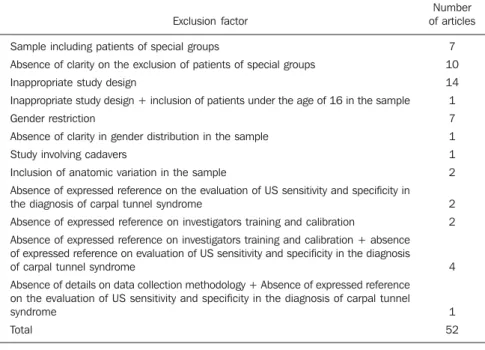
se-lected articles, 52 were excluded, some of them because they met some of the exclu-sion criteria elected for the present study and others because they did not meet ap-propriately the inclusion criteria (Table 1). Considering that only one(16) among the
68 articles included in Series 1 detailed the calculation utilized for obtaining the sample size, the absence of such inclusion criterion was not considered as an exclud-ing factor for the other articles. Apart from this latter criterion, ten articles have appro-priately met all the other inclusion criteria. However six other articles were included as over inclusion by the third investigator, to avoid selection bias. The Series 2 result-ing from the second selection comprised 16 articles.
The third selection was aimed at assess-ing the methodological quality of the ar-ticles, following three criteria: randomiza-tion, masking of investigators and descrip-tion of losses and exclusions. Of the 16 ar-ticles resulting from the second selection, four were included in Series 3.
In the fourth selection, the quality of the four articles in Series 3 was analyzed by means of the CASP tool. All the articles selected in Series 3 were included in Series 4. It is important to comment on the refer-ence standard utilized by the authors in the selected studies. As far as carpal tunnel syndrome is concerned, there is no
refer-ence standard unanimously accepted by the authors; some of them have utilized clini-cal data while others utilized electroneuro-graphy, and others an association of both reference standards. In the answers to the answering the questions of the CASP tool, all of these reference standards were con-sidered. Among the four selected articles, three authors considered the association of clinical data and electroneurography as a reference standard(16-18) and one considered
electroneurography alone(19). Table 2
sum-marizes the evidences in the articles se-lected in Series 4.
All the ultrasonography studies were performed with high resolution equipment (multifrequency transducers ranging from 5 to 13 MHz). In three studies, the observ-ers were rheumatologists(17–19) while in
one(16), radiologists, all of them
experi-enced in the method. In three studies(16,18,19)
the reliability was evaluated among the readers, with a good correlation being ob-served among them, by obtaining the intraclass correlation coefficients(study 16: 0.71–0.90; study 18: 0.93; study 19: 0.912– 0.987), highlighting the fact that the utili-zation of a standardized technique is suf-ficient to achieve a good reliability, even with observers with a baseline knowledge on ultrasonography, a relevant aspect in the generalization of the use of such method as a diagnostic evidence of carpal tunnel syn-drome(19). All examinations were
per-formed with the patients sitting in front of the investigator, with flexed elbows and hands in the supine position, resting on a rigid surface.
In all the studies, except the one devel-oped by Wong et al.(16), ROC (receiver
op-erating characteristics) curves were utilized in order to explore the relationship between sensitivity and specificity of the different described sonographic measurements and to define the best cutoff point for such measurements. Wong et al.(16) have utilized
a linear regression software (Answer Tree, version 2.1; SPSS, Chicago, IL, USA) to evaluate such relationship.
In the last phase, all the four articles selected in Series 4 were hierarchized ac-cording to the level of evidence and respec-tive grade of recommendation of the scien-tific publication as proposed by Phillips et al.(15). Considering the fact that such Series
Table 1 Reasons for exclusion of articles from Series 1.
Exclusion factor Sample including patients of special groups
Absence of clarity on the exclusion of patients of special groups Inappropriate study design
Inappropriate study design + inclusion of patients under the age of 16 in the sample Gender restriction
Absence of clarity in gender distribution in the sample Study involving cadavers
Inclusion of anatomic variation in the sample
Absence of expressed reference on the evaluation of US sensitivity and specificity in the diagnosis of carpal tunnel syndrome
Absence of expressed reference on investigators training and calibration
Absence of expressed reference on investigators training and calibration + absence of expressed reference on evaluation of US sensitivity and specificity in the diagnosis of carpal tunnel syndrome
Absence of details on data collection methodology + Absence of expressed reference on the evaluation of US sensitivity and specificity in the diagnosis of carpal tunnel syndrome
Total
Number of articles
7 10 14 1 7 1 1 2 2 2
4
included prospective, unicenter cohort studies with a good reference standard, the four articles obtained an evidence level “1b” and grade of recommendation “A”, i.e., those studies present excellent evi-dence levels to routinely recommend the conduct proposed in the articles.
DISCUSSION
The diagnosis of carpal tunnel syn-drome has always raised debate among clinical physicians, neurologists, surgeons, electroneurographers and radiologists, with no diagnostic criteria uniformity. What is the sonographic method accuracy in the diagnosis of carpal tunnel syndrome? The answer to this question would be highly beneficial for patients and would help to address the medical and legal problems arising from clinical, surgical legal
require-ments. In the current days, the best option for the decision making related to preven-tion, diagnosis and treatment of disorders is the practice of medicine based on evi-dences.
The present study was developed in the search for the best scientific evidence re-lated to the sonographic diagnosis of car-pal tunnel syndrome. The utilization of the proposed method demonstrated how diffi-cult the task was, requiring researchers availability, patience and skill for search-ing in databases, selectsearch-ing and qualitatively analyzing the articles. The utilized model, in which two researchers worked indepen-dently in each phase of the process, with consensus meetings and, in the case of doubts, solution by a third more experi-enced researcher, demonstrated to be an indispensable requirement for the process completion.
The search for articles in the electronic databases, with different forms of interac-tion with the user, has demonstrated to be the most important phase of the research, taking a greater length of time to be com-pleted. The number of articles found was not significant to justify the utilization of a bibliographic management software (such as Endnote). Of the 68 articles se-lected in the first phase, 52 (76.4%) psented methodological shortcomings re-lated to sampling, study design, diagnostic criteria and results presentation, causing the exclusion of such articles.
After the application of the mentioned inclusion and exclusion criteria, the meth-odological quality of the articles selected in phase 2 (16 articles) was performed by means of a first objective filtering to mea-sure the sample randomization, investiga-tors masking, losses and exclusions. No Table 2 Summary of evidences in the articles selected at Series 4.
Authors Wong et al.(16)
Ziswiler et al.(17)
Naranjo et al.(18)
Peiteado-López et al.(19)
Wrists/ patients 195/120
101/71
105/68
75/42
Ultrasonographic diagnostic criterion / statistical analysis AMN proximal to the tunnel, AMNe+ and AMNs (direct method). Statistical analysis: regression analysis. Evalua-tion of best cutoff point
Larger AMN between the proximal level of the carpal tunnel and its outlet (di-rect method). Statistical analysis: ROC curves. Logistic regression. Relation-ship with ENG (Spearman’s correlation coefficient) / table 2 × 2
AMNe+, AMNm and AMNs (direct method). FLI and BR. Statistical analy-sis: tables 2 × 2. ROC curves / logistic regression. Pearson’s correlation coef-ficient
AMNe+, AMNm, AMNs (direct meth-od). FLI and BR. Statistical analysis: ROC curves. Logistic regression. Odds ratio
Results
Right hand: cutoff point AMN proximal to the tunnel = 9.0 mm2; cutoff point AMNs = 12 mm2. Left hand: cutoff point AMN proximal to the tunnel > 10.0 mm2. Righ hand + left hand: cutoff point AMN prox-imal to the tunnel = 10 mm2; cutoff point AMNs > 12 mm2
No difference between right hand and left hand (CI 95%). No difference between genders (CI 95%). Inversely proportional ratio between AMN and SCV (CI 95%) and directly proportional between AMN and DML (CI 95%). AMN < 8 mm2: satisfactory condition to exclude CTS (negative likelihood ratio = 0.13). AMN > 12 mm2: excellent CTS diagnostic condition (positive likelihood ratio = 19,9)
Correlation between ultrasonography and clinical tests. Accuracy of different sonographic criteria for CTS diagnosis. Mean AMN values: AMNm = 12.4 mm2 – the whole patients group; AMNm = 10.08 mm2 – normal ENG; AMNm = 13.13 mm2 – abnormal ENG; AMNm = 11.14 mm2 – mild CTS; AMNm = 12.5 mm2 – moderate CTS; AMNm = 14.34 mm2 – severe CTS
Significant correlation between in-creased AMN and symptoms severity (CI 95%). Significant negative association between increased AMN and velocity of nervous conduction (CI 95%)
Sensitivity / specificity / accuracy AMN right hand: S = 94%; Sp = 65%; FP = 12; FN = 19%. AMN left hand: S = 83%; Sp = 73%; FP = 15%; FN = 31%. Right hand + left hand: S = 86%; Sp = 74%; FP = 115; FN = 33%
AMN:
Cutoff point = 9.0 mm2: S = 86%; Sp = 70%. Cutoff point = 10 mm2: S = 82%; Sp = 87%. Cutoff point = 11 mm2: S = 54%; Sp = 96%
AMNm:
Cutoff = 9.7 mm2: S = 86.3%; Sp = 48%; A = 77.1%; PPV = 100%. No difference of S and Sp: (right hand × left hand / men × women)
Best cutoff point: AMNs = 9.5 mm2 (S = 88%; Sp = 67%); AMN > 14 mm2 (Sp = 100%, inclusion CTS); AMN < 7 mm2 (S = 100%, CTS ruling out)
specific scale was utilized. Instead, a tool developed by the authors of the present study was utilized as an adaptation of the criterion of patients masking because of the difficulty in achieving a double-blind masking in studies aimed at measuring the accuracy of a diagnostic method in which the index test (ultrasonography) and the test utilized one as a reference standard (elec-troneurography) are difficult to be dis-guised. This first filtering resulted in the selection of four articles. The main exclu-sion criterion was the omisexclu-sion regarding the sample randomization, with such crite-rion, isolatedly or associated with others, responsible for the exclusion of ten articles. The remaining exclusions occurred be-cause of inappropriate investigators mask-ing. Aiming at a more critical and deep analysis of the selected articles, the authors utilized a specific tool developed by CASP for evaluation of papers approaching diag-nostic methods. As a result, the four articles selected in the first filtering (16, 17, 18 and 19) were included in the final series.
It is necessary to draw a parallel be-tween the selected articles, discussing and evaluating the methodological similarities and differences among them. All the stud-ies were prospective, randomized and were developed with investigators masking. It is important to highlight that the studies de-veloped by Wong et al.(16) and Ziswiler et
al.(17) were the first prospective studies with
the objective of measuring the diagnostic value of ultrasonography in carpal tunnel syndrome. In the four selected articles, 476 wrists were evaluated in a universe of 301 patients. The ratio between women and men in the studied samples ranged between 1.8(17) and 7.4(19). In the remaining studies,
such ratio ranged between 4.4(16) and
4.7(18), practically the variation observed in
the literature review data. The mean age in the samples ranged between 47 and 51 years, in agreement with data in the litera-ture, which demonstrates that the incidence and prevalence of carpal tunnel syndrome is higher above the age of 40.
It was observed that the cross sectional measurement of the median nerve (AMN) was the most relevant criterion for the sonographic diagnosis of carpal tunnel syn-drome and that, in the selected studies, the
most frequent cutoff value was between 9 and 10 mm2 (sensitivity = 82–86.3%; specificity = 48–87%). Another relevant information to be taken into consideration is the fact that the authors found cutoff values for the AMN at which the diagno-sis of carpal tunnel syndrome can be ruled out (AMN < 8 mm2) or ratified (AMN > 13/ 14 mm2), with no need of complementary electroneurography.
CONCLUSIONS
The authors of all the studies concluded that the sonographic evaluation of patients with clinical suspicion of carpal tunnel syndrome can be performed as a first line test, with a good cost-benefit ratio, reduc-ing the need of electroneurographic stud-ies for such patients. Also, it is important to observe that ultrasonography can diag-nose associated disorders and neural ana-tomic variations, in addition to the advan-tages of being a dynamic method easy to perform, at a relatively low cost as compared with electroneurography. The electroneuro-graphic evaluation would be indicated in the case of symptomatic patients with nega-tive diagnosis by ultrasonography.
All the authors were unanimous in re-lation to the necessity of controlled and ran-domized prospective studies with larger samples for better understanding the actual value of ultrasonography in the diagnosis of carpal tunnel syndrome.
According to the hierarchization of the evidence level of the selected studies (1b), the guidances contained in such articles can be considered as recommended evidences for application in the daily practice of de-cision making for professionals of differ-ent areas involved in the diagnosis of idio-pathic or work-related carpal tunnel syn-drome.
Acknowledgements
Special thanks to Conselho Nacional de Desenvolvimento Científico e Tecnológico (CNPq) (National Council for Scientific and Technological Development).
REFERENCES
1. Atroshi I, Gummesson C, Johnsson R, et al. Prevalence of carpal tunnel syndrome in a gen-eral population. JAMA. 1999;282:153–8.
2. Prick JJW, Blaauw G, Vredeveld JW, et al. Results of carpal tunnel release. Eur J Neurol. 2003;10: 733–6.
3. Stoller DW. Magnetic resonance imaging in or-thopaedics & sports medicine. 3rd ed. Baltimore, MD: Lippincott Williams & Wilkins; 2007. 4. Resnick DL, Kang HS, Pretterklieber ML.
Inter-nal derangements of joints. 2nd ed. Philadelphia, PA: WB Saunders; 2006.
5. Papaioannou T, Rushworth G, Atar D, et al. Car-pal canal stenosis in men with idiopathic carCar-pal tunnel syndrome. Clin Orthop. 1992;(285):210–3. 6. Fernandes JL, Viana SL. Diagnóstico por imagem em reumatologia. Rio de Janeiro, RJ: Guanabara Koogan; 2007.
7. Aroori S, Spence RA. Carpal tunnel syndrome. Ulster Med J. 2008;77:6–17.
8. Koyuncuoglu HR, Kutluhan S, Yesildag A, et al. The value of ultrasonographic measurement in carpal tunnel syndrome in patients with negative electrodiagnostic tests. Eur J Radiol. 2005;56: 365–9.
9. Machado DA, Martins WP. Síndrome do túnel do carpo. EURP. 2009;1:136–40.
10. [No authors listed]. Practice parameter for carpal tunnel syndrome (summary statement). Report of the Quality Standards Subcommittee of the American Academy of Neurology. Neurology. 1993;43:2406–9.
11. Marinho VCC. Systematic reviews of controlled trials in general and oral health care. Braz J Oral Sci. 2003;2:215–26.
12. Jadad AR, Moore RA, Carroll D, et al. Assessing the quality of reports of randomized clinical tri-als: is blinding necessary? Control Clin Trials. 1996;17:1–12.
13. Higgins JPT, Green S. Cochrane Handbook for Systematic Reviews of Interventions Version 5.0.2. [updated September 2009]. The Cochrane Collaboration, 2009. [cited 2010 Jan 17]. Avail-able from: www.cochrane-handbook.org 14. Critical Appraisal Skills Programme (CASP).
Diagnostic test studies. [cited 2010 Jan 18]. Avail-able from: http://www.sph.nhs.uk/what-we-do/ public-health-workforce/resources/critical-ap-praisals-skills-programme
15. Phillips B, Ball C, Sackett D, et al. Oxford Cen-tre for Evidence-based Medicine – Levels of evi-dence. Grades of recommendation. [cited 2010 Feb 18]. Available from: http://www.cebm.net/ index.aspx?o=1025
16. Wong SM, Griffith JF, Hui ACF, et al. Carpal tun-nel syndrome: diagnostic usefulness of sonog-raphy. Radiology. 2004;232:93–9.
17. Ziswiler HR, Reichenbach S, Vögelin E, et al. Diagnostic value of sonography in patients with suspected carpal tunnel syndrome: a prospective study. Arthritis Rheum. 2005;52:304–11. 18. Naranjo A, Ojeda S, Mendoza D, et al. What is
the diagnostic value of ultrasonography compared to physical evaluation in patients with idiopathic carpal tunnel syndrome? Clin Exp Rheumatol. 2007;25:853–9.