Letters to the Editor
Radiol Bras. 2015 Jul/Ago;48(4):263–270
266
Beatriz Regina Alvares1, Aline Satomi Yumioka1, Isabela
Gusson Galdino dos Santos1
1. Faculdade de Ciências Médicas da Universidade Estadual de Campi-nas (FCM-Unicamp), CampiCampi-nas, SP, Brazil. Mailing Address: Dra. Beatriz Regina Alvares. Rua Alberto de Salvo, 238, Barão Geraldo. Campinas, SP, Brazil, 13084-759. E-mail: [email protected].
http://dx.doi.org/10.1590/0100-3984.2014.0134 is the most common symptom, usually occurring as a result from
inflammation or ileal volvulus(1,4). Meckel’s diverticulum rupture is rarely found in neonates, occurring in less than 10% and mani-festing at radiography as pneumoperitoneum(1). In such situations, the differential diagnosis should be made with necrotizing entero-colitis, since this disease is responsible for 41% of cases of neona-tal pneumoperitoneum(4).
In the present case, there was a clinical suspicion of necrotiz-ing enterocolitis, but this hypothesis was ruled out as the pres-ence of a perforated MD was intraoperatively confirmed.
The causes of MD include inflammatory reaction, mucosal ulceration and defective muscular layer of the diverticulum(1,2). Rarely, MD perforation may occur as a result from umbilical cath-eterization by means of an umbilical vein connection with the MD via umbilical cord(6). In the present case, catheterization of umbili-cal vein and artery was performed with two hours of life; but the late symptoms onset and the exploratory laparotomy demonstrated that the catheterization was not related to the MD perforation.
Hirschsprung’s disease may also predispose to MD perfora-tion due to delayed passage of meconium, determining increased pressure upstream of the diverticulum(5). Such a condition occurs with typical symptoms of bowel obstruction, abdominal pain and bilious vomiting(5). In the present case, despite the symptoms of bowel obstruction and abdominal discomfort at palpation, bilious vomiting was not observed. Furthermore, the histopathological analysis of the surgical specimen ruled out the hypothesis of Hirschsprung’s disease.
Finally, MD should be included as a diagnostic hypothesis in the absence of other factors that might justify the presence of pneumoperitoneum in a neonate. Such a complication is confirmed by means of a surgical procedure.
REFERENCES
1. Aguayo P, Fraser JD, St Peter SD, et al. Perforated Meckel’s diverticulum in a micropremature infant and review of the literature. Pediatr Surg Int. 2009;25:539–41.
2. Chang YT, Lin JY, Huang YS. Spontaneous perforation of Meckel’s di-verticulum without peritonitis in a newborn: report of a case. Surg To-day. 2006;36:1114–7.
3. Gandy BJ, Byrne P, Lees G. Neotatal Meckel’s diverticular inflammation with perforation. J Pediatr Surg. 1997;32:750–1.
4. Oyachi N, Takano K, Hasuda N, et al. Perforation of Meckel’s diverticu-lum manifesting as aseptic peritonitis in a neonate: report of a case. Surg Today. 2007;37:881–3.
5. Skelly BL, Ervine E, Bisharat M, et al. Small bowel skip segment Hirschprung’s disease presenting with perforated Meckel’s diverticulum. Pediatr Surg Int. 2012;28:645–8.
6. Costa S, De Carolis MP, Savarese I, et al. An unusual complication of umbilical catheterization. Eur J Pediatr. 2008;167:1467–9.
Desmoplastic fibroma with perineural spread: conventional and diffusion-weighted magnetic resonance imaging findings
Fibroma desmoplásico com disseminação perineural: achados nas sequências convencionais de ressonância magnética e na difusão
Dear Editor,
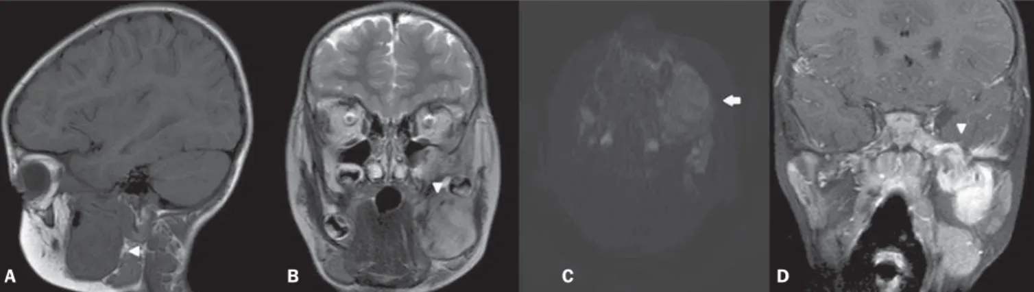
A male, three-year-old child with morphostructural alteration developed over the last year in the region of the mandible at left, presenting with recent onset of pain, with no other associated complaints. Laboratory tests did not demonstrate any alteration and magnetic resonance imaging (MRI) (Figure 1) showed a le-sion with predominant iso/hyposignal on T1-weighted image, hypersignal on T2-weighted image with subtle low signal inten-sity foci, absence of signal loss on susceptibility-weighted
se-quences and absence of diffusion restriction. After gadolinium injection, exuberant enhancement was observed in addition to perineural dissemination through the third division of the trigemi-nal nerve. Histopathological atrigemi-nalysis revealed spindle cells with-out atypias and pleomorphism, besides areas with acellular fibrous connective tissue, with immunohistochemical negative for S100, and positivity for vimentin and SMA, with Ki-67 < 5%. Such find-ings are compatible with desmoplastic fibromas. The patient was submitted to incomplete surgical excision supplemented with radiotherapy.
Desmoplastic fibroma is an extremely rare, benign bone tu-mor with aggressive and usually insidious behavior, representing 0.1% of all primary bone tumors(1–5). The mandible is the most affected site, particularly in its posterior portion, corresponding to 22% of cases(1,2,4), followed by the metaphyseal region of long
Figure 1. A: Sagittal, T1-weighted image showing lesion with hyposignal affecting the mandible (arrowhead). B: Coronal, T2-weighted sequence showing heteroge-neous lesion with subtle hypersignal intermingled with foci of low signal intensity (arrowhead). C: Axial, functional diffusion-weighted sequence does not demonstrate diffusion restriction (arrow). D: Contrast-enhanced coronal, T1-weighted sequence with fat suppression demonstrating exuberant gadolinium enhancement and notice-able perineural dissemination in the third division of the trigeminal nerve (arrowhead).
Letters to the Editor
Radiol Bras. 2015 Jul/Ago;48(4):263–270
267
Bruno Niemeyer de Freitas Ribeiro1, Tiago Medina
Salata2, Lívia de Oliveira Antunes2, Edson Marchiori3 1. Instituto Estadual do Cérebro Paulo Niemeyer, Rio de Janeiro, RJ, Brazil. 2. Hospital Casa de Portugal / 3D Diagnóstico por Imagem, Rio de Janeiro, RJ, Brazil. 3. Universidade Federal do Rio de Janeiro (UFRJ), Rio de Janeiro, RJ, Brazil. Mailing Address: Dr. Bruno Niemeyer de Freitas Ribeiro. Instituto Estadual do Cérebro Paulo Niemeyer – Ser-viço de Radiologia. Rua do Rezende, 156, Centro. Rio de Janeiro, RJ, Brazil, 20231-092. E-mail: [email protected]. bones. Desmoplastic fibromas may occur at any age range,
al-though its higher incidence is observed at the first three decades of life(1–3,6). Despite conflicting data, it seems there is no predi-lection for sex(2,6). Local recurrence is frequently observed in cases where complete resection is not.Clinically, the patients are either asymptomatic or may present with pain, edema, joint effusion and pathological fracture(1–6). The differential diagnosis should con-sider rhabdomyosarcoma, fibrosarcoma, giant cell tumor, among others. Despite the imaging methods usefulness in the lesion delimitation, the diagnosis is histopathological.
At MRI, most lesions present with iso/hyposignal on T1-weighted images and low signal intensity on T2-T1-weighted im-ages(1,3–6), but there are reports of lesions with hypersignal on T2-weighted images(1–3,6). The enhancement may be variable, and according to some authors, such variation may be a result of the cellular content of the lesion(3,4). In the present case, there was homogeneous iso/hyposignal on T1-weighted images and subtle hypersignal on T2-weighted images, with foci of low signal inten-sity. After gadolinium injection, marked contrast enhancement, with noticeable perineural dissemination through the third divi-sion of the trigeminal nerve were observed. Such aspects on T2-weighted sequences, and the presence of perineural dissemina-tion are not commonly observed as compared with the typical imaging pattern described at MRI.
Reports on diffusion in desmoplastic fibromas were not found in the literature. In the present case, areas of diffusion restriction were not observed. Recent studies highlight the use of diffusion-weighted imaging in the evaluation of head and neck lesions, showing that apparent diffusion coefficient < 1.22 × 10⁻³
mm2 /s are suggestive of malignancy(7). In the present case, the value for apparent diffusion coefficient was 1.45 × 10⁻³
mm2
/s, corroborat-ing the previously described findcorroborat-ings.
The authors conclude that the diagnosis of desmoplastic fi-bromas should be considered in patients under the age of 30 pre-senting with tumor particularly located in the mandible, and that such a hypothesis cannot be ruled out in case of less noticeable foci of hyposignal on T2-weighted images.
REFERENCES
1. Woods TR, Cohen DM, Islam MN, et al. Desmoplastic fibroma of the mandible: a series of three cases and review of literature. Head Neck Pathol. 2015;9:196–204.
2. Nedopil A, Raab P, Rudert M. Desmoplastic fibroma: a case report with three years of clinical and radiographic observation and review of the literature. The Open Orthopaedics Journal. 2013;7:40–6.
3. Kim OH, Kim SJ, Kim JY, et al. Desmoplastic fibroma of bone in a toe: radiographic and MRI findings. Korean J Radiol. 2013;14:963–7. 4. Kang DM, Juhng SK, Sohn YJ, et al. Imaging findings of desmoplastic
fibroma rarely involving the clavicle: case report. Korean J Radiol. 2014; 15:130–3.
5. Frick MA, Sundaram M, Unni KK. Imaging findings in desmoplastic fibroma of bone: distinctive T2 characteristics. AJR Am J Roentgenol. 2005;184:1762–7.
6. Moorjani V, Stockton V. Desmoplastic fibroma with perineural extension. AJR Am J Roentgenol. 2005;185:1498–9.
7. Gonçalves FG, Ovalle JP, Grieb DFJ, et al. Diffusion in the head and neck: an assessment beyond the anatomy. Radiol Bras. 2011;44:308–14.
http://dx.doi.org/10.1590/0100-3984.2014.0135
Creutzfeldt-Jakob dementia
Demência por doença de Creutzfeldt-Jakob
Dear Editor,
A 72-year-old woman with rapidly progressive dementia, be-havioral changes and apraxia of gait for seven months, extrapyra-midal signs and diffuse myoclonus. Electroencephalography
dem-onstrated periodic electric activity with high amplitude acute phase waves diffusely distributed over the cortex. The cerebrospinal fluid was normal. Magnetic resonance imaging (MRI) was performed (Figure 1).
The association of clinical, radiological, electroencephalic or cerebrospinal fluid findings (presence of 14-3-3 brain protein in diseased patient for less than two years – absent in this case),
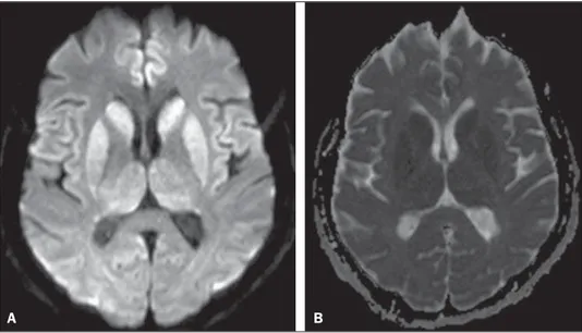
Figure 1.A: Axial magnetic resonance imaging of the skull demonstrating foci of hypersignal at diffusion-weighted sequences in the heads of the caudate nuclei, putamina, thalami and medial occipitotemporal gyri. B: At the ADC mapping, the low signal intensity in the same region confirms the diffusion restriction.