João Gualberto de Cerqueira Luz(a)
Rogerio Bonfante Moraes(a) Ricardo Pimenta D’Ávila(b) Marcos Kazuo Yamamoto(a)
(a) Department of Maxillofacial Surgery, Prosthodontics and Traumatology, School of Dentistry, Universidade de São Paulo - USP, São Paulo, SP, Brazil.
(b)Hospital Municipal Dr. Arthur Ribeiro de Saboya, São Paulo, SP, Brazil.
Corresponding Author: João Gualberto de Cerqueira Luz E-mail: [email protected]
retreatment of mandibular fractures
Abstract: The purpose of this retrospective study was to evaluate con-tributing factors in patients requiring surgical retreatment of mandibular fractures. Of all the patients with mandibular fractures who were treated using internal ixation at a trauma hospital over a seven-year period, 20 patients (4.7%) required a second surgery and thus composed the “reop-erated” group. The control group comprised 42 consecutive patients with mandibular fractures who were treated at the same clinic and who healed without complications. Medical charts were reviewed for gender, age, substance abuse history, dental condition, etiology, location of fracture, degree of fragmentation, fracture exposure, teeth in the fracture line, associated facial fractures, polytrauma, time elapsed between trauma and initial treatment, surgical approach and ixation system. Statistical analyses were performed using the Statistical Package for Social Sciences (SPSS) version 20.0; descriptive statistics and the chi-squared test were used to determine differences between groups. Signiicant differences in substance abuse (p = 0.006), dental condition (p < 0.001), location of fracture (p = 0.010), degree of fragmentation (p = 0.003) and fracture ex-posure (p < 0.001) were found. With regard to age and time elapsed be-tween trauma and initial treatment, older patients (31.4 years, SD = 11.1) and a delay in fracture repair (19.1 days, SD = 18.7) were more likely to be associated with reoperation. It was concluded that substance abuse, age, dental condition, location of fracture, degree of fragmentation, frac-ture exposure and the time between trauma and initial treatment should be considered contributing factors to the occurrence of complications that require surgical retreatment of mandibular fractures.
Descriptors: Mandibular Fractures; Jaw Fixation Techniques; Postoperative Complications; Retrospective Studies.
Introduction
The ideal method for treating mandibular fractures is rigid or stable internal ixation using plates or miniplates.1,2 Rigid or stable ixation, or
simply internal ixation, is a more cost-effective treatment than non-rigid methods, in part due to the decreased probability of postoperative com-plications,2 that could necessitate further surgery. The consequences of
retreatment include greater patient morbidity, further hospitalization and hospital costs and a longer period of absence from work, which leads to high social costs.3 Therefore, all efforts should be made to avoid
compli-cations during treatment of mandibular fractures.4
Declaration of Interests: The authors certify that they have no commercial or associative interest that represents a conflict of interest in connection with the manuscript.
Submitted: Sep 20, 2012
Infection is a common initial complication fol-lowing the surgical treatment of mandibular frac-tures, which, in more severe cases, may progress to osteomyelitis and pseudoarthrosis.4,5 Other
compli-cations requiring reoperation include the nonunion or abnormal union of bone tissue and soft tissue infection associated with screw loosening or plate exposure. Such complications may require further ixation, exploratory surgery to remove the ixation materials, the removal of bone sequestration and re-fracture.4,6-8
There is no consensus in the literature on the most prevalent factors associated with postoperative complications following the treatment of mandibu-lar fractures. Factors including the location of the fracture, its complexity, the presence of a tooth in the fracture line, teeth with extensive caries, peri-odontal disease and exposed fractures have been reported by a number of studies.9,10 Patient habits
such as smoking, alcohol consumption, drug abuse, noncompliance and poor oral hygiene are addition-al important contributing factors that may lead to a requirement for further surgery.6 Moreover, age,
gender, preexisting medical status and treatment tactics are considered contributing factors to the oc-currence of infection or the abnormal union of man-dibular fractures.10,11
The purpose of this study was to evaluate pos-sible contributing factors in patients requiring a sur-gical retreatment of mandibular fractures that were originally treated using internal ixations compared with a sample of patients with mandibular fractures that were not associated with complications but that were treated using the same method.
Methodology
This study was approved by the local hu-man research ethics committee (process no. 0179.0.162.017-09). A retrospective study was con-ducted using information from the medical charts of patients treated at the Oral and Maxillofacial Surgery Clinic of the Hospital M. Dr. Arthur R. de Saboya, which is a trauma hospital that provides coverage for the southern area of São Paulo, SP, Brazil. The charts of patients treated between 2002 and 2009 were reviewed for mandibular fractures
treated with internal ixation. Of the 364 patients identiied, 20 (4.7%) developed postoperative com-plications and required surgical retreatment (the “reoperated group”). For the purposes of compari-son, the control group comprised a randomly se-lected sample of 42 consecutive cases of mandibular fractures treated with internal ixation at the same clinic and during the same period, but that healed without complications (termed the “uncomplicated” group).
The complications that led to surgical retreat-ment in the reoperated group were nonunion (50%
of cases), soft tissue infection associated with loos-ened screws or plate exposure (35%), osteomyeli-tis (10%) and malunion (5%). The procedures per-formed were new ixation (30% of cases), removal of bone sequestration and new ixation (20%), surgical exploration and removal of ixation material (35%), removal of bone sequestration (10%) and refracture and ixation (5%).
All charts were reviewed and the following data were recorded:
• age;
• gender;
• major health conditions, such as diabetes, hu-man immunodeiciency virus status, anemia and cancer;
• substance abuse history (alcohol, drugs and smoking);
• dental condition (dentate, partially edentulous or completely edentulous); and
• the etiology and location of the fracture.
The fractures were classiied based on the degree of fragmentation:
• single,
• multiple or
• comminuted.
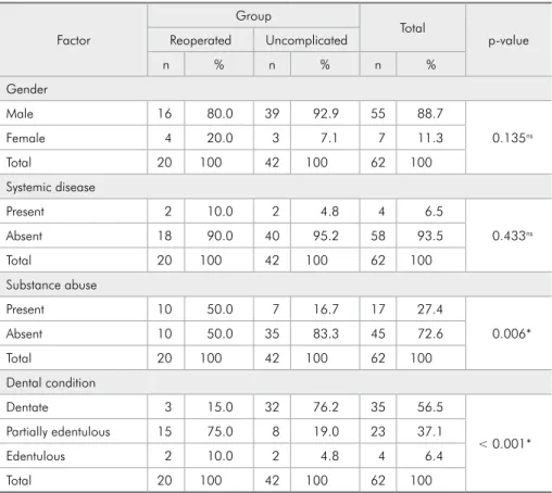
the uncomplicated (92.9%) group were males, with no statistically signiicant difference between groups (p = 0.135). Only a few cases of major systemic dis-ease were noted in the reoperated (10%) and un-complicated (4.8%) groups (p = 0.433). Half of the patients in the reoperated group (50%) and 16.7%
of the uncomplicated group reported having abused drugs; this difference was statistically signiicant (p = 0.006). With regard to dental status, 75% of the patients in the reoperated group were partially edentulous, whereas dentate patients predominated in the uncomplicated group (76.2%) (p < 0.001). Table 1 shows the patient characteristics of each group.
The predominant etiologies in the reoperated group were aggression (30%) and motor vehicle ac-cidents (30%), followed by gunshot injuries (25%). The predominant etiology in the uncomplicated group was motor vehicle accidents (28.6%), followed by aggression (26.2%) and falls (16.7%). However, no signiicant difference in etiology between groups or extra-oral) and the ixation system used in the
initial surgery were recorded. All patients received cefazolin during the postoperative period because this antibiotic is used as standard practice in the hospital in question. No maxillomandibular ixa-tion was used during the postoperative period.
The data were tabulated, and chi-squared tests were used for analyses. All categorical variables were analyzed to determine differences between groups. Conidence intervals were calculated for the quantitative variables (age and time elapsed between the trauma and the initial treatment), and means outside these intervals indicated signiicant differ-ences between the groups. The level of signiicance was set to 5% (p < 0.050) for all statistical analyses. The Statistical Package for Social Sciences (SPSS) version 20.0 (IBM Software Group, Chicago, USA) was used for all analyses.
Results
Most patients in both the reoperated (80%) and
Factor
Group
Total
p-value Reoperated Uncomplicated
n % n % n %
Gender
Male 16 80.0 39 92.9 55 88.7
0.135ns
Female 4 20.0 3 7.1 7 11.3
Total 20 100 42 100 62 100
Systemic disease
Present 2 10.0 2 4.8 4 6.5
0.433ns
Absent 18 90.0 40 95.2 58 93.5
Total 20 100 42 100 62 100
Substance abuse
Present 10 50.0 7 16.7 17 27.4
0.006*
Absent 10 50.0 35 83.3 45 72.6
Total 20 100 42 100 62 100
Dental condition
Dentate 3 15.0 32 76.2 35 56.5
< 0.001*
Partially edentulous 15 75.0 8 19.0 23 37.1
Edentulous 2 10.0 2 4.8 4 6.4
Total 20 100 42 100 62 100
*significant; ns non-significant.
was found (p = 0.109). Table 2 displays the distribu-tion of mandibular fractures based on etiology. The predominant location of the fracture in the reoper-ated group was in the mandibular body (43.8%), which was followed by the symphysis (28.1%) and angle (25%), whereas the predominant location in the uncomplicated group was in the condyle (31%), which was followed by the symphysis (26.8%) and body (22.5%) (p = 0.010). Table 3 displays the
dis-tribution of mandibular fractures based on location. The mean age was 31.4 years (standard deviation [SD] = 11.1) in the reoperated group and 26.4 years (SD = 7.4) in the uncomplicated group; this differ-ence was statistically signiicant. The mean time elapsed between the trauma and the initial treat-ment was 19.1 days (SD = 18.7) in the reoperated group and 13.5 days (SD = 9.0) in the uncomplicat-ed group; this difference was also statistically
sig-Etiology
Group
Total
p-value Reoperated Uncomplicated
n % n % n %
Aggressions 6 30.0 11 26.2 17 27.4
0.109ns
Motor vehicle accidents 6 30.0 12 28.6 18 29.0
Gunshot wounds 5 25.0 4 9.5 9 14.5
Falls 1 5.0 7 16.7 8 12.9
Bicycle 0 0.0 5 11.9 5 8.1
Work 1 5.0 0 0.0 1 1.6
Sports 0 0.0 3 7.1 3 4.8
Not available 1 5.0 0 0.0 1 1.6
Total 20 100 42 100 62 100
ns non-significant.
Location
Group
Total
p-value Reoperated Uncomplicated
n % n % n %
Symphysis 9 28.1 19 26.8 28 27.2
0.010*
Body 14 43.8 16 22.5 30 29.1
Angle 8 25.0 14 19.7 22 21.4
Condyle 1 3.1 22 31.0 23 22.3
Total 32 100 71 100 103 100
*significant.
Table 2 - Distribution of the cases of mandibular fracture based on etiology and group.
Table 3 - Distribution of the sites of mandibular fractures based on location and group.
Factor Group n Mean SD
95% confidence
interval Difference between
groups Lower
limit Upper limit
Age Reoperated 20 31.4 11.1 26.5 36.2 Significant
Uncomplicated 42 26.4 7.4 24.2 28.6
Time elapsed Reoperated 18 19.1 18.7 10.4 27.7 Significant
Uncomplicated 42 13.5 9.0 10.8 16.2
niicant. Data from 2 cases in the reoperated group were not available. In the reoperated group, many patients who waited a longer time had polytrauma. Table 4 displays the mean age and the time elapsed between the trauma and the initial treatment in both groups.
With regard to the degree of fragmentation, there was a predominance of multiple (35%) and comminuted (35%) fractures, followed by single
(30%) fractures in the reoperated group, and a pre-dominance of single (71.5%) fractures in the un-complicated group (p = 0.003). Fracture exposure occurred in the majority of cases (80%) in the reop-erated group and was absent in the majority of cases (78.6%) in the uncomplicated group (p < 0.001). The majority of cases in both the reoperated (65%) and uncomplicated (52.4%) groups (p = 0.349) showed teeth in the fracture line. Some cases of associated
Factor
Group
Total
p-value Reoperated Uncomplicated
n % n % n %
Degree of fragmentation
Single 6 30.0 30 71.5 36 58.1
0.003*
Multiple 7 35.0 3 7.1 10 16.1
Comminuted 7 35.0 9 21.4 16 25.8
Total 20 100 42 100 62 100
Fracture exposure
Present 16 80.0 9 21.4 25 40.3
< 0.001*
Absent 4 20.0 33 78.6 37 59.7
Total 20 100 42 100 62 100
Teeth in the fracture line
Present 13 65.0 22 52.4 35 56.4
0.349ns
Absent 7 35.0 20 47.6 27 43.6
Total 20 100 42 100 62 100
Associated facial fractures
Present 4 20.0 4 9.5 8 12.9
0.250ns
Absent 16 80.0 38 90.5 54 87.1
Total 20 100 42 100 62 100
Polytrauma
Present 6 30.0 7 16.7 13 21.0
0.228ns
Absent 14 70.0 35 83.3 49 79.0
Total 20 100 42 100 62 100
Surgical approach
Intra-oral 5 25.0 19 45.2 24 38.7
0.126ns
Extra-oral 15 75.0 23 54.8 38 61.3
Total 20 100 42 100 62 100
Initial fixation system
2.0 mm 11 55.0 22 52.4 33 53.2
0.847ns
2.4 mm 9 45.0 20 47.6 29 46.8
Total 20 100 42 100 62 100
*significant; ns non-significant.
facial fractures were found in both the reoperated (20%) and uncomplicated (9.5%) groups (p = 0.250). Some polytrauma occurred in the reoperated (30%) and uncomplicated (16.7%) groups (p = 0.228). The most frequent surgical access during the initial sur-gery was extra-oral in both the reoperated (75%) and uncomplicated (54.8%) groups (p = 0.126). An initial ixation using the 2.0-mm system predomi-nated in both the reoperated (55%) and uncompli-cated (52.4%) groups (p = 0.847). Table 5 displays the fracture characteristics and the initial surgical ixation in each group.
Discussion
This study evaluated factors in patients that may contribute to the surgical retreatment of mandibular fractures that were initially treated using internal ixation compared with mandibular fractures with-out complications and found signiicant differences between groups in patient characteristics, fracture characteristics and surgical treatment. Recognizing these contributing factors may help prevent the re-quirement for retreatment. It has been reported that patient factors contribute more to complications than do iatrogenic factors in the treatment of man-dibular fractures.12
Of the patient demographics, signiicant differ-ences between groups were found in terms of sub-stance abuse, age and dental condition. It has been demonstrated previously that alcohol abuse, smok-ing and drug use are contributsmok-ing factors to the de-velopment of postoperative infection.4,13-16 Nicotine
reduces vascularization, which is associated with a slower repair process, at bone repair sites.8 Patients
who smoke and drink alcohol often fail to comply with treatment, which elevates the rate of complica-tions.5 In this study, a signiicant difference in age
was found between the groups, with a higher mean age (31.4 years; SD: 11.1 years) in the reoperated group. Age has been associated with the abnormal union of mandibular fractures, which is likely due to a decline in general health.10,11 Moreover, a study
addressing infection following mandibular fractures showed that older patients were more likely to ex-hibit this complication.12 There was also a
predomi-nance of partially edentulous patients in the
reoper-ated group. This inding is consistent with a previous study that showed a greater frequency of partially edentulous individuals in a series of mandibular fractures showing nonunion.4 The possibility that
malocclusion may induce postoperative instability in partially edentulous individuals should be consid-ered, and occlusal analysis should be included in the preoperative evaluation of these patients.17
With regard to fracture characteristics, sig-niicant differences were found between groups in terms of the location, degree of fragmentation and fracture exposure. There were more cases with complications in the body region in the reoperated group than in the uncomplicated group. This loca-tion has been shown to be a frequent site of non-union that requires symptomatic plate removal.4,9
Impaired bone supply due to bone atrophy in par-tially edentulous patients and mucoperiosteal strip-ping have both been described to underlie complica-tions in this location.4 There was a predominance
of comminuted and multiple fractures in the reoper-ated group. A signiicant correlation has previous-ly been demonstrated between the development of complications and the degree of fracture fragmen-tation.9,10,18,19 Multiple and comminuted fractures of
the mandible can induce stability deicits, which can lead to nonunion.4 In such cases, the use of
stron-ger materials for osteosynthesis is suggested to avoid complications.20 This inding was demonstrated
here by the higher number of multiple or comminut-ed fractures in the group with complications that re-quired reoperation. Complications in the treatment of mandibular fractures are related more to the se-verity of the fracture than to the type of treatment employed.18 Moreover, there was a predominance of
intra-oral exposure in the reoperated group. An ex-posed fracture presents a great risk of infection, and more rigid systems are indicated in the treatment of such cases.1
Early fracture reduction has been associated with a reduced rate of complications,4 and delayed medical
care has been reported to be a strong predictor of the development of infection.10,18 However, there is
no robust evidence of the effect of immediate treat-ment of mandibular fractures on minimizing repair complications compared with later treatment.15,21 A
diverse set of opinions indicate that other factors, such as type of injury, dental status, medical sta-tus and adequacy of ixation, may inluence these studied complications.4 The mean period of time
between the trauma and the initial treatment in the uncomplicated group was governed by the routine of the clinic. Although the waiting time was longer, this clinic routine did not cause further issues in the uncomplicated group. Outpatient triage with an elective repair of isolated mandibular fractures has
been reported to be more cost-effective than admis-sion with inpatient management.22
Conclusion
This retrospective study evaluated possible con-tributing factors to additional surgeries in patients treated for mandibular fractures with an initial in-ternal ixation compared with a group of patients without complications. Signiicant differences be-tween these groups were found in terms of sub-stance abuse, age, dental condition, location of the fracture, the degree of fragmentation, fracture ex-posure and the time elapsed between the trauma and the initial treatment. Thus, these factors should be considered to contribute to the occurrence of complications that then require surgical retreatment of mandibular fractures.
References
1. Moraes RB, Landes CA, Luz JGC. Fixation of mandibular fractures with plates or miniplates: prospective study. Minerva Stomatol. 2010 Apr;59(4):159-66.
2. Dodson TB, Pfeffle RC. Cost-effectiveness analysis of open reduction/nonrigid fixation and open reduction/rigid fixation to treat mandibular fractures. Oral Surg Oral Med Oral Pathol Oral Radiol Endod. 1995 Jul;80(1):5-11.
3. Assael LA. Treatment of mandibular angle fractures: plate and screw fixation. J Oral Maxillofac Surg. 1994 Jul;52(7):757-61. 4. Mathog RH, Toma V, Clayman L, Wolf S. Nonunion of the
mandible: an analysis of contributing factors. J Oral Maxil-lofac Surg. 2000 Jul;58(7):746-52.
5. Iizuka T, Lindqvist C, Hallikainen D, Paukku P. Infection after rigid internal fixation of mandibular fractures: a clinical and radiologic study. J Oral Maxillofac Surg. 1991 Jun;49(6):585-93.
6. Lamphier J, Ziccardi V, Ruvo A, Janel M. Complications of mandibular fractures in an urban teaching center. J Oral Maxillofac Surg. 2003 Jul;61(7):745-9.
7. Vega LG. Reoperative mandibular trauma: management of posttraumatic mandibular deformities. Oral Maxillofac Surg Clin North Am. 2011 Feb;23(1):47-61.
8. Yamamoto MK, D’Ávila RP, Luz JGC. Evaluation of surgical retreatment of mandibular fractures. J Craniomaxillofac Surg. 2013 Jan;41(1):42-6.
9. Chaushu G, Manor Y, Shoshani Y, Taicher S. Risk factors contributing to symptomatic plate removal in maxillofacial trauma patients. Plast Reconstr Surg. 2000 Feb;105(2):521-5. 10. Malanchuk VO, Kopchak AV. Risk factors for develop-ment of infection in patients with mandibular fractures
lo-cated in the tooth-bearing area. J Craniomaxillofac Surg. 2007 Jan;35(1):57-62.
11. Li Z, Zhang W, Li ZB, Li JR. Abnormal union of mandibular fractures: a review of 84 cases. J Oral Maxillofacial Surg. 2006 Aug;64(8):1225-31.
12. Hindawi YH, Oakley GM, Kinsella CR Jr, Cray JJ, Lind-say K, Scifres AM. Antibiotic duration and postoperative infection rates in mandibular fractures. J Craniofac Surg. 2011 Jul;22(4):1375-7.
13. Kirkpatrick D, Gandhi R, Van Sickels JE. Infections associated with locking reconstruction plates: a retrospective review. J Oral Maxillofac Surg. 2003 Apr;61(4):462-6.
14. Serena-Gómez E, Passeri LA. Complications of mandible frac-tures related to substance abuse. J Oral Maxillofac Surg. 2008 Oct;66(10):2028-34.
15. Hermund NU, Hillerup S, Kofod T, Schwartz O, Andreasen JO. Effect of early or delayed treatment upon healing of man-dibular fractures: a systematic literature review. Dent Trau-matol. 2008 Feb;24(1):22-6.
16. Mehra P, Van Heukelom E, Cottrell DA. Rigid internal fixa-tion of infected mandibular fractures. J Oral Maxillofac Surg. 2009 May;67(5):1046-51.
17. Libersa P, Roze D, Dumousseau T. Spontaneous mandibular fracture in a partially edentulous patient: case report. J Can Dent Assoc. 2003 Jul-Aug;69(7):428-30.
19. Ellis E 3rd, Muniz O, Anand K. Treatment considerations for comminuted mandibular fractures. J Oral Maxillofac Surg. 2003 Aug;61(8):861-70.
20. Feller KU, Schneider M, Hlawitschka M, Pfeifer G, Lauer G, Eckelt U. Analysis of complications in fractures of the man-dibular angle – a study with finite element computation and evaluation of data of 277 patients. J Craniomaxillofac Surg. 2003 Oct;31(5):290-5.
21. Furr AM, Schweinfurth JM, May WL. Factors associated with long-term complications after repair of mandibular fractures. Laryngoscope 2006 Mar;116(3):427-30.