w w w . r b o . o r g . b r
Original
Article
Effects
of
nutritional
supplementation
with
l
-arginine
on
repair
of
injuries
due
to
muscle
strain:
experimental
study
on
rats
夽
Lauren
Izabel
Medeiros
Couto,
William
Luiz
Wuicik,
Ivan
Kuhn,
Juan
Rodolfo
Vilela
Capriotti,
João
Carlos
Repka
∗DepartmentofOrthopedicsandTraumatology,HospitalAngelinaCaron,CampinaGrandedoSul,PR,Brazil
a
r
t
i
c
l
e
i
n
f
o
Articlehistory: Received16April2014 Accepted11August2014 Availableonline26July2015
Keywords: Muscles/injury Arginine Regeneration Rats
a
b
s
t
r
a
c
t
Objective:Toevaluatetheinfluenceoforalsupplementationwitharginineonregeneration ofinjuriesduetostrainingoftheanteriortibialmuscleofrats.
Methods:Twenty-fourWistarratsofweight492.5±50.45gwereused.Injurieswereinduced throughstrainingtheanteriortibialmuscles.Theratswereseparatedintothreegroups ofeightratseach.Intheuntreatedgroup(UTG),afterinductionofinjuries,theratswere observedfor24h.Inthesimulationgroup(SG)andtheargininegroup(AG)respectively, theratsreceivedisotonicsalinesolutionandargininesolutionviadirectgavage,overa seven-dayperiod.Attheendoftheperiod,bloodsampleswerecollectedforserum eval-uations ofcreatinekinase(CK),lactic dehydrogenase(LDH),aspartateaminotransferase (AST)andC-reactiveprotein(CRP).Therightandleftanteriortibialmuscleswereresected forhistopathologicalevaluationsonthemuscleinjuries,investigatingedema,hemorrhage anddisorganizationormorphometricalterationofthemusclefibers.Thetissuerepairwas investigatedintermsofproliferationofadiposetissue,angiogenesisandcollagenfibers. TheANOVAandStudent’stmethodswereusedandp≤0.05wastakentobestatistically significant.
Results:Intheserumevaluations,theAGshowedlowerCKassayvaluesandhigherAST values. Inthehistopathological evaluation,theUTGpresentededemaandhemorrhage compatiblewithinjuriesduetostrain;theSGpresentededemaandhemorrhagewith pro-liferationofadiposetissueandcollagenfibers;andtheAGpresentednotonlythefindings oftheSGbutalso,especially,intenseangiogenesis.
Conclusion: Oralsupplementation withargininedidnot causeany significant metabolic alterationsthatwouldcontraindicateitsuseanditinducedangiogenesisduringtherepair ofmusclesinjuredduetostrain.
©2015SociedadeBrasileiradeOrtopediaeTraumatologia.PublishedbyElsevierEditora Ltda.Allrightsreserved.
夽
WorkperformedwithintheTeachingandResearchCoordinationSector,HospitalAngelinaCaron,CampinaGrandedoSul,Paraná, Brazil.
∗ Correspondingauthor.
E-mails:[email protected],[email protected](J.C.Repka). http://dx.doi.org/10.1016/j.rboe.2015.07.004
Efeitos
da
suplementac¸ão
nutricional
com
L-arginina
no
reparo
de
lesões
por
estiramento
muscular.
Estudo
experimental
em
ratos
Palavras-chave: Músculos/lesão Arginina Regenerac¸ão Ratos
r
e
s
u
m
o
Objetivo: Avaliarainfluênciadasuplementac¸ãooralcomargininanaregenerac¸ãodelesão porestiramentodomúsculotibialanteriorderatos.
Método: Usaram-se24ratosWistar(492,5±50,45gramas),induzidoscomlesãopor estira-mentodosmúsculostibiaisanterioreseseparadosemtrêsgruposcomoitoratoscada.No gruponãotratado(GNT),apósainduc¸ãodaslesões,osratosforamobservadospor24horas, nosgrupossimulac¸ão(GS)earginina(GA)receberam,porgavagemdiariamente, respec-tivamentesoluc¸ãosalinaisotônicaesoluc¸ãodearginina,durantesetedias.Aotérminodos períodosforamcoletadasamostrasdesangueparaasavaliac¸õesséricasdecreatina-quinase (CK),desidrogenase lática(LDH),aspartato-aminotransferase(AST)e proteínaCreativa
(PCR).Foramressecadososmúsculostibiaisanteriores(direitoseesquerdos)paraavaliac¸ões histopatológicasdaslesõesmuscularesepesquisadeedema,hemorragia,desorganizac¸ão oualterac¸ão morfométricadasfibras musculares.Efoi feitaa reparac¸ãotecidual, para pesquisadaproliferac¸ãodetecidoadiposo,angiogêneseefibrascolágenas.Empregaram-se ostestesANOVAetdeStudentcomp≤0,05parasignificac¸ãoestatística.
Resultados: Nasavaliac¸õesséricasoGAmostrouvaloresmenoresnasdosagensdeCPKe maioresnasdosagensdeAST.Nasavaliac¸õeshistopatológicas,noGNTforamevidenciados edemaehemorragiacompatíveiscomlesõesporestiramento,noGSedema,hemorragia comproliferac¸ãodetecidoadiposoefibrascolágenasenoGA.AlémdosachadosdoGS destacou-seintensaangiogênese.
Conclusão: Asuplementac¸ãooralcomargininanãocausoualterac¸õesmetabólicas impor-tantesquecontraindiquemseuusoeinduziuangiogêneseduranteareparac¸ãodelesões muscularesporestiramento.
©2015SociedadeBrasileiradeOrtopediaeTraumatologia.PublicadoporElsevier EditoraLtda.Todososdireitosreservados.
Introduction
Physical activity is one of the ways of delaying the developmentofchronicnon-transmissible diseases and an increasinglylarge numberofstudies havecontributed new knowledgeontheacuteandchroniceffectsofphysical exer-cise,therebydemonstratingthebenefitstohealththatcome fromexercise.1 Inthelightofthis evidence,along withthe
spreadofgymsandsportscentersandnewpossibilitiesfor practicing sports,occurrencesofdifferentforms oftrauma through excessive demands formuscle strength have also increased,especiallyduetomanybadpracticesorpractices thatarenotguidedbyprofessionalswithinthisfield.2,3Muscle
traumaaccountsforhighnumbersofinjuriesinprofessional andrecreationalsportsandmayoccurthroughvarious mech-anisms.Consequently,therehasbeenaproportionalincrease inthenumberofstudiesrelatingnotonlytotheprocessof muscleregeneration,butalsotonewtherapeuticoptionsfor thevariousinjuriesthataffectthemusculoskeletalsystem.4
Immobilizationisgenerallythemethodofchoicefortreating theseinjuries,althoughthishastheimplicationofstructural alterations suchasatrophy, proliferation ofconnective tis-sue,fibrosis,lossofmuscleextensibilityandresistance,and also metabolicdisorders.5,6 The therapeutic methods used
includecombinationsofimmobilization,lowtemperatureat the site, compression and elevation, ultrasound and laser
rays.7,8Revascularizationisadeterminingfactorfor
regenera-tionofthemusclefiberafterinjury,9sincethisenablesaccess
to nutrients and oxygenationfrom vesselsin the adjacent tissues,whichisfundamentaltotissuerepair.10This
revascu-larizationoccursbymeansofproliferationofendothelialcells, stimulated bymeansofgrowth factorssuchasbasic fibro-blasticgrowthfactor(bFGF)andvascularendothelialgrowth factor.11Arginineisabasicaminoacidandisaprecursorfor
synthesizingnitric oxide,whichisamolecule ofgreat bio-logical importance,amongother molecules.12 Traditionally,
this hasbeen considered tobeanon-essential aminoacid foradultsandchildrenbecauseoftheorganism’scapacityto synthesizeit.13However,undercertainstressfulconditions,
itsconsumptionincreasessuchthatthisexceedsthecapacity forendogenousproductionofarginine.Insuchsituations argi-nine becomesaconditionally essentialaminoacid.14 Nitric
acid isinvolved inalarge varietyofbiologicalfunctions.15
Itfunctionsasavasoactive regulator,promotesendothelial relaxationwithconsequentvasodilatation,andthusincreases the blood flowtotheinjured tissues.16 Italsoperformsan
importantroleintheimmunologicalresponsethrough medi-atingcytotoxicityandnonspecificdefensemechanismsinthe host.17 Currently,muscleinjuriesformagroupofdisorders
ofteninefficient.Itiscommonforathleteswithsuchinjuries torequirelongperiodsawayfromtheiractivitiesbeforethey canfullyreturntothem,andinsomecasessequelaemayform partofthefinalresult.18Inthislight,thepresentstudyhad
theobjectiveofevaluatingtheinfluenceoforal supplemen-tationwithl-arginineonregenerationofmuscleinjuriesdue
tostrain,inducedintheanteriortibialmusclesofrats.
Materials
and
methods
Thisstudywasapprovedbytheethicscommitteefor exper-imentalresearch(reportno.023/12),inaccordancewiththe protocoloftheinstitutionatwhichitwasconducted. Twenty-fouradultratsofWistarlineagewereused(Rattusnorvegicus, albinus),ofmeanweight492.5±50.45g,originatingfromthe vivariumoftheFederalUniversityofParaná(UFPR).Theywere keptinaspecificenvironmentwithautomaticallycontrolled temperature(20±4◦C)andhumidity,andwithlightanddark cycles of 12h each. They received specific feed (Nuvilab®,
QuimitiaS/A)andwateradlibitum.
Studydesign
Theratsweresubjectedtotractiontoperformpassive strain-ing of the anterior tibial muscle ofthe right hind leg, for 45min.Thelefthindlegwaskeptintactasacontrol. Follow-ingthis,theratsweredividedintothreegroupsofeightrats each.Twenty-fourhoursaftertractionperiod,theuntreated group(UTG)underwentcardiacpunctureunderanesthesiain ordertocollect blood inavolumesufficient toinduce car-diorespiratoryarrest.Thisblood wassubsequentlyusedfor biochemicalevaluations.Afterdeathhadbeen verified,the rightandleftanteriormuscleswereresectedfor histopatho-logical evaluations. The simulation group (SG) underwent dailyoraltreatmentwithanisotonicsalinesolutionforseven days.Theargininegroup(AG)underwentdailyoraltreatment withanargininesolutionindosesof3g,dilutedinisotonic salinesolution,forsevendays.Ontheseventhdayafterthe proceduretoinduceinjuriesthroughmusclestrain,theratsin theSGandAGunderwentthesameproceduresasintheUTG, underanesthesia.
Theratswereanesthetizedbothtoinducemuscleinjury through straining and to collect blood. For this purpose, theratswere firstsedatedthroughinhalationofhalothane (Tanohalo®; Cristália)ina closedcircuit and then weighed
onanelectronicscale(Coleman®).Followingthis,theywere
anesthetizedusinganassociationof100mg/kgofketamine (Ketamin®; Cristália) and 10mg/kg of xylazine (Calmiun®;
AgnerUnião)intraperitoneally,whichensuresanesthesiafor aminimumof4h.
Inductionofmuscleinjury
Itwasstandardizedthattraction wouldonlybeperformed on the right hind leg. The apparatus used had previously been described19 and had been specially constructed for
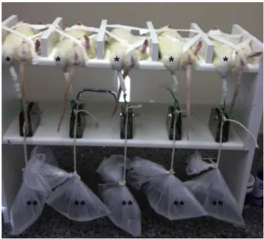
thepurposeofinducingmuscleinjurythroughstraining,as demonstrated in Fig. 1. In this, groups of five rats under anesthesiawereindividuallyfixedindorsaldecubitustothe
Fig.1– Detailsofthesystemusedforinducingstraininthe anteriortibialmusclesofratsunderanesthesia:(*)right hindlegsundertraction;(**)pulleysenablingsuspensionof plasticbagscontainingwater.
apparatususingadhesivetape,withthedorsumofthepaw oftherighthindlegattachedtoastringusingadhesivetape. Thisstringpassedoverapulleyandafreelyhangingplastic bagcontainingavolumeofwatercorrespondingto150%ofthe weightoftherespectiveratwasattachedtotheotherendof thestring.Inthismanner,plantarflexionwasperformedfor 45min,whichcausedaninjuryduetostrainingofthe ante-rior tibialmuscle.Afterinductionofthe muscleinjury, the ratsweretransferredtoaspecificenvironment.TheUTGrats werekepttherefor24handtheSGandAGratsforsevendays. Afterthesemaintenanceperiodsandthenutritional supple-mentationdescribedearlier,bloodcollectionwasperformed, followed byresection ofthe anteriortibialmusclesofboth ofthehindlegs,forserumbiochemicalandhistopathological evaluations.
Supplementationwitharginine
Anamountof420gofl-arginine(Merck®)wasused,diluted
withisotonicsalinesolutionsufficientlytoyield112ml,which wasthensterilizedbymeansoffiltrationthroughMF mem-branes (SCWP304F0Millipore®).Toadministerthis,the rats
weresedatedbymeansofinhalationofhalothane(Tanohalo®;
Cristália)inaclosedcircuit.Followingthis,0.8mlofthe solu-tion(whichcontained3gofl-arginine)wasadministeredonce
adayforsevendays,alwaysatthesametimeofday.20
Biochemicalevaluations
120.8±27
129.8±8.1
188±41.7
0 50 100 150 200 250
UTG SG AG
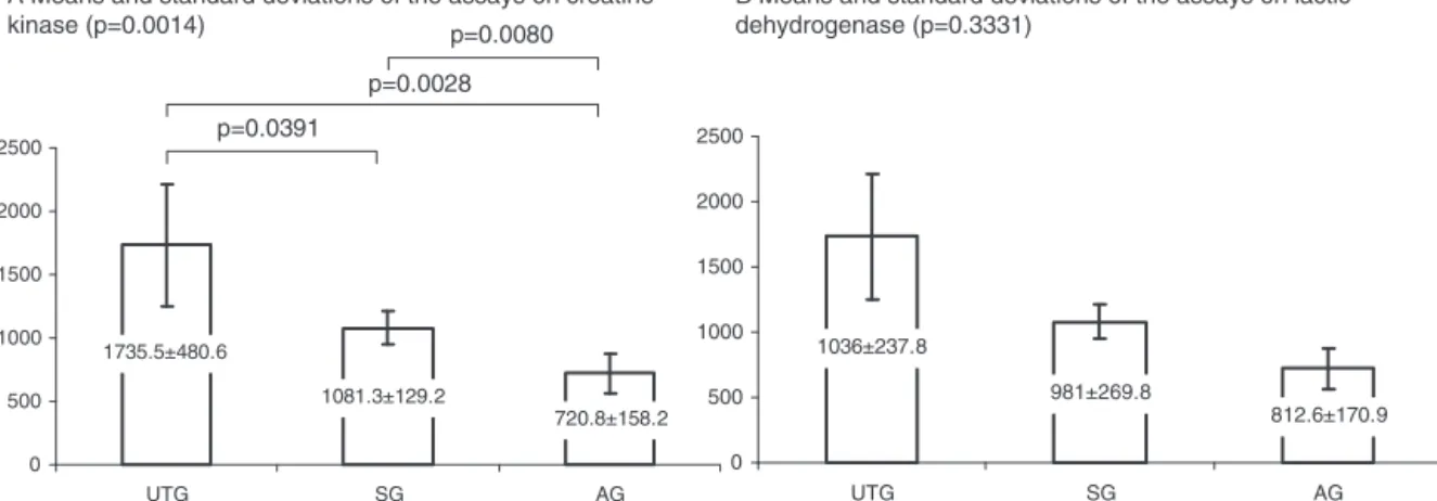
A Means and standard deviations of the assays on creatine kinase (p=0.0014)
B Means and standard deviations of the assays on lactic dehydrogenase (p=0.3331)
C Means and standard deviations of the assays on aspartate aminotransferase (p=0.0063)
D Means and standard deviations of the assays on C-reactive protein (p=0.1149)
p=0.0391
p=0.0028 p=0.0080
p=0.021 p=0.0131
p=0.6221
1735.5±480.6
1081.3±129.2
720.8±158.2
0 500 1000 1500 2000 2500
UTG SG AG
1036±237.8
981±269.8
812.6±170.9
0 500 1000 1500 2000 2500
UTG SG AG
0.0425±480.6
0.055±0.0191
0.03±0.0173
0.00 0.01 0.02 0.03 0.04 0.05 0.06 0.07 0.08
UTG SG AG
Fig.2–Graphsrepresentingthemeansandstandarddeviationsofthebiochemicalassays,comparingthethreegroups: UTG(untreatedandsubjectedtomuscletraction),SG(simulation)andAG(arginine).
Histologicalevaluations
The histological processing was standardized for all the samples from the anterior tibial muscle and began with fixingthemusclesinformalin,followedbycuttingof stan-dardizedsections inplanestransversal and longitudinalto the muscle fibers. Automated histological processing was thenperformed, followed byHarrishematoxylinand eosin staining. The histological slides were analyzed under an optical microscope and were described histopathologically at magnifications of 50, 100 and 400 by two pathologists independently, withemphasison findings compatible with lesions due to strain, such as edema, hemorrhage and morphometric disorganization or alteration of the mus-cle fibers. To show the tissue repair, histological patterns consisting of proliferation of adipose tissue, angiogene-sis and muscle fibers were sought. The samples from the anterior tibial muscles of the left hind leg, which had not been subjected to traction, were taken to be controls.21
Statisticalevaluations
TheANOVAandStudentttestswereused,andthevalueof 0.05 wastaken todefine statistical significanceamongthe variablesevaluated.
Results
Biochemicalevaluations
In the biochemical evaluations, there was a difference betweenthegroupswithregardtothemeansfromtheassays oncreatinekinase(p=0.0014)andaspartateaminotransferase (p=0.0150).Regarding the means from theassays on lactic dehydrogenase(p=0.3331)andC-reactiveprotein(p=0.1149), therewasnodifferencebetweenthegroups(Fig.2).Itcanbe seenfromdetailAofFig.2thatthemeanfromtheassayon creatinekinaseintheUTGwassignificantgreaterthan the meansintheSGandAG,andthatthemeanintheAGwas significantlylowerthanthemeanintheSG.IndetailC,itcan beseenthattherewasasignificantdifferencebetweenUTG andSG,butnotbetweenSGandAG.
Histologicalevaluations
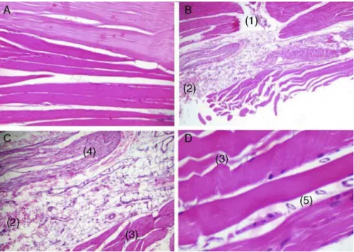
Fig.3–Photomicrographsofhistologicalsectionsthroughstriatedskeletalmuscletissueofthesamplesfromtheanterior tibialmuscleofrats,stainedwithHarrishematoxylinandeosinHarris.Detail[A]:longitudinalsection(×40)through
non-tractionedcontrolmuscle,withoutalterations.Detail[B]:longitudinalsection(×40)throughtractionedmusclefrom
untreatedrat(UTG),showingtearingofmusclefibers(1),edemaandextravasationofredbloodcells(hemorrhage)among themusclefibers(2).Detail[C]:longitudinalsection(×40)throughtractionedmusclefromuntreatedrat(UTG),showing
edemaandextravasationofredbloodcells(hemorrhage)amongthemusclefibers(2),tortuousmusclefibers(3)and hypereosinophilicfiberswithlossofstriations,characterizinghyalinedegeneration(4).Detail[D]:longitudinalsection (×100)throughtractionedmusclefromrattreatedwitharginine(AG),withevidenceofintenseangiogenesisamongthe
musclefibers(5),edemaandtortuousfibers.
hypereosinophilia with loss ofstriations, which character-izedhyalinedegeneration.Inthesamplesfromthetractioned muscles from the rats treated with arginine (AG), intense angiogenesisbetweenthemusclefiberscouldbeseen,along withedemaandtortuousfibers.
Discussion
Experimentalstudiescontributetowardelucidatingthemain aspectsofthe muscle regenerationprocess because ofthe high degreeofmorphological similarity ofmusculoskeletal tissuesamongmammals,onthesignsofinjuryandmuscle regeneration.22 Inthe present study,the samplesfrom the
anteriortibialmusclesoftheratsintheUTGwereevaluated earlyon,i.e.24haftertheinjuryhadbeeninduced,onorder toestablishthenatureofthehistologicalpatterncompatible withmuscleinjuryduetostrain,asshownbytheoccurrences ofedemaandhemorrhageinallthesamples.Thisconfirmed theeffectivenessofthemethodchosenfortheinduction. Dif-ferentexperimentalmethodsforinducingmuscleinjuryhave beenused,includingbruising,23electrostimulation,24
physi-calexercise,25injectionsofmyotoxins24 anddenervation.26
Inthepresentstudy,itwasdecidedtouseastrainmodelin whichhistopathologicalalterationssimilartothoseobserved inhumanswouldbeinduced.19Thedosesofanestheticused
duringinductionofthelesionswerecalculatedsuchthatthese wouldkeeptheanimalsunderanesthesiafor4handwould
avoiduseofanti-inflammatorydrugsorsedativesthatmight interferewiththemusclerepairprocess.Regardingthe histo-logicalresultsfromthisstudy,theUTGpresentedhistological findingscompatiblewithinjuriesduetostrain,asdescribedin detailsBandCofFig.3,inwhichedemaandhemorrhageare shownamongthemusclefibers.Thevasodilatationpromoted by nitric oxideafter metabolization ofl-arginine has been
seentoresultinincreasedmuscleperfusionanddiminished glucose consumption by the skeletal muscles, thus caus-ingdiminishedmusclefatigueandinducingimprovementof physicalperformance.27Adoseof3gofarginineadministered
orallywasindicatedinastudyonhumansthatdemonstrated improved resistance to fatigue upon great effort21 and in
anotherthatshowedincreasedstrengthandmusclemassin individuals undergoinga weighttraining program.28 In the
present study,therewerenointercurrences duringtheoral treatmentwitharginine.FromFig.2,itcanbeseenthatthere wasasignificantdecreaseinserumcreatinekinaselevelsin theratsthatweretreated.Thisenzymehasbeenusedasan indicatorofthestressimposedontheskeletalmusculature, resultingfromintensephysicalactivity,andalsoasafactor formonitoringthetrainingload.29IntheUTG,themean
crea-tinekinaselevelwas1375±480.6U/L,whichwassignificantly greater than the means inthe SGand AG. This groupnot onlyhadthefunctionofdemonstratingtheeffectivenessof themodelused,19butalsoshowedconsequentincreasesin
out24hafterthemusclestrainwashalted,whiletheassays ontheSGandAGwerecarriedoutsevendaysafterwards.A significantdifference(p=0.0080)wasobservedbetweenthese groups,whichshowsthatl-argininemayhaveanreparative
effectwithregardtothestressimposedontheanteriortibial musclethroughtheprovocativetraction.Thus,argininemay beapotentialtreatmentfortheselesions,giventhatintense angiogenesiswasobservedinthehistologicalevaluationson theratsthatweretreatedwith3gofarginineorallyforseven days.Thiseffectwasalreadyknownfrommuscletrainingwith weights,inwhichadministrationofarginineprovidesgreater resistanceand gains in mass,and alsocontributes toward diminishingthepercentageofbodyfat.28 Inevaluatingthe
aspartateaminotransferaselevels,itwasobservedthatthere wasasignificantincreaseintheserumlevelsintheratsofthe AG(Fig.2).Anincreaseintheconcentrationofcytosolic pro-teinsinthecirculationafterexercisereflectsmuscleinjury. The proteins evaluated in these situations are frequently creatinekinase(CK),lactatedehydrogenase(LDH),aspartate aminotransferaseandmyoglobin,whichareusuallyincapable ofcrossingtheplasmamembrane.30Themetabolicprocesses
relatingtopphysicalactivityareimprovedthroughusing argi-nine,sincethisprovidesimprovementsinbloodperfusionin musclesandthusinducesgreaterreleaseofnutrients.Inthis manner,themusclesbecomecapableofproducingenergyfor longertimes,alongwithoxygen,whichretardsthe anaerobio-sisoftheprocess.Italsofavorseliminationoftoxicsubstances thataccumulatewhilephysicalactivityisbeingpracticedand makesmusclerecoveryeasier.28
Conclusion
Useofarginineadministeredorallydidnotcausesignificant metabolicalterationsthatwouldcontraindicateits useand itinducedangiogenesisduringtherepairofmusclesinjured throughstrain.
Conflicts
of
interest
Theauthorsdeclarenoconflictsofinterest.
r
e
f
e
r
e
n
c
e
s
1. AraújoDSMS,AraújoCGS.Aptidãofísica,saúdeequalidade devidarelacionadaàsaúdeemadultos.RevBrasMed Esporte.2000;6(5):194–203.
2. ThompsonD,WilliamsC,KingsleyM,NicholasCW,Lakomy HK,McArdleF,etal.Musclesorenessanddamageparameters afterprolongedintermittentshuttle-runningfollowingacute vitaminCsupplementationD.IntJSportsMed.
2001;22(1):68–75.
3. ThompsonD,NicholasCW,WilliamsC.Muscularsoreness followingprolongedintermittenthigh-intensityshuttle running.JSportsSci.1999;17(5):387–95.
4. CarazzatoJG.Lesõesmusculoesqueléticaseseutratamento. RevBrasOrtop.1994;29(10):723–8.
5. BoothFW.Timecourseofmuscularatrophyduring immobilizationofhindlimbsinrats.JApplPhysiol. 1977;43(4):656–61.
6.TuckerKR,SeiderMJ,BoothFW.Proteinsynthesisratesin atrophiedgastrocnemiusmusclesafterlimbimmobilization. JApplPhysiol.1981;51(1):73–7.
7.JärvinenTA,KääriäinenM,JärvinenM,KalimoH.Muscle straininjuries.CurrOpinRheumatol.2000;12(2):155–61. 8.SeneGL,ShimanoAC,PicadoCHF.Recuperac¸ãomecânica
muscularcomlaser.ActaOrtopBras.2008;17(2):46–9. 9.LefaucheurJP,SébilleA.Thecellulareventsofinjuredmuscle
regenerationdependonthenatureoftheinjury. NeuromusculDisord.1995;5(6):501–9.
10.KauhanenS,SalmiA,vonBoguslawskiK,Asko-SeljavaaraS, LeivoI.Satellitecellproliferation,reinnervation,and revascularizationinhumanfreemicrovascularmuscleflaps.J SurgRes.2003;115(2):191–9.
11.MenetreyJ,KasemkijwattanaC,DayCS,BoschP,VogtM,Fu FH,etal.Growthfactorsimprovemusclehealinginvivo.J BoneJointSurgBr.2000;82(1):131–7.
12.ChiarlaC,GiovanniniI,SiegelJH.Plasmaargininaand correlationsintraumaandsepsis.AminoAcids. 2006;30(1):81–6.
13.HardyI,AlanyR,RussellB,HardyG.Antimicrobialeffectsof arginineandnitrogenoxidesandtheirpotentialrolein sepsis.CurrOpinClinNutrMetabCare.2006;9(3):225–32. 14.PanM,ChoudryHA,EplerMJ,MengQ,KarinchA,LinC,etal.
Argininetransportincatabolicdiseasestates.JNutr.2004;134 Suppl.10:2826–9.
15.LuikingYC,PoezeM,RamsayG,DeutzNE.Reducedcitrulline productioninsepsisisrelatedtodiminisheddenovoarginine andnitricoxideproduction.AmJClinNutr.2009;89(1): 142–52.
16.SamelS,KeeseM,LanigS,KleczkaM,GretzN,HafnerM,etal. Supplementationandinhibitionofnitricoxidesynthesis influencesbacterialtransittimeduringbacterial translocationinrats.Shock.2003;19(4):378–82. 17.BogdanC.Nitricoxideandtheimmuneresponse.Nat
Immunol.2001;2(10):907–16.
18.VieiraDFF,GuarnieroR,VazCES,SantanaPJ.Efeitoda utilizac¸ãodeumcentrifugadodemedulaósseano tratamentodelesãomuscular:estudoexperimentalem coelhos.RevBrasOrtop.2011;46(6):718–25.
19.NikolaouPK,MacDonaldBL,GlissonRR,SeaberAV,GarretWE Jr.Biomechanicalandhistologicalevaluationofmuscleafter controlledstraininjury.AmJSportsMed.1987;15(1):9–14. 20.SantosR,PachecoMTT,MartinsRAB,VillaverdeAB,GianaHE,
BaptistaF.Studyoftheeffectoforaladministrationof
l-arginineonmuscularperformanceinhealthvolunteers.An
isokineticstudy.IsokineticExercSci.2002;10(3):153–8. 21.CarlsonBM,FaulknerJA.Theregenerationofskeletalmuscle
fibersfollowinginjury:areview.MedSciSportsExerc. 1983;15(3):187–98.
22.TidballJG.Inflammatorycellresponsetoacutemuscleinjury. MedSciSportsExerc.1995;27(7):1022–32.
23.MinamotoVB,BunhoSR,SalviniTF.Regeneratedratskeletal muscleafterperiodiccontusions.BrazJMedBiolRes. 2001;34(11):1447–52.
24.HillM,WernigA,GoldspinkG.Musclesatellite(stem)cell activationduringlocaltissueinjuryandrepair.JAnat. 2003;203(1):89–99.
25.SerrãoFV,FoersterB,SpadaS,MoralesMM,Monteiro-PedroV, TannúsA,etal.Functionalchangesofhumanquadriceps muscleinjuredbyeccentricexercise.BrazJMedBiolRes. 2003;36(6):781–6.
26.Jakubiec-PukaA,CiechomskaI,MorgaJ,MatusiakA.Contents ofmyosinheavychainsindenervatedslowandfastratleg muscles.CompBiochemPhysiol.1999;122(3):355–62.
27.McConellGK,HuynhNN,Lee-YoungRS,CannyBJ,WadleyGD.
l-Arginineinfusionincreasesglucoseclearanceduring
28.GerseliA,BarrosTL,BarrosDFL,LimaM.Investigac¸ãodos efeitosdasuplementac¸ãooraldeargininanoaumentode forc¸aemassamuscular.RevBrasMedEsporte.
2007;13(2):129–32.
29.CoelhoDB,MorandiRF,MeloMAA,Silami-GarciaE.Cinética dacreatinaquinaseemjogadoresdefutebolrofissionalem
umatemporadacompetitiva.RevBrasCineantropom DesempenhoHum.2011;13(3):189–94.
30.StupkaN,LowtherS,ChorneykoK,BourgeoisJM,HogbenC, TarnopolskyMA.Genderdifferencesinmuscleinflammation aftereccentricexercise.JApplPhysiol.2000;89(6):