Printed in Brazil - ©2005 Sociedade Brasileira de Química 0103 - 5053 $6.00+0.00
Article
* e-mail: [email protected]
Voltammetric Behavior of Nitrofurazone and its Hydroxymethyl Prodrug with
Potential Anti-Chagas Activity
Mauro Aquiles La-Scalea*,a,b, Carla Maria de Souza Menezesa, Murilo Sérgio da Silva Juliãob,c,
Man Chin Chungd, Sílvia Helena Pires Serranob and Elizabeth Igne Ferreiraa
a
Faculdade de Ciências Farmacêuticas, Universidade de São Paulo, Av. Prof. Lineu Prestes, 580, 05508-900 São Paulo - SP, Brazil
b
Instituto de Química, Universidade de São Paulo, CP 26077, 05599-970 São Paulo - SP, Brazil
c
Centro de Ciências Exatas e Tecnológicas, Universidade Estadual Vale do Acarajú, Av. da Universidade, 850, 62040-370 Fortaleza - CE, Brazil
d
Faculdade de Ciências Farmacêuticas, Universidade Estadual Paulista, CP 502, 14801-902 Araraquara - SP, Brazil
A doença de Chagas é um grave problema de saúde pública para a América Latina, situação agravada pela inexistência de quimioterapia eficiente. Os dois fármacos comercialmente encontrados, benznidazol e nifurtimox, são eficazes apenas na fase aguda da doença. A nitrofurazona é ativa contra
Trypanosoma cruzi, entretanto, a alta toxicidade impede seu uso na parasitose. A hidroximetilnitrofurazona é um pró-fármaco da nitrofurazona, que apresenta maior atividade contra
Trypanosoma cruzi, além de ser menos tóxico. Estudou-se o comportamento voltamétrico da nitrofurazona por voltametria cíclica, varredura linear e pulso diferencial, comparando-o ao do metronidazol e do cloranfenicol. Os coeficientes de difusão dos três fármacos foram estimados aplicando-se a equação de Wilke-Chang. Este artigo também apresenta o estudo do derivado hidroximetilnitrofurazona por voltametria cíclica. A redução de nitrofurazona é pH-dependente e em meio ácido o derivado hidroxilamínico, envolvendo quatro elétrons, é o principal produto formado. Em meio alcalino e com prévio tratamento do eletrodo de carbono vítreo, a redução de nitrofurazona ocorre em duas etapas: a primeira envolve um elétron para formar o nitro-radical aniônico e a segunda etapa corresponde à formação da hidroxilamina. Hidroximetilnitrofurazona possui comportamento voltamétrico semelhante e, de forma análoga, apresentou a mesma eletroatividade e capacidade de estabilização do nitro-radical, indicando que a modificação molecular de nitrofurazona não introduziu alterações no seu comportamento voltamétrico. Uma breve discussão das diferenças de atividade biológica entre os compostos também é apresentada.
Chagas’ disease is a serious health problem for Latin America. The situation is worsened by the lack of efficient chemotherapy. The two available commercial drugs, benznidazole and nifurtimox,
are more effective in the acute phase of the disease. Nitrofurazone is active against Trypanosoma
cruzi, however its high toxicity precludes its current use in parasitosis. Hydroxymethylnitrofurazone
is a prodrug of nitrofurazone. It is more active against Trypanosoma cruzi than nitrofurazone,
besides being less toxic. This work shows the voltammetric behavior of nitrofurazone and a comparison with those of metronidazole and chloramphenicol using cyclic, linear sweep and differential pulse voltammetries. For these drugs also the prediction of the diffusion coefficients using Wilke-Chang equation was performed. The reduction of nitrofurazone is pH-dependent and in acidic medium the hydroxylamine derivative, involving four electrons, is the principal product formed. In aqueous-alkaline medium and with a glassy carbon electrode pre-treatment the reduction of nitrofurazone occurs in two steps, the first involving one electron to form the nitro-radical anion and the second corresponding to the hydroxylamine derivative formation. Hydroxymethylnitrofurazone presented the same voltammetric behavior and electroactivity, indicating that the molecular modification performed in nitrofurazone did not change its capacity to be reduced. A brief discussion regarding the differences in biological activity between the two compounds is also presented.
Introduction
Chagas’ disease affects about one quarter of the population of Latin America. According to the World Health Organization, there are about 120 million people living in risk of contracting parasitosis and 16 to 18 million people infected with the parasite.1 In Brazil, where about 6 million
people are infected, the main problem with treatment is the resistance of Trypanosoma cruzi to nifurtimox.2
Furthermore, benznidazole (2-nitroimidazole) is the only drug marketed in Brazil used for the chemotherapy of Chagas’ disease. Both drugs are only effective in the acute phase of the disease.2
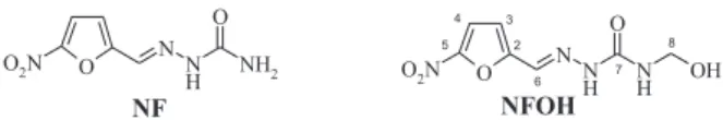
New drug candidates have been proposed for Chagas’ disease chemotherapy and other nitroheterocyclic compounds have been tested as antichagasic drugs to face this serious health problem for Latin America. Nitrofurazone (5-nitro-2-furaldehyde semicarbazone, Figure 1) was synthesized based on the knowledge that furoic acid, as well as its derivatives, demonstrates antimicrobial activity, being active against Gram-positive and Gram-negative bacteria.3 However, its high toxicity has precluded its use
in systemic infections.4
In contrast, a report has shown that nitrofurazone is also able to destroy T. cruzi through trypanothione reductase inhibition, this enzyme is found in the parasite but not in the host.5 Based on this finding, and considering
the mechanism of action of primaquine which may act by increasing the oxidative stress in the parasite due to free radicals formation,6 mutual prodrugs of both drugs were
previously synthesized as Mannich bases and shown to be active in vitro in LLC-MK2 cell cultures infected with T. cruzi trypomastigotes.6 Hydroxymethylnitrofurazone
(Figure 1), a new potential prodrug against Chagas’ disease,7,8 is the intermediate of these compounds. Initial
results showed that this derivative is more active against
T. cruzi than nitrofurazone, in addition to being less toxic.7,8
The biological action of nitroheterocyclic drugs is dependent on the reduction of the nitro group. The nitro-radical anion and hydroxylamine derivative are the main intermediate products responsible for the cytotoxic action of some of the nitroheterocyclic compounds.9,10 The
pharmacological properties of these compounds have been quantitatively related to reduction of the redox couple
R-NO2/R-NO2• ¯ and hydroxylamine formation.10,11
Structure-activity relationship studies15 demonstrated that
the reduction potential correlates with the antimicrobial activity of nitroheterocyclic compounds. Some of these compounds have a trypanocidal action due to either the ability of flavoproteins to reduce the nitrocompound to the nitro-radical or the formation of superoxide and hydrogen peroxide, as a consequence of the electron transfer from the nitro-radical to the molecular oxygen.9,10,16
On the other hand, results of electrochemical studies with some 5-nitrofurane and 5-nitrothiophene derivatives were not conclusive enough to explain the differences in trypanocidal activity. All the derivatives presented values close to that of the reduction potential, which indicates similar eletcroactivities.17
The electrochemical reduction of nitroheterocyclic compounds follows a complex mechanism. Theoretically, the nitro group can receive up to six electrons in the complete reduction to the amine derivative.9,18 Under
anaerobic conditions or low oxygen pressure, the reduction process is similar to that observed for nitrobenzene.18 A
total of two electrons and two protons is involved in the formation of the nitroso (R-NO) intermediate, two more electrons and protons result in the hydroxylamine (R-NHOH):13,14,18
R-NO2 + e– R-NO 2
• ¯ (1)
R-NO2• ¯ + 2H+ R-NO 2H2
• + (2)
R-NO2H2• + + e– R-N(OH)
2 (3)
R-N(OH)2 R-NO + H2O (4)
R-NO + 2e– + 2H+ R-NHOH (5)
The addition of two more electrons results in formation of the amine:
R-NHOH + 2e– + 2H+ R-NH
2 + H2O (6)
Using cyclic voltammetry and aprotic media, nitro-radical stabilization can be observed.12-14 The cyclic
voltammetry studies for nitro-radical formation show that the reduction mechanism depends significantly on the solvent and support electrolyte. Currently, the use of aprotic solvents provokes an expressive decrease in proton availability in the medium, favoring the stabilization of the R-NO2• ¯ radical.12 Many reports on the electrochemistry
behavior of nitrofurans can be found in the literature.14,19-22
The voltammetric behavior of nitrofurazone and its electrochemical determination in pharmaceutical samples have also been registered.23-27
In this study, the voltammetric behavior of nitrofurazone, and its hydroxymethyl derivative, on a
Figure 1. Molecular structures of nitrofurazone (NF) and hydroxymethylnitrofurazone (NFOH).
O O2N NN
H O
NH
2 NF
O O2N N N
H O
N H
OH NFOH
3 2 4 5
6 7
glassy carbon electrode in aqueous media is reported. A comparison with voltammetric behavior of metronidazole (MTZ) and chloramphenicol (CFN) also is presented.
Moreover, a discussion about the biological activity of these compounds is also surveyed from the voltammetric results obtained.
Experimental
Chemicals
Stock solutions (0.01 mol L-1) of nitrofurazone
(Avocado Company) and hydroxymethylnitrofurazone were prepared through direct dissolution in deionizade water and ethanol (1:1) using an ultrasonic bath; metronidazole (Rhodia Farma Lda.) and chloramphenicol (Sigma Chemical Co.) through dissolution only in deionizade water. The pH study was accomplished with universal buffer starting from the mixture of phosphoric, acetic and boric acids with NaOH.28 All solutions were
prepared using analytical grade reagents from Merck and purified water from a Barnsted Nanopure UV system.
Hydroxymethylnitrofurazone synthesis
Hydroxymethylnitrofurazone is not available commercially and its obtainment followed procedure described previously.7,8 The synthesis of NFOH was carried
out in alkaline medium. Nitrofurazone (5.0 mmol), K2CO3 (5.0 mmol), water (50 mL) and 18 mL of formaldehyde were mixed. The reaction was carried out for 49 hours at room temperature, monitored using TLC, following the mobile phase: chloroform:methanol:acetic acid, 85:10:5, v/v/v. The suspension formed was then filtered. The resulting product was washed with methanol and recrystallized from methanol/water. Yield 56%. Melting point: 150-154 oC. Elemental analysis: Calc. C 36.84; H
3.51, N 24.56%; Found: C 37.20; H 3.26; N 24.09%. 1H
NMR (DMSO-d6) δ 7.81 (s, 1H, CH), 7.77-7.75 (d, 1H, Het-H; J 3.9), 7.68-7.61 (t, 1H, NH; J 6.3), 7.22-7.20 (d, 1H, Het-H; J 3.9), 4.62-4.59 (d, 1H, CH2; J 6.6); 13C NMR
(DMSO-d6) δ 127.89 (C2), 112.72 (C3), 115.17 (C4), 151.36 (C5), 152.86 (C6), 154.60 (C7), 63.22 (C8).
Electrochemical assays
The cyclic, linear sweep and differential pulse voltammograms were recorded using an Autolab PGSTAT 20 potentiostat/galvanostat from Eco-Chimie, Utrecht, Netherlands, coupled to a 20 mL cell with a system of three electrodes: a glassy carbon (GCE, Metrohm, ∅ = 2 mm) as
the working electrode, Ag/AgCl as the reference and Pt as the auxiliary electrode. The acquisition and treatment of data were performed using the GPES 4.3 program (Eco-Chimie). Dissolved air was removed from the solutions by bubbling with nitrogen for 10 minutes. The pH control was measured with a Metrohm 654 pH-meter and the combined-glass electrode at room temperature.
Pre-treatment of the glassy carbon electrode
The glassy carbon electrode was manually polished with 0.3 mm alumina suspension on metalographic paper (Arotec S/A, Brazil). The nitro-radical studies were performed using the same polishing procedure, followed by sonication (10 min) in ethanol, using an ultrasonic bath (40 kHz), and rinsed with water.
Prediction of diffusion coefficients
The diffusion coefficient predictions in aqueous phase with infinite dilution were carried out using the Wilke-Chang equation:29
where D is the diffusion coefficient of the solute in water (cm2 s-1); η is the viscosity of water (centipoise) at the
temperature of interest (η = 0.8937 at 25 °C),30 M is the
molar mass of water (g mol-1), T is the temperature (K), x is
the association parameter of water and V is the Le Bas molar volume of the solute (cm3 mol-1).
The molar volume of the solute was calculated starting from the ratio, empirically determined,31 between molecular
volume (VW) and the Le Bas molar volume (V):
VW = 1.06 V
The VW values (Table 1) were calculated by molecular modeling study using the AM1 semi-empirical method32
The association parameter x is a dimensionless empirical factor characteristic for water (solvent). In this work x = 2.5331 was used. All the predictions and graphic
studies were made using de Origin 7.5 software (OriginLab Corp.).
Results and Discussion
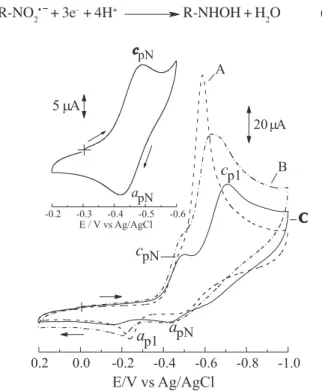
Voltammetric reduction of nitrofurazone
The physiological pH (7.45) was used as a reference for the initial voltammetric tests. The cyclic voltammograms recorded for NF solution showed two irreversible reduction waves and one anodic peak (Figure 2). The first reduction peak (Ecp1 = -0.469 V) corresponds to the reduction of the nitro group to the hydroxylamine derivative (peak 1), involving four protons and four electrons, as depicted below.
R-NO2 + 4e– + 4H+ R-NHOH + H
2O (7)
The affirmation above could be confirmed by the comparison of the voltammetric results obtained with NF, regarding MTZ and CFN. Both drugs are reduced involving four electrons.12,21 Figure 3 shows the linear sweep
voltammograms of the studied drugs. This comparison was possible since diffusion coefficient (D) predictions showed these three compounds have similar sizes. Diffusion coefficients depend on the solvent viscosity (water at 25 0C) and molar volume of solute. The empirical
correlation between V and VW values presented excellent linear relationship, enabling the determination of the molar volume from the volumes calculated by AM1.31
Table 1 shows the results obtained by linear sweep voltammetry (LSV) for the studied drugs. NF is more electroactive than MTZ and CFN. However, the current values are much more closely related and this is a good indication that the three drugs are reduced involving the same number of electrons (n). The results correspond to the highest values obtained after successive GCE polishing. The current function (If = Icp1/n1/2C
O) in LSV
depends on n3/2 and D1/2.33 Thus, the current function
relationship between these drugs (If NF/If MTZ and
If NF/If CFN) present similar values to that obtained by the diffusion coefficient relationship. This suggests that the
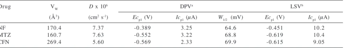
Table 1. Voltammetric results for reduction of the studied drugs at pH 7.45 using GCE
Drug VW D x 106 DPVa LSVb
(Å3) (cm2 s-1) Ec
p1 (V) Icp1 (µA) W1/2 (mV) Ecp1 (V) Icp1 (µA)
NF 170.4 7.37 -0.389 3.25 64.6 -0.451 10.2
MTZ 160.7 7.63 -0.552 3.22 68.8 -0.619 10.4
CFN 269.4 5.60 -0.569 2.33 69.9 -0.615 9.05
a [Drug] = 0.1 mmol L-1; ν = 5 mV s-1; ∆E = 50 mV; medium results obtained from 5 runs; b Linear sweep voltammetry: experimental conditions presented in Figure 3.
Figure 2. Cyclic voltammogram registered in universal buffer pH 7.45 and 1.0 mmol L-1 NF solution, scan rate = 0.1 V s-1; working
electrode:GCE without sonication pre-treatment; ionic strength = 0.6 mol L-1.
Figure 3. Linear sweep voltammograms registered in universal buffer pH 7.45; (A) 1.0 mmol L-1 NF; (B) 1.0 mmol L-1 MTZ; (C) 1.0 mmol
L-1 CFN. Scan rate = 0.01 V s-1; working electrode: GCE without
NF reduction involves the same number of electrons as those for MTZ and CFN reductions: n = 4.
In order to better understand the reduction mechanism of NF, similar experiments are presented using differential pulse voltammetry (DPV, Table 1). For solid electrodes DPV introduces advantages in the study of organic compounds, since they frequently lead to electrode adsorption. Differential pulse methodology can better discriminate effects that are kept constant before and after the pulse application.34 The width of the peak at half height
is defined as: W1/2 = 3.52 RT/nF.34 The proximity of the W 1/2
values for NF, MTZ and CFN allows us to confirm that these drugs have similar reduction processes. The other parameters (Ecp1 and Icp1) follow the behavior above described for LSV. The differences of current values can be attributed to the different values of D.
From these results, we can conclude that the second reduction wave shown in the Figure 2 corresponds to amine derivative formation, involving two more electrons and two protons (equation 6), with a peak potential (Ecp2) at -1.03 V. In the same cyclic voltammogram an anodic peak (Eap1) was also observed at 0.130 V, related to hydroxylamine oxidation to the nitroso derivative. The nitroso-hydroxylamine couple was not detected at low scan rate values. In these experimental conditions, only CFN has demonstrated this wave (Ea = 0.0260 V, Ec = -0.126 V). For NF, 1.0 mmol L-1, the record of this semi-reversible
wave was favored in acidic medium (pH 2). For high scan rate (2.0 V s-1) Ea
p1 was registered at 0.510 V and Ecp3 =
0.430 V with current values of 8.60 µA and 4.90 µA, respectively. The hydroxylamine oxidation occurs according to the following reaction:
R-NHOH R-NO + 2e– + 2H+ (8)
In contrast, previously results24 have shown that NF, in a
solvent-electrolyte system containing pyridine and formic acid with tetramethyl-ammonium chloride,produces just one reduction wave involving six electrons to the amine derivative. Thus, the electrolyte composition appears to be a determinant for the reduction process of NF.
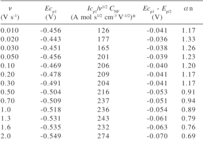
Table 2 presents the cyclic voltammetric results of NF at pH 7.45. The scan rate variation from 0.01 to 2.0 V s-1
increases the height of the reduction wave and Ecp1 is shifted towards negative potentials. The relationship between cathodic current values (Icp1) and ν1/2 is linear,
indicating that the electrodic reaction is diffusion controlled. However, is important to highlight that before each scan the GCE was efficiently polished with alumina, otherwise the current decreased by about by 35 per cent after the first scan. The application of the current function
values (Icp1/ν1/2 C
NF, ν≤ 0.3 V s
-1) and D value of NF
(Table 1) in the Randles-Ševčik equation33,34
(Icp1 = -2.69x10⁵ ν3/2 A D1/2 C NF ν
1/2) can be a good
approximation for the number of electrons calculation. From the data of Table 2 a total of four electrons was estimated, corroborating the results presented above.
The αn values were obtained from the voltammetric equation for the irreversible electrochemical reaction (Ep/2
– Ep,c = 48/αn).33,34 The αn values suggest the involvement
of two electrons in the rate-determining step of the electrode reaction, being, therefore, the formation of nitroso compound the slowest step of the first reduction wave. The reduction potential reflects the transfer of the first or second electron and identical behavior was previously registered for CFN21 and MTZ.12 The significant decrease
of αn values indicates that NF irreversible reduction can be evidenced at higher scan rates. This is because the potential application is faster than the charge transfer process involved in the rate-determining step of the electrode reaction.
Effect of pH on nitrofurazone reduction
The voltammetric reduction of NF was investigated using a GCE in the range 2.32 ≤ pH ≤ 12.1. The Ecp1 value for the NF reduction wave is shifted to negative values with increasing pH, indicating the existence of an acid-base equilibrium close to the electrochemical reaction, confirming that H+ ions are involved in the reduction
process (equation 7), as shown in the Ecp1vs. pH plots, Figure 4. The NF reduction is pH-dependent with
Ecp1 = - 0.206 V – 0.0349 pH. The value of the proton numbers (p) was estimated as 0.71, indicating that one
Table 2. Cyclic voltammetric results for reduction of 1.0 mmol L-1
NF at pH 7.45 using GCE
ν Ecp1 Icp1/ν1/2C
NF Ecp1 - Ep/2 αn
(V s-1) (V) (A mol s1/2 cm-3 V-1/2)* (V)
0.010 -0.456 126 -0.041 1.17
0.020 -0.443 177 -0.036 1.33
0.030 -0.451 165 -0.038 1.26
0.050 -0.456 201 -0.039 1.23
0.10 -0.469 206 -0.040 1.20
0.20 -0.478 209 -0.041 1.17
0.30 -0.491 204 -0.041 1.17
0.50 -0.504 216 -0.053 0.91
0.70 -0.509 237 -0.051 0.94
1.0 -0.518 236 -0.054 0.89
1.3 -0.531 243 -0.061 0.79
1.6 -0.535 232 -0.063 0.76
2.0 -0.549 274 -0.070 0.69
proton is involved in the rate-determining step of the reaction in the pH range studied. Probably, this result is due to the occurrence of a fast protonation step preceding charge transfer. The H+ ion, involved in the
rate-determining reduction step, corresponds to a second slow protonation reaction of the nitro group, which is further reduced to the nitroso intermediate. This reasoning has also been applied to the electrochemical behavior of other nitroheterocyclic drugs.12,21,24
On the other hand, it is also possible to make an analysis in which the Ecp1 vs pH plots presents two linear regions, in acidic medium (2.32 ≤ pH ≤ 7.45) with
Ecp1 = -0.188 V – 0.0384 pH and in alkaline medium (7.69 ≤ pH ≤ 12.1) with Ecp1 = -0.275 V – 0.0280 pH. Although the difference between both regions is very small, this can indicate distinct processes are occurring on the electrode. This could be proved with the surface treatment of the GCE that will be discussed in the next section.
The anodic peak registered (peak 2) is also pH-dependent on the pH range studied. In alkaline medium, the oxidation is facilitated and the slope of the first linear relationship (Eap1 = 0.569 V – 0.0593 pH) indicates the involvement of the same number of electrons and protons (Figure 4).
Voltammetric generation of nitro-radical anion from nitrofurazone
At pH > 8 a change of the reduction process of NF was observed, because a shoulder around -0.5 V preceding the main reduction wave has been registered. In this
experimental conditions this behavior change may be related to the reversible reduction of the nitro group to the nitro-radical, involving one electron.
Figure 5 presents the cyclic voltammograms of NF 1.0 mmol L-1 at pH 12.1. The C voltammogram clearly
shows the unfolding of the NF reduction wave and the appearance of a peak with EcpN = -0.490 V. This reduction peak (equation 1) has a corresponding oxidation peak at -0.419 V, as can be observed from the inset in Figure 5 and corresponds to the nitro-radical formation in a step involving 1e. In the sequence, the nitro-radical is reduced to the hydroxylamine derivative (Ecp1 = -0.708V), according to the following mechanism:
R-NO2• ¯ + 3e- + 4H+ R-NHOH + H
2O (9)
The differences among voltammograms (A, B, C) of Figure 5 are related to the procedures adopted for the cleanup of the GCE surface. After the polishing using alumina the GCE was submitted to sonication in deionized water (curve B) or ethanol (curve C) for complete elimination of the alumina from the electrode surface.
The sonication of the GCE in ethanol was the best condition for detection of the R-NO2/R-NO2• ¯ couple, and Figure 4. Plot of Epvs. pH using GCE as a working electrode
without sonication pre-treatment: () Ecp1 value for the NF re-duction wave; (S) Eap1 values corresponding to hydroxylamine oxidation of the nitroso derivative. [NF] = 1.0 mmol L-1;
0.1 mol L-1 <ionic strength < 1.0 mol L-1.
Figure 5. Cyclic voltammograms recorded in universal buffer pH 12.1 and 1.0 mmol L-1 NF solution. Scan rate = 1.0 V s-1. A (dashed
line): GCE polished with 0.3 µm alumina; B (dash-doted line): GCE polished with 0.3 µm alumina and sonicated in an ultrasonic bath with deionized water; C (solid line): GCE polished with 0.3 mm alumina and sonicated in an ultrasonic bath with ethanol. Inset: Cyclic voltammogram corresponding to the R-NO2/R-NO2• ¯ couple
for 1.0 mmol L-1 NF solution in pH 12.1 using a GCE polished with
shows that the electrode surface plays an important role in the nitro-radical stabilization. The sonication with ethanol may modify the electrode surface, even though ethanol is a hydrophilic solvent. Despite the long duration of the sonication (10 min) the frequency was low (40 kHz). Longer sonication times do not produce significant differences in this electrochemical behavior. Rubinstein35 also registered
effects of the GCE immersion in methanol in an ultrasonic bath to study of the nitrobenzene reduction. The demonstration of the voltammetric behavior of NF as well as the differences provoked by sonication on the electrode surface and the use of the others solvents for the GCE pre-treatment has been aim of the our workgroup and further studies are planned, since previous reports have36,37 related
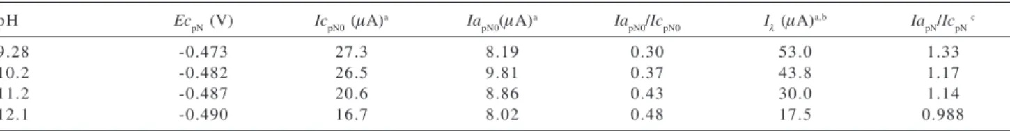
the importance of sonication on GCE surface activation. Table 3 shows the voltammetric results for the R-NO2/ R-NO2• ¯couple. The reduction peak, Ec
pN, shifted towards
a more negative potential, indicating the existence of a protonation equilibrium before the charge transfer process. Thus, a decrease in the values of IcpN0 was observed suggesting the stabilization of the R-NO2H+/R-NO
2H2 •at
the lowest pH values.12 Similarly, a linear increase of the
anodic current values with pH would be expected, however, we observed a maximum anodic current value at pH 10.2. The current ratio values (IapN0/IcpN0), corresponding to the R-NO2/R-NO2• ¯ couple, were established using currents
measured with respect to the zero current axis. These results differ from unity, however, we may deduce that the stability of the nitro-radical increased at high pH due to the difficulty in the protonation process. These results are compatible with those previously recorded.23
It is also possible to observe from Figure 5 and by the results of Table 3 that the switching potential currents (Iλ) are higher than the IcpN0 values in all studied cases, showing a strong interference of the second reduction step, corresponding to hydroxylamine formation. The choice of the switching potential (Eλ = -0.6 V) is important for the recorder of the anodic wave. The best separation (218 mV) between both reduction peaks, EcpN and Ecp1, was obtained at pH 12.1. However, the current anodic measurement was jeopardized due to the base line hard correction. Thus, the
IapN/IcpN values calculated using the Nicholson method38
were higher than the unity, except at pH 12.1, which incapacitated the kinetics study of nitro-radical disproportionation.39
Since the effect of pH on the protonation process of the species is more complex, shifting the reduction wave to more negative potentials, this situation occurs when protonation process precedes the electron transfer.24 This
explains the large distance between both reduction peaks at pH 12.1. The use of mixed medium can improve this separation.22,23 On the other hand, the nitro-radical
generation in aqueous media can be more useful for further studies with biological targets, in which the experimental conditions should be as close as possible to the biological system.
Similarly to other nitroheterocyclic drugs,13,19,22,23 the
current ratio (IapN0/IcpN0) increases towards unity as a function of the scan rate, a typical behavior of an irreversible chemical reaction following a charge transfer step.9,39 We have assumed that NF undergoes the same
mechanism as that proposed by other authors,13,19,22,23 as is
depicted in the reaction mechanism below:
2R-NO2• ¯ + 2H+ R-NO
2 + R-NO + H2O (10)
The generation and electrochemical stabilization of nitro-radical anion in the absence of aprotic solvents enable a reactivity comparison between nitrofurazone and its prodrug. The possible differences in voltammetric behavior of these drugs can be correlated with the pharmacological activities.8,9
Voltammetric reduction of hydroxymethylnitrofurazone
NFOH is a prodrug of NF, that is expected to be released from Mannich bases with primaquine peptides.6-8 The
hydroxymethylation renders NF more hydrophilic. However, further studies are needed to demonstrate whether this property improves the transport across cell/parasite membrane.8
The voltammetric reduction of NFOH presented the
Table 3. The pH effect on voltammetric parameters for the R-NO2/R-NO2• ¯ couple from NF reduction. [NF] = 1.0 mmol L-1; ν = 1.0 V s-1;
switching potential (Eλ) = - 0.6 V
pH EcpN (V) IcpN0 (µA)a Ia
pN0(µA)
a Ia
pN0/IcpN0 Iλ (µA)a,b IapN/IcpN c
9.28 -0.473 27.3 8.19 0.30 53.0 1.33
10.2 -0.482 26.5 9.81 0.37 43.8 1.17
11.2 -0.487 20.6 8.86 0.43 30.0 1.14
12.1 -0.490 16.7 8.02 0.48 17.5 0.988
a current value measured with respect to the zero current axis; b current value corresponding to the switching potential; c current ration calculated
same NF reduction mechanism at all pHs studied. The reduction wave corresponds to the hydroxylamine derivative formation (Ecp1’ = - 0.469 V) involving four electrons. The amine formation was also registered at a slightly less negative potential, Ecp2’ = - 0.951 V.
Table 4 presents the potential peak values at different pH. The Ecp1’ values of NFOH show little difference, except at pH 7.45 where both compounds presented exactly the same value. These results indicate that both compounds have similar eletcroactivity, in other words, the molecular modification performed in NF did not change its capacity to be reduced.
Maintaining the same experimental conditions, i.e., polishing the GCE surface followed by activation in the ultrasonic bath using ethanol, NFOH was also reduced to R-NO2• ¯ in a step involving one electron. At pH 12.1, and
a scan rate of 1.0 V s-1, the NFOH presented two reduction
waves. The potential value for the first peak is - 0.500 V and the second peak of the hydroxylamine derivative is - 0.704 V (Figure 6), demonstrating exactly the same
Table 4. The peak potentials for NF and NFOH at different pH values. n = 0.1 V s-1
pH NFEcp1 (V) NFOHEcp1’ (V)
2.32 -0.270 -0.257
6.21 -0.425 -0.420
7.45 -0.469 -0.469
10.2 -0.566 -0.535
12.1 -0.611 -0.553
Figure 6. Cyclic voltammograms recorded in universal buffer pH 12.1 and 1.0 mmol L-1 NF solution. GCE polished with 0.3 mm
alumina and sonicated in an ultrasonic bath with ethanol and scan rate = 1.0 V s-1; ionic strength = 1.0 mol L-1. Dashed line is the cyclic
voltammogram corresponding to the R-NO2/R-NO2• ¯ couple.
voltammetric behavior as NF. The quantitative aspects of the electrochemical nitro-radical generation from NFOH will be the goal of further studies.
As already mentioned, NFOH was synthesized according to the theory that Mannich bases release hydroxymethyl derivatives that are converted to the starting drug. NFOH, however, was shown to be more active than benznidazole and nitrofurazone against trypomastigote and amastigote forms of T. cruzi.8 Furthermore, its toxicity
was much lower than NF itself, since this prodrug proved to be less mutagenic (about four times) than the parent drug.29 These properties account for the high interest in
studying this prodrug under a multidisciplinary viewpoint. Since the bioactivity and, probably, the toxicity of nitroheterocyclic compounds in protozoa is currently related to the R-NO2 reduction, the voltammetric studies could explain the higher activity and lower toxicity of a derivative if a significant change in its potential reduction was observed. As NFOH behavior paralleled that of NF, the hydroxymethylation did not change the reduction profile of the latter. Thus, differences of physicochemical properties may be involved in the biological activity and in the toxicity of NFOH to that of NF.
Conclusion
NF was been reduced on a glassy carbon electrode, producing, in acid medium, only one reduction wave involving four electrons due to the hydroxylamine derivative formation. The activation of the glassy carbon electrode, using polishing with alumina suspension followed by sonication in ethanol, enabled the nitro-radical stabilization in alkaline medium. The study of the nitro-radical anion in aqueous media will allow us to study biological targets. NFOH presented the same voltammetric behavior as that of NF, indicating that both compounds have similar electroactivity. On the other hand, NFOH was shown to be more active and less toxic, in comparison to NF. Whilst the voltammetric study was unable to explain the biological differences between both compounds, voltammetry may be a useful complementary technique for further studies of biological mechanisms.
Acknowledgements
We thank FAPESP for the financial support (process no.
01/01192-3 and 03/10763-0) and for the posdoctor fellowship to M. A. La-Scalea (process no. 01/09418-0),
Capes-Prodoc for the fellowship to C. M. S. Menezes (process no. 00019-03-8) and to Richard Compton, from
References
1. WHO; Chagas Disease: Strategic Direction for Research. Disease Burden and Epidemiological Trends, http:// www.who.int/tdr/diseases/chagas/direction.htm, accessed in September 2004.
2. Coura, J.R.; de Castro, S.L.; Mem. Inst. Oswaldo Cruz2002,
97, 3.
3. Dodd, M.C.; Stillman, W.B.; J. Pharmacol. Exp.Ther.1944,
82, 11.
4. Henderson, G.B.; Ulrich, P.; Fairlamb, A.H.; Rosenberg, I.; Pereira, M.; Sela, M.; Cerami, A.; Proc. Natl. Acad. Sci. USA
1988, 85, 5374.
5. Korolkovas, A.; Dicionário Terapêutico GuanabaraEd. 2000/
2001, Guanabara Koogan: Rio de Janiero, 2002.
6. Gonçalves, M.T.; Chung, M.C.; Colli, W.; Miranda, M.T.M.; Ferreira, E.I.; Rev. Soc. Bras. Med. Trop.1994, 27, 164. 7. Chung, M.C.; Miranda, M.T.M.; Ferreira, E.I.; Abstracts of
211th American Chemical Society Meeting, New Orleans, USA, 1996.
8. Chung, M.C.; Martinelli, T.F.; Gonçalves, M.T.; Güido, R.V.C.; Varanda, E.A.; Polli, M.C.; Botelho, K.C.; Colli, W.; Miranda, M.T.M.; Ferreira, E.I.; Bioorg. Med. Chem. 2003, 11, 4779. 9. Edwards, D.I. In Comprehensive Medicinal Chemistry. The
Rational Design, Mechanistic Study & Therapeutic Application
of Chemical Compounds; Hansch, C.; Sammes, P.G.; Taylor J.B., eds; Pergamon Press: Oxford, 1990, p. 725-751. 10. Viodé, C.; Albuquerque, C.N.; Chauvière, G.; Houée-Lévin,
C.; Périe, J.; New J. Chem.1997, 21, 1331.
11. Guissani, A.; Henry, Y.; Lougmani, N.; Hickel, B.; Free Rad.
Biol. Med.1990, 8, 173.
12. La-Scalea, M.A.; Serrano, S.H.P.; Gutz, I.G.R.; J. Braz. Chem.
Soc.1999, 10, 127.
13. Tocher, J.H.; Gen. Pharmacol.1997, 28, 485.
14. Núñez-Vergara, L.J.; Squella, J.A.; Aldunate, J.; Letelier, M.E.; Bollo, S.; Repetto, Y.; Morello, A.; Spencer, P.L.;
Bioelectrochem. Bioenerg.1997, 43, 151.
15. Rozenski, J.; De Ranter, C.J.; Verplanken, H.; Quant.
Struct.-Act. Relat.1995, 14, 134.
16. Martinez-Merino, V.; Cerecetto, H.; Bioorg. Med. Chem.2001,
9, 1025.
17. Cerecetto, H.; Di Maio, R.; González, M.; Risso, M.; Sagrera, G.; Seoane, G.; Denicola, A.; Peluffo, G.; Quijano, C.; Stoppani, A.O.M.; Paulino, M.; Olea-Azar, C.; Basombrío, M.A.; Eur. J.
Med. Chem.2000, 35, 343.
18. Zuman, P.; Microchem. J.1997, 57, 4.
19. Núñez-Vergara, L.J.; Aldunate, J.; Letelier, M.E.; Bollo, S.; Repetto, Y.; Morello, A.; Spencer, P.L.; Squella, J.A.;
Bioelectrochem. Bioenerg.1995, 38, 355.
20. Squella, J.A.; Lemus, I.; Lonza, G.; Núñez-Vergara, L.J.; Bol. Soc. Chil. Quim.1991, 36, 109.
21. Morales, A.; Toral, M.I.; Richter, P.; Analyst1984, 109, 633. 22. Merino, M.; Carbajo, J.; Núñez-Vergara, L.J.; Squella, J.A.;
Bol. Soc. Chil. Quim.2000, 45, 97.
23. Symons, T.; Tocher, J.H.; Tocher, D.A.; Edwards, D.I.; Free
Rad. Res. Comms.1991, 14, 33.
24. Morales, A.; Richter, P.; Toral, M.I.; Analyst1987, 112, 971. 25. Khodari; M.; Mansour, H.; Mersal, G.A.M.; J. Pharm. Biomed.
Anal.1999, 20, 579.
26. Reday, C.S.; Reddy, S.J.; Electroanalysis1992, 4, 595. 27. Gode, K.D.; Analyst1985, 110, 1373.
28. Lurie, Ju.; Handbook of Analytical Chemistry, Mir Publishers: Moscow, 1978, p. 263.
29. Wilke, C.R; Chang, P.; A.I.Ch.E. J. 1955, 1, 264.
30. Schramke, J.A.; Murphy, S.F.; Doucette, W.J.; Hintze, W.D.;
Chemosphere 1999, 38, 2381.
31. La-Scalea, M.A.; Menezes, C.M.S.; Ferreira, E.I.; Abstracts
Book of 27ª Annual Reunion of Brazilian Chemical Society,
2004, abstract FQ-097.
32. Dewar, M.J.S.; Zoebisch, E.G.; Heally, E.F.; Stweart, J.J.P.; J. Am. Chem. Soc.1985, 107, 3902.
33. Brown, E.R.; Sandifer, J.R. In Physical Methods of Chemistry, Electrochemical Methods 2, Rossiter, B.W.; Hamillton, J.F., eds.; Wiley: New York, 1986, p. 273.
34. Brett, C.M.A.; Brett, A.M.O.; Electrochemistry. Principles,
Methods, and Applications, Oxford University Press: Oxford, 1996.
35. Rubinstein, I.; J. Electroanal. Chem.1985, 183, 379. 36. Zhang, H.; Coury Jr., L.A.; Anal. Chem.1993, 65, 1552. 37. Marken, F.; Kumbhat, S.; Sanders, G.H.W.; Compton, R.G.; J.
Electroanal. Chem.1996,414, 95.
38. Nicholson, R.S.; Anal. Chem.1966, 38, 1406.
39. Olmstead, M.L.; Nicholson, R.S.; Anal. Chem.1969, 41, 862.
Received: September 29, 2004
Published on the web: May 20, 2005