One hundred million years of
interhemispheric communication:
the history of the corpus callosum
Departamento de Psiquiatría y Centro de Investigaciones Médicas, Facultad de Medicina, Pontificia Universidad Católica de Chile, and Millenium Nucleus for Integrative Neuroscience, Santiago, Chile F. Aboitiz and
J. Montiel
Abstract
Analysis of regional corpus callosum fiber composition reveals that callosal regions connecting primary and secondary sensory areas tend to have higher proportions of coarse-diameter, highly myelinated fibers than callosal regions connecting so-called higher-order areas. This suggests that in primary/secondary sensory areas there are strong timing constraints for interhemispheric communication, which may be related to the process of midline fusion of the two sensory hemifields across the hemispheres. We postulate that the evolutionary origin of the corpus callosum in placental mammals is related to the mechanism of midline fusion in the sensory cortices, which only in mammals receive a topographically organized representation of the sensory surfaces. The early corpus callosum may have also served as a sub-strate for growth of fibers connecting higher-order areas, which possibly participated in the propagation of neuronal ensembles of synchronized activity between the hemispheres. However, as brains became much larger, the increasingly longer interhemispheric dis-tance may have worked as a constraint for efficient callosal transmis-sion. Callosal fiber composition tends to be quite uniform across species with different brain sizes, suggesting that the delay in callosal transmission is longer in bigger brains. There is only a small subset of large-diameter callosal fibers whose size increases with increasing interhemispheric distance. These limitations in interhemispheric con-nectivity may have favored the development of brain lateralization in some species like humans.
Correspondence
F. Aboitiz
Departamento de Psiquiatría, Facultad de Medicina Pontificia Universidad Católica Marcoleta 387, 2º piso Casilla 114-D Santiago 1 Chile
Fax: +56-2-665-1951 E-mail: [email protected]
Presented at the XVII Annual Meeting of the Federação de Sociedades de Biologia Experimental, Salvador, BA, Brazil, August 28-31, 2002.
Research partially supported by Fondecyt (No. 1970294) and by the Millenium Nucleus for Integrative Neuroscience.
Received July 18, 2002 Accepted November 26, 2002
Key words
·Commissures ·Evolution
·Interhemispheric transfer ·Lateralization
·Synchronization
Introduction
The corpus callosum is a unique feature of the brain of placental mammals, so much so that we may state that it can be a diagnos-tic character of placentals just like the very placenta is. This structure was first described
unique to placentals, since the dorsal com-missure originated mainly from the hippo-campal formation, while the corpus callo-sum had no relation to the hippocampus. The absence of any kind of callosal primordia in nonplacental mammals implied a sudden evolutionary origin of the corpus callosum, with no structures ancestral to it. This was unexpected according to Darwins concept of evolution by the successive accumulation of small changes. In fact, even Huxley, a champion of gradualism, would admit in 1863 that the origin of the corpus callosum signified a major leap in evolution. In 1865, some thirty years later than Owen, Flower (2) and later Smith (3) argued that both the corpus callosum of placentals and the dorsal commissure of marsupials contained fibers connecting the anterior part of the mesial hemisphere and consequently were partly homologues. This observation would agree with the concept of a gradual origin of the corpus callosum, since fibers that originally traveled through the dorsal commissure might have found a different route across the hemi-spheres. However, Flower went perhaps too far in naming the dorsal commissure of mar-supials as the corpus callosum, and propos-ing that the corpus callosum could be found even in reptiles and other lower verte-brates. Indeed, when saying this, he was actually speaking of the dorsal hippocampal commissure. For this reason, Flower later became strongly discredited (3). Neverthe-less, Flower may have been right in that some neocortical fibers might cross through the hippocampal commissure in marsupials, which would explain its large size.
Aside from these historical considerations, the corpus callosum remains an evolutionary puzzle. Mutations producing the absence of the corpus callosum are not uncommon, and if there were no adaptive value in this structure it would be common to find placental lineages in which the corpus callosum had been lost. In addition, it is surprising that few dramatic long-term effects beside the callosal
discon-nection syndrome are seen after section of the corpus callosum in humans and in other ani-mals (4,5). Therefore, the question arises about the adaptive value of the largest fiber tract in the placental nervous system. Which circum-stances were involved in the origin of the corpus callosum, and why was it maintained in the history of placental mammals? Further-more, is the corpus callosum related to the emergence of brain lateralization in humans and to conscious experience? (4) In this paper, we will discuss our studies on the comparative fiber composition of the corpus callosum, which in our view may provide important insights into the problems raised above.
Fiber composition of the corpus callosum
highly myelinated fibers of more than 3 µm in diameter. However, in other mammals such as carnivores, rodents and ungulates, we did not observe regional differences in fiber size, at least in the posterior corpus callosum (10). This may partly reflect the more diffuse topographic arrangement of different cortical areas in the corpus callo-sum of these species (see Ref. 10,11).
Functions of different callosal fibers
The uneven distribution of fiber types along the corpus callosum suggests impor-tant functional differences in interhemi-spheric communication between different types of cortical areas. First, it is important to recall that many callosal projections are ho-motopic, that is, they connect equivalent regions between the two hemispheres (12). This is not to say that there are no hetero-topic callosal projections (i.e., connecting different areas across the hemispheres). In the visual cortex, callosal cells and fibers connecting lower-order areas (V1, V2, V3) with a visuotopic organization tend to be concentrated on the borders between these areas (Figure 2). More specifically, many callosal-projecting cells and their terminals are located in a stripe corresponding to the representation of the visual fields midline (Figure 3); these cells can be found both in superficial and deep cortical layers (12,13). Since each hemisphere contains a represen-tation of the contralateral visual hemifield, callosal fibers have been proposed to con-nect the two hemirepresentations of the whole visual field at the level of the midline. On the border between the primary and secondary visual areas of the cat and other mammals there is a band containing two mirror images (one in V1 and the other in V2) of the central, binocular visual field; each image has an ipsilateral and a contralateral repre-sentation of the central visual field (14-16). In this region, many callosal cells connect points of the contralateral visual
representa-Figure 1. Cross-section of the human corpus callosum indicating the representation of different cortical regions(top). Regional differences in fiber composition along the corpus callosum (larger circles indicate larger fiber diameters) (bottom). A, auditory fibers; F, frontal fibers; M, motor cortex fibers; Ss, somatosensory fibers; T/P, temporoparietal fibers; V, visual fibers.
Figure 2. Diagrams indicating the distribution of callosal fibers in different cortical areas of the cat. Top, Fibers originating at the 17/18 border project to several regions representing visually equivalent points along the midline (0 degree). Bottom, Fibers connecting higher-order areas (ectosylvian cortex, ES) originate modified from and project to the whole contralateral area. CC, corpus callosum; SSI, suprasylvian cortex. Modified from Ref. 17.
0º
0º 0º
0º
0º 0º
0º 0º
0º
0º 0º
SSI
CC
SSI
ES ES
ES
SSI SSI
ES
0º
CC
19
18
17 17 19
18
19 18
17
19
18
17 1/3
1/3
1/5
F
M
Ss A
tion of one hemisphere with regions repre-senting the same or a neighboring visual position in the ipsilateral representation of the other hemisphere (see Figure 3). In addi-tion, in V1 and V2, a few cells in deep layer V have been found to project through the corpus callosum from regions well within the respective areas, representing peripheral regions of the visual field (18). The explana-tion for these cells is not clear yet, but may be related to anticipation of trajectories of
fast-moving objects from one visual field to the other and to integration of large-scale fea-tures across the visual field, such as optic flow during locomotion (see below; 19,20). In the somatosensory cortex, S1, and the motor cortex, M1, callosal-projecting cells have been described to be restricted to a narrow band representing the body midline (see Ref. 12). However, more recent find-ings indicate that in S1 there are a substantial number of neurons with bilateral receptive fields in the representations of the hand and feet, which could participate in integrating information necessary for the cooperative actions of the two hands (21). These bilateral cells are probably connected to the other hemisphere via the corpus callosum. In pri-mary and secondary auditory areas of the rat, in which different tones are represented as stripes across the respective areas, callosal cells have been described to be segregated in relation to the tonotopic representation, al-though there are some discrepancies in the details of this array (22-26).
Callosal projections in higher-order cor-tical regions have been mostly studied in the prefrontal and temporoparietal visual areas. These regions tend to be connected inter-hemispherically by poorly myelinated, thin-diameter, slow-conducting fibers (8,9). There, callosal cells tend to be distributed all over the respective cortical regions, inter-digitating with ipsilateral corticocortical pro-jections and with thalamic propro-jections in a manner resembling the ocular-dominance array of the primary visual cortex. These higher-order cortical areas tend to have a poor visuotopic organization. Their cells have large receptive fields and respond better to stimulus properties such as color, shape or direction of movement than to strict position in the visual field (12).
Visual callosal fibers: midline fusion and depth perception
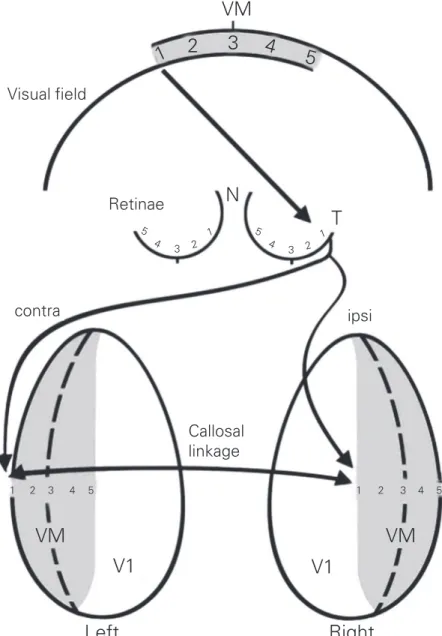
The concentration of fast-conducting, Figure 3. In V1, callosal fibers connect contralateral ocular dominance columns with
ipsilat-eral ocular dominance columns, representing visually equivalent points. N, nasal; T, tempo-ral; VM, visual midline. Modified from Ref. 15.
Visual field
Retinae
contra ipsi
Callosal linkage
VM
V1
V1
VM
1
2
3
4
5
5 4
3 2
1 5
4
3 2
1
5 4 3 2
1 1 2 3 4 5
N
T
VM
large-diameter callosal fibers in primary/sec-ondary sensorimotor areas might reflect the importance of performing the fusion of the two sensory and motor spatial hemirepre-sentations across the hemispheres. This is especially important in two sensory systems: the auditory system, which may use binaural cells to make up a cortical sound localization system, and the visual system, in which cen-tral vision involves high-resolution process-ing and is related to aspects of depth percep-tion. In fact, the callosal regions correspond-ing to the primary and secondary auditory and visual areas have the largest fiber diam-eters of the corpus callosum (9).
The role of the corpus callosum in central vision and depth perception has been de-bated for some time. Reports indicate that damage to the posterior corpus callosum produces inability to judge depth in patients with damage of the optic chiasm, and section of the corpus callosum may reduce the pro-portions of binocular cells (27-29). In addi-tion, experimentally induced early strabis-mus leads to an expanded callosal receiving zone in areas 17 and 18; in these conditions callosal cells also show decreased binocu-larity, decreased ability to respond to fast-moving stimuli, a small receptive field size, and poor orientation selectivity (19). Mid-line visual deficits (30) and impairment of interhemispheric depth comparisons when using head movements to determine relative depth (31) have been reported in acallosal humans. However, other reports indicate that the corpus callosum is not critical for depth perception, and that stereoscopic vision is mostly determined by the interactions be-tween the thalamocortical crossed and un-crossed pathways in the visual cortex of each hemisphere (32-34). It is possible that the corpus callosum is involved in more subtle mechanisms of depth perception than stere-opsis (which is specifically defined as bin-ocular disparity), such as relative motion or parallax, that is, using the differences in relative motion of near and far objects to
judge depth (31). In this context, it has been suggested that visual callosal fibers partici-pate in predicting trajectories of moving ob-jects across the midline, and in the genera-tion of binding mechanisms in the central visual field (19,20).
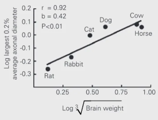
Recently, we studied the fiber composi-tion of the posterior corpus callosum in dif-ferent species which differ in brain size, namely the mouse, the rabbit, the cat, the dog, the cow and the horse (10). We found that, despite a quite conservative cross-spe-cies distribution of fiber sizes, a small popu-lation of large-diameter fibers increased their caliber in species with larger brains. Further-more, in the cat and the dog, both species with frontal vision, the largest fiber diam-eters were larger than expected from their brain size, as compared to species with more lateral vision (see Figure 4). This is consist-ent with the hypothesis that the large callosal fibers are related to aspects of central vision.
Origin of the corpus callosum
In reptiles, topographically organized sen-sory maps are restricted to mesencephalic levels (35), for which there is a well-devel-oped tectal commissure (Figure 5). In these animals, the sensory projections to the telen-cephalon lose their topographic organiza-tion. Furthermore, interhemispheric fibers, represented by the anterior commissure and the hippocampal commissure, are quite scarce in reptiles. In contrast, in the mammalian
Log largest 0.2%
average axonal diameter
0.2
0.1
0.0
-0.1
-0.2
-0.3
0.25 0.50 0.75 1.00
Log 3 Brain weight
Dog Cat
Rabbit
Rat
Horse r = 0.92
b = 0.42 P<0.01
Cow
integrate the two topographic hemirepresen-tations of the sensory surface across the midline (Figure 5; 39). Another important early callosal function may have been bi-manual coordination (21), but in our view this may have involved the corpus callosum at later stages of cortical evolution, after the topographic maps were already established and the cerebral cortex had begun to exert a significant control in motor behavior via the corticospinal tract.
In early mammals, there may have been strong selection for a system performing midline fusion in the neocortical sensory representations. There were two different telencephalon, topographically organized
sensory representations take place in the multilaminar neocortex (36,37). This coin-cides with the development of a strong com-missural system in monotremes, marsupials and placentals (38).
Based on the correspondence between the origin of topographic representations in the cerebral cortex and the origin of inter-hemispheric fibers, and on the observation that the most important timing constraints for interhemispheric transmission may be in primary and secondary sensory areas, we suggest that the origin of the mammalian corpus callosum is related to the need to
Visual Field
L R
DCX VCX
SC OT
Reptiles Mammals
L R L
AC R
L R
TC
TC Figure 5. Topographic
represen-tation of the visual fields on both sides of the brain in reptiles and mammals. In reptiles, topo-graphic visual projections are lo-calized in the mesencephalic op-tic tectum (OT), while the telen-cephalic dorsal cortex (DCX) has a very poor visuotopic organiza-tion. In mammals, the superior colliculus (SC, homologous to the OT) has a visuotopic organi-zation, but the most important topographic representation is in the primary visual cortex (VCX, homologous to the DCX). The tectal commissure (TC) con-nects the two hemirepresenta-tions of the visual field in the mesencephalon of reptiles and mammals, while in the mamma-lian visual cortex both hemirep-resentations are connected via the anterior commissure (AC, marsupials and monotremes) or the corpus callosum (CC, placentals). L, left; R, right.
pathways that interhemispheric fibers could use to travel across the midline: the anterior commissure and the hippocampal commis-sure. One pathway was followed by mono-tremes and marsupials, in which many axons run to the other hemisphere through the ante-rior commissure. Consequently, this struc-ture is notably enlarged in relation to other mammals and reptiles. This is not to say that in this group there may not be interhemi-spheric fibers running via the hippocampal commissure. However, the route via the an-terior commissure has the disadvantage that fibers must run a long distance from the dorsally located cerebral cortex to the ven-tral anterior commissure, which may pro-duce an unwanted delay in interhemispheric transmission - especially in species with large brains. In fact, in large-brained marsupials like the kangaroo, interhemispheric cortical fibers form the so-called fasciculus aberrans, which descends from the subcor-tical white matter along the internal capsule and then reaches the anterior commissure, thus significantly shortening the traveling distance (Figure 6; 38). A more efficient solution to interhemispheric communication evolved in placental mammals (Figure 6) with the origin of the corpus callosum. Onto-genetically and phyloOnto-genetically, the corpus callosum originates from the hippocampal commissure and gradually separates from it, moving to a position dorsal to the hippocam-pal formation (38). In this position, axons travel a much shorter distance between the hemispheres than if they were to cross ven-trally through the anterior commissure. In our view, interhemispheric fibers permitted the integration of both hemirepresentations of the sensory surface in the cerebral cortex,
Hippocampal commissure
external capsule
rhinal fissure
Hippocampal commissure
Hippocampal commissure EUTHERIAN FOREBRAIN
anterior commissure internal
capsule
anterior commissure corpus callosum
external capsule
rhinal fissure
caudate putamen
internal capsule
anterior commissure
METATHERIAN FOREBRAIN (Polyprotodontia)
rhinal fissure external capsule
METATHERIAN FOREBRAIN (Diprotodontia)
Neocortex Paleocortex
while the corpus callosum originated in pla-cental mammals as a strategy to minimize interhemispheric transmission time for fi-bers connecting primary and secondary sen-sory areas.
In present day placentals, only a small contingent of callosal fibers connect primary and secondary areas. Most callosal fibers connect higher-order areas, which have a poor topographic organization. How did these fibers become so abundant if the primary function of the corpus callosum relates to midline fusion and bimanual coordination? One possibility is that they crossed the hemi-spheres just because they could. Despite its underlying molecular complexity, the ability to cross the telencephalic midline may be shared by many cortical axons, in fact many more than those that are finally retained in the adult. The number of callosal fibers is dramatically reduced by 75% during the peri-natal period of the primate (41), and perhaps in similar proportions in other mammals. Furthermore, some callosal axons have been found to re-cross to the ipsilateral hemi-sphere through the anterior commissure (42). These axons retract their bicommissural ex-tension early in the postnatal period. This indicates that there is not much specificity in terms of the kinds of fibers that will cross the midline. In other words, once a way was open for axons to cross to the other hemi-sphere, the axons began to use this pathway to colonize new regions. Although this may help explain why the fibers connecting higher-order areas were initially able to grow to the contralateral hemisphere, it leaves open the question of why these fibers were maintained in evolution.
Factors favoring the expansion of the corpus callosum: interhemispheric synchrony
In recent years, synchronicity in neu-ronal firing has been proposed as a general mechanism for neuronal processing (43-45).
Small- or large-scale groups of intercon-nected neurons can show synchronous oscil-lations in their firing activity, which enables them to work as a unified whole for a period of time. This mechanism has been proposed to account for basic aspects of perception such as binding (that is, the process by which the information of color, movement and form become integrated into unified percepts) and for more general phenomena like conscious-ness. In the cat, callosal-dependent inter-hemispheric synchrony has been observed during visual stimulation (46), and in hu-mans, synchronized activity in the gamma band (40 Hz) has been found to spread across the hemispheres during a visual recognition task (47), which may be mediated by the corpus callosum. Furthermore, analyses of the morphology and diameter of callosal axonal branches suggest that the architec-ture of callosal axons is suitable to promote the synchronous activation of multiple tar-gets in the opposite hemisphere (16). It would not be surprising that processes such as depth perception or binding in the central visual field depend on the generation of synchro-nous ensembles of neuronal activity in the two hemispheres, for which callosal fibers may be fundamental (20).
fa-cilitated through a mechanism of interhemi-spheric reciprocal interactions. In a way, this may be similar to the callosal propagation of an epileptic focus; interhemispheric recipro-cal excitation works as a facilitatory mech-anism for the seizure, which spreads to the ipsilateral territory in both hemispheres. In fact, in patients callosotomy has proved to be an effective treatment to prevent the ex-pansion of epileptic activity (4). It is there-fore possible that interhemispheric connec-tions may be advantageous for the spread of normal synchronous activity in the cerebral cortex.
Relation to brain lateralization
Some years ago, a controversial hypo-thesis suggested that in large-brained spe-cies, interhemispheric transmission time would be increased due to the longer dis-tances needed to reach the contralateral hemi-sphere; this would promote interhemispheric isolation and consequently the development of brain lateralization (48). We and others have determined that callosal fiber composi-tion tends to be conservative across species (10,49), indicating that interhemispheric transmission time can be significantly im-paired in large-brained species (50). There is only a small contingent of fibers of very wide diameter whose caliber increases with increasing brain weight, but their increase in maximal diameter may not be sufficient to fully compensate for the increasingly long interhemispheric distances (10). Further-more, across species the proportion of cal-losal fibers in relation to brain size or to the estimated numbers of cortical cells tends to decrease with increasing brain weight (10,51, 52), thereby reducing the degree of inter-hemispheric connectivity. Overall, this evi-dence suggests that reduced callosal trans-mission is related to increased brain size, which is consistent with the hypothesis of Ringo et al. (48).
This discussion is also relevant to the
issue of interhemispheric synchrony. One important requisite for the development of synchronous ensembles, especially large-scale ones, is a high transmission velocity across distant regions in the brain (43). In small-brained mammals this may not be a significant problem because the axonal dis-tances are short, but in larger-brained ani-mals like humans, the interhemispheric dis-tances may become so large as to somewhat impair callosal transmission. It is possible that in large-brained animals, synchronous activity mediated by the corpus callosum can be achieved only by specialized chan-nels provided by a few fast-conducting fi-bers (53). In this context, it is of interest that in all callosal regions the proportions of large-diameter fibers in the corpus callosum tend to increase with age (54,55). This may imply that these fibers participate in the gen-eration of interhemispheric ensembles which become stabilized during late ontogeny. In any case, the size-related constraints for in-terhemispheric synchrony may have well fa-cilitated the development of intrahemispheric ensembles, which may have promoted the development of brain lateralization. In this sense, although the corpus callosum may have contributed to the interhemispheric spread of synchronous ensembles in small mammals, it is possible that a point was reached in which, beyond a given brain size, problems appeared for the transmission of some types of information between the hemi-spheres. Even considering that interhemi-spheric synchronous activity is possible in humans and other animals, the question still remains as to whether in our species there are certain subtle limiting factors in the gen-eration of interhemispheric synchrony. These may have facilitated the development of strong, intrahemispheric neuronal loops, thus promoting hemispheric independence and the establishment of brain lateralization.
docu-mented in several studies (9,56,57). How-ever, it is not clear if in humans increased brain lateralization is caused by a decrease in interhemispheric communication or vice versa. One possibility is that the two factors reinforce each other in development, that is, an initially increased anatomical brain asym-metry may produce a larger than normal retraction of callosal axons, which in turn may promote interhemispheric isolation, thus strengthening the incipient brain laterality (39,58).
Final comment
In this article we have intended to pro-vide a scenario for the origins and evolution of the corpus callosum in placental mam-mals. Interhemispheric fibers possibly origi-nated in mammals as a consequence of the development of the isocortex and of topo-graphic maps of the sensory and motor sur-faces in it. In monotremes and marsupials, fibers travel to the other hemisphere via the anterior commissure and the hippocampal commissure. In placental mammals, the cor-pus callosum originates above the hippo-campal commissure as the main tract con-necting the left and right cortices, and may provide a shortcut for interhemispheric axons to the contralateral hemisphere.
Commis-sural fibers may have first participated in binding mechanisms related to midline fu-sion and bimanual coordination, an ability observed in many so-called primitive mam-mals. The lack of specificity by which corti-cal axons may cross through the corpus corti- cal-losum allowed many kinds of fibers con-necting diverse areas of the cerebral cortex, especially through heterotopic connections, which contributed to the spread of neuronal ensembles in different aspects of cognitive processing. In species with large brain size like humans, constraints may have appeared limiting conduction speed and the amount of interhemispheric fibers, thus favoring intra-hemispheric processing and the development of brain lateralization. Although some of these ideas may not be testable, there are proposals that may provide ideas for future research. For example, it will be interesting to compare the tectal commissure of reptiles and the visual callosal fibers of mammals, as well as the role of the large-diameter visual fibers in central vision. Furthermore, the role of the corpus callosum in the generation of large-scale neuronal ensembles remains to be demonstrated, and further studies are needed to ascertain the role of interhemi-spheric communication in the development of brain lateralization.
References
1. Huxley TH (1863). Man’s Place in Nature. McMillan, London, UK. 2. Flower WH (1865). On the commissures of the cerebral
hemi-spheres of the Marsupialia and Monotremata as compared with those of placental mammals. Philosophical Transactionsof the Royal Societyof London, 23: 633-651.
3. Smith E (1910). Some problems relating to the evolution of the brain. Lancet, 1: 1-6.
4. Gazzaniga MS (2000). Cerebral specialization and interhemispheric communication. Does the corpus callosum enable the human condi-tion? Brain, 123: 1293-1326.
5. Schalomon PM & Wahlsten D (2002). Wheel running behavior is impaired by both surgical section and genetic absence of the mouse corpus callosum. Brain Research Bulletin, 57: 27-33.
6. Rockland KS & Pandya DN (1986). Topography of occipital lobe commissural connections in the rhesus monkey. Brain Research,
365: 174-178.
7. Kim JH, Ellman A & Juraska JM (1996). A re-examination of sex differences in axon density and number in the splenium of the rat corpus callosum. Brain Research, 740: 47-56.
8. LaMantia AS & Rakic P (1990). Cytological and quantitative charac-teristics of four cerebral commissures in the rhesus monkey. Jour-nal of Comparative Neurology, 291: 520-537.
9. Aboitiz F, Scheibel AB, Fisher RS & Zaidel E (1992). Fiber composi-tion of the human corpus callosum. Brain Research, 598: 143-153. 10. Olivares R, Montiel J & Aboitiz F (2001). Species differences and
similarities in the fine structure of the mammalian corpus callosum.
Brain, Behavior and Evolution, 57: 98-105.
12. Innocenti GM (1986). General organization of callosal connections in the cerebral cortex. In: Jones EG & Peters A (Editors), Cerebral Cortex. Vol. 5. Plenum Press, New York, NY, USA, 291-354. 13. Innocenti GM, Aggoun-Zouaoui D & Lehmann P (1995). Cellular
aspects of callosal connections and their development. Neuropsy-chologia, 33: 961-988.
14. Olavarría JF (1996). Non-mirror symmetric patterns of callosal link-ages in areas 17 and 18 in cat visual cortex. Journal of Comparative Neurology, 366: 643-655.
15. Olavarría JF (2001). Callosal connections correlate preferentially with ipsilateral cortical domains in cat areas 17 and 18, and with contralat-eral domains in the 17/18 transition zone. Journal of Comparative Neurology, 433: 441-457.
16. Houzel JC & Milleret C (1999). Visual inter-hemispheric processing: constraints and potentialities set by axonal morphology. Journal of Physiology, 93: 271-284.
17. Berlucchi G (1972). Anatomical and physiological aspects of visual function of the corpus callosum. Brain Research, 214: 239-259. 18. Olavarría JF & Van Sluyters RC (1983). Widespread callosal
connec-tions in infragranular visual cortex of the rat. Brain Research, 279: 233-237.
19. Milleret C & Houzel JC (2001). Visual interhemispheric transfer to areas 17 and 18 in cats with convergent strabismus. European Journal of Neuroscience, 13: 137-152.
20. Houzel JC, Carvalho ML & Lent R (2002). Interhemispheric connec-tions between primary visual areas: beyond the midline rule. Brazil-ian Journal of Medical and Biological Research, 35: 1441-1453. 21. Iwamura Y (2000). Bilateral receptive field neurons and callosal
connections in the somatosensory cortex. Philosophical Transac-tions of the Royal Society of London, Series B. Biological Sciences, 355: 267-273.
22. Imig TJ & Bruge JF (1978). Sources and terminations of callosal axons related to binaural and frequency maps in primary auditory cortex of the cat. Journal of Comparative Neurology, 182: 637-660. 23. Pallas SL, Littman T & Moore DR (1999). Cross-modal reorganization of callosal connectivity in auditory cortex without altering thalamo-cortical projections. Proceedings of the National Academy of Sci-ences, USA, 96: 8751-8756.
24. Pallas SL (2001). Intrinsic and extrinsic factors that shape neocortical specification. Trends in Neurosciences, 24: 417-425.
25. Scheich H, Heil P & Langner G (1993). Functional organization of auditory cortex in the Mongolian gerbil (Meriones unguiculatus). II. Tonotopic 2-deoxyglucose. European Journal of Neuroscience, 5: 898-914.
26. Thomas H, Tillein J, Heil P & Scheich H (1993). Functional organiza-tion of auditory cortex in the Mongolian gerbil (Meriones unguicula-tus). I. Electrophysiological mapping of frequency representation and distinction of fields. European Journal of Neuroscience, 5: 882-897.
27. Elberger AJ & Smith EL (1985). The critical period for corpus callo-sum section to affect cortical binocularity. Experimental Brain Re-search, 57: 213-223.
28. Gardner JC & Cynader MS (1987). Mechanisms for binocular depth sensitivity along the vertical meridian of the visual field. Brain Re-search, 413: 60-74.
29. Hubel DH (1988). Eye, Brain and Vision. Scientific American Library, New York, NY, USA, 156-157.
30. Jeeves MA (1991). Stereo perception in callosal agenesis and partial callosotomy. Neuropsychologia, 29: 19-34.
31. Rivest J, Cavanagh P & Lassonde M (1994). Interhemispheric depth judgment. Neuropsychologia, 32: 69-76.
32. Lepore F, Ptito M & Lassonde M (1986). Stereoperception in cats following section of the corpus callosum and/or the optic chiasma.
Experimental Brain Research, 61: 258-264.
33. Ptito M, Lepore F & Guillemot JP (1991). Stereopsis in the cat: behavioral demonstration and underlying mechanisms. Neuropsy-chologia, 29: 443-464.
34. Cowey A & Wilkinson F (1991). The role of the corpus callosum and extra striate visual areas in stereoacuity in macaque monkeys. Neu-ropsychologia, 29: 465-479.
35. Ten Donkelaar HJ (1998). Reptiles. In: Nieuwenhuys R, Ten Donkelaar HJ & Nicholson C (Editors), The Central Nervous System of Vertebrates. Springer-Verlag, Berlin, Germany.
36. Aboitiz F (1999). Evolution of isocortical organization. A tentative scenario including roles of reelin, p35/cdk5 and the subplate zone.
Cerebral Cortex, 9: 655-661.
37. Aboitiz F, Morales D & Montiel J (2001). The inverted neurogenetic gradient of the mammalian isocortex: development and evolution.
Brain Research Reviews, 38: 129-139.
38. Abbie AA (1939). The origin of the corpus callosum and the fate of the structures related to it. Journal of Comparative Neurology, 70: 9-44.
39. Aboitiz F & Ide A (1998). Anatomical asymmetries in language-related cortex and their relation to callosal function. In: Stemmer E & Whitaker H (Editors), Handbook of Neurolinguistics. Academic Press, New York, NY, USA, 393-404.
40. Shang F, Ashwell KWS, Marotte LR & Waite PME (1997). Develop-ment of commissural neurons in the Wallaby (Macropus eugenii).
Journal of Comparative Neurology, 387: 507-523.
41. LaMantia AS & Rakic P (1990). Axon overproduction and elimination in the corpus callosum of the developing rhesus monkey. Journal of Neuroscience, 10: 2156-2175.
42. Hedin-Pereira C, Uziel D & Lent R (1992). Bicommissural neurones in the cerebral cortex of developing hamsters. NeuroReport, 3: 873-876.
43. Singer W (1999). Neuronal synchrony: a versatile code for the defini-tion of reladefini-tions? Neuron, 24: 49-65, 111-125.
44. Varela FJ, Lachaux J-P, Rodríguez E & Martinerie J (2001). The brainweb: phase synchronization and large-scale integration. Nature Reviews. Neuroscience, 2: 229-239.
45. Engel AK, Fries P & Singer W (2001). Dynamic predictions: oscilla-tions and synchrony in top-down processing. Nature Reviews. Neu-roscience, 2: 704-716.
46. Engel AK, König P, Kreiter AK & Singer W (1991). Interhemispheric synchronization of oscillatory neural responses in cat visual cortex.
Science, 252: 1177-1179.
47. Rodríguez E, George N, Lachaux J-P, Martinerie J, Renault B & Varela FJ (1999). Perception’s shadow: long-distance gamma band synchronization of human brain activity. Nature, 397: 430-433. 48. Ringo JL, Doty RW, Demeter S & Simard PY (1994). Time is of the
essence: a conjecture that hemispheric specialization arises from interhemispheric conduction delay. Cerebral Cortex, 4: 331-343. 49. Schüz A & Preissl H (1996). Basic connectivity of the cerebral cortex
and some considerations on the corpus callosum. Neuroscience and Biobehavioral Reviews, 20: 567-570.
50. Aboitiz F, Montiel J & López J (2002). A hypothesis on the early evolution of the develoment of the isocortex. Brain Research Bulle-tin, 57: 481-483.
51. Rilling JK & Insel TR (1999). Differential expansion of neural projec-tion systems in primate brain evoluprojec-tion. NeuroReport, 10: 1453-1459.
intraspecies morphometric analysis of the corpus callosum. Brain, Behavior and Evolution, 55: 37-43.
53. Aboitiz F, López J & Montiel J (2002). Long distance communication in the human brain: timing constraints for interhemispheric syn-chrony and the origin of brain lateralization. Biological Research (in press).
54. Aboitiz F, Rodríguez E, Olivares R & Zaidel E (1996). Age-related changes in fiber composition of the human corpus callosum. Neuro-Report, 7: 1761-1764.
55. Godlewski A (1991). Morphometry of myelin fibers in corpus callo-sum and optic nerve of aging rats. Journal für Hirnforschung, 83:
568-581.
56. Dorion AA, Chantome M, Hasboun D, Zouaoui A, Marsault C, Capron C & Duyme M (2000). Hemispheric asymmetry and corpus callosum morphometry: a magnetic resonance imaging study. Neuroscience Research, 36: 9-13.
57. Witelson SF & Goldsmith CH (1991). The relationship of hand prefer-ence to anatomy of the corpus callosum in men. Brain Research, 545: 175-182.