Effect of Fe
3+Doping in the Photocatalytic Properties of BaSnO3 Perovskite
Kleber Figueiredo Mouraa, Laís Chantellea, Débora Rosendoa, Elson Longob, Ieda Maria Garcia dos Santosa*
Received: December 12, 2016; Revised: July 07, 2017; Accepted: July 12, 2017
In the last ten years, stannates with perovskite structure have been tested as photocatalysts. In spite
of the ability of perovskite materials to accommodate different cations in its structure, evaluation of
doped stannates is not a common task in the photocatalysis area. In this work, Fe3+ doped BaSnO 3 was
synthesized by the modified Pechini method, with calcination between 300 and 800°C/4 h. The powder precursor was characterized by thermogravimetry after partial elimination of carbon. Characterization after the second calcination step was done by X-ray diffraction, Raman spectroscopy and UV-visible
spectroscopy. Materials were tested in the photocatalytic discoloration of the Remazol Golden Yellow
azo dye under UVC irradiation. Higher photocatalytic efficiency was observed under acid media. As no
meaningful adsorption was observed at this condition we believe that an indirect mechanism prevails. Fe3+ doping decreased the band gap and favored the photocatalytic reaction, which may be assigned
to the formation of intermediate levels inside the band gap.
Keywords: Perovskite, Fe-doped, polymeric precursor method, photocatalysis, RNL.
*e-mail: [email protected]
1. Introduction
Alkaline earth stannates (MSnO3, M = Ba, Sr, Ca) with perovskite structure have become alternative materials to the technological sector due to its applications as dielectric components. Its use as photocatalyst has also been reported, especially for water splitting1-3 and for the photodegradation
of organic dyes, with emphasis in the SrSnO34,5. Previous results of our research group indicated that BaSnO3 has a higher photocatalytic activity than SrSnO3 for the degradation
of an azo-dye, the golden yellow remazol (RNL)6.
BaSnO3 has been studied in various applications in recent years but it is not widely explored as photocatalyst. In spite
of its band gap of 3.1 eV, a small activity is usually reported,
being assigned to a high electron-hole recombination rate7.
This drawback may be overcome by the use of nanostructured
materials, as reported by Moshtaghi et al.8,9, who attained a high activity in the photodegradation of organic dyes. A high activity may also be attained, using the perovskite ability to form solid solutions resulting in defects, which can therefore improve its photocatalytic properties10-12. For instance, the
solid solution BaSn0.2Pb0.8O3 has been evaluated by Borse et al. showing a high activity for the photo-oxidation of water10.
Literature also reports the use of La3+/2+, Ni3+/2+, Fe3+/2+, Cu2+/+,
Co3+/2+ as dopant into oxides as TiO
2, ZnTiO3, ZnO 13-17.
Fe3+ has been used as TiO
2 dopant in several studies
18-23,
behaving as electron scavenger which suppress electron-hole
recombination improving the photocatalytic efficiency. Fe3+
has also been used as BaSnO3 dopant and leads to formation
of multi energy levels below the conduction band edge. Charge
balance is obtained by formation of oxygen vacancies, besides oxidation or reduction of Fe3+, which contribute to the perovskite
stabilization24-26. The magnetic properties of this Fe-doped perovskite have been studied, but, up to our knowledge, its use
as photocatalyst has not been reported yet. The heterogeneous catalysis offers technical and environmental advantages over
homogeneous catalysis, allows the recycling of the solid catalyst
over its useful life and minimizes the generation of effluents.
Several solids have been proposed as potential catalysts for
photodegradation of textile dyes. The performance of these
materials as catalysts is naturally related to the nature of the acid or basic sites found in these materials.
In the present work, Fe-doped BaSnO3 was synthesized by
the modified Pechini method and applied in the photoactivity
degradation of RNL azo-dye.
2. Experimental
2.1 Synthesis of photocatalysts
Fe3+-doped BaSnO
3 (0; 0.05 and 0.1 in mol) was
synthesized by the modified Pechini method, similarly to the
methodology described Lucena et al.27. After tin dissolution
in a 0.1 mol.L-1 nitric acid aqueous solution cooled with an
ice bath, citric acid (C6H8O7.H2O, Cargill - 99.5 %) was
added into the solution. The pH of the solution was adjusted to 3 by adding ammonium hydroxide (NH4OH, Vetec - 28-30 %). Solutions of iron citrate or barium citrate were
prepared from the respective nitrates, Ba(NO3)2 (Vetec - 99 aNúcleo de Pesquisa e Extensão - NPE, Laboratório de Combustíveis e Materiais - LACOM,
Departamento de Química, Universidade Federal da Paraíba, Campus 1, CEP 58059-900, João Pessoa, PB, Brazil.
bCentro de Desenvolvimento de Materiais Funcionais - CDMF, Laboratório Interdisciplinar de
Moura et al.
318 Materials Research
%), Fe(NO3)3.9H2O (Vetec - 99.5 %), and added into the tin
citrate solution under a slow agitation at 25°C for 12 h. A
molar ratio citric acid:metal of 3:1 was used for all of the
citrates. Ethylene glycol (Vetec - 99.5 %) was added into the solution at 90°C to promote its polymerization. The mass ratio of citric acid to ethylene glycol was 60:40.
The polymeric resins were calcined at 250°C/2 h,
deagglomerated, dry milled in a Spex mill, and sieved (100
mesh) to obtain the powder precursors. A heat treatment under
an oxygen atmosphere (O2) was performed at a temperature
of 300°C /6 h at a heating rate of 1°C min-1 under a flow of
1000 cm3 min-1 to partially eliminate the organic material.
The materials were calcined from 300 to 800°C/4 h under stagnated air atmosphere at a rate of 10°C min-1.
The precursors obtained after heat treatment under
oxygen atmosphere were characterized by thermogravimetry
(TG) and its derivative curve (DTG) using a SDT-2960 thermobalance (TA Instruments) with a heating rate of 10°C
min-1 up to 1000°C under synthetic air with a flow of 100
mL min-1 using alumina crucibles. The samples calcined at
300-800°C were characterized by X-ray diffraction (XRD) using an XRD-6000 Shimadzu diffractometer with Cu Kα radiation between 10° and 80°, using a step size of 0.02° and a step time of 2 s. The values of the lattice parameters were obtained with the Rede93 software using the least
squares method. Micro-Raman spectra were obtained in the region of 100-1000 cm-1 by an InVia spectrophotometer
from Renishaw, using an Ar laser (514 nm) with a power of 20 mW and an objective lens of 50x. UV-vis spectra were obtained by reflectance mode using an UV-2550 Shimadzu spectrophotometer, in the 190-900 nm range.
2.2 Photodegradation reaction
During the photocatalytic test, 15.0 mL of a RNL solution
with a concentration of 10 mg L-1 and pH = 3 and 6 was
placed in a Petri plate with 10 mg of the photocatalyst. The suspensions were irradiated for 1, 2 and 4 h with a Super Niko UVC lamp (0.5-1.0mW), model ZG-30T8. After the reaction, the suspensions were centrifuged at 5000 rpm for 30 min at room temperature before being filtered. The solutions were analyzed by UV-Vis spectroscopy using a SHIMADZU UV-2550 spectrometer, in the range of 300 to 700 nm. The dye discoloration percentage was determined by the measured absorbance at λ = 411 nm, which was
assigned to the azo group.
3. Results and Discussion
3.1 Synthesis and characterization of Fe-doped
BaSnO
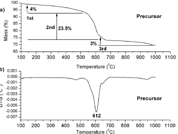
3Figure 1 shows the TG and DTG curves of the precursor. Three thermal decomposition steps were observed in the TG curves. In the first step, water and gases adsorbed on the powder
surfaces were eliminated; the second step was assigned to the decomposition of the Ba(NO3)2 between 460 and 680°C; the third step was assigned to the carbonate decomposition28.
Similar behaviors were obtained by Udawatte et. al.28 and Li et. al.29 for the synthesis of BaSnO3 using BaCO3, SnO2
and BaCl2 as precursors.
The X-ray diffraction (XRD) patterns of the materials obtained after heating between 300 and 800 °C are shown
in Figure 2.
The planes were indexed according to ICDD 01-074-1300 (BaSnO3), 00-041-1445 (SnO2), 00-045-1471
(BaCO3), 00-024-0053 [Ba(NO3)2] and 00-0010891 (Sn).
For the precursor's heat treated at 300 °C, peaks assigned to
tetragonal SnO2, Ba(NO3)2, Sn and BaCO3 were observed.
After heat treatment at 400 °C, higher intensity peaks were
observed for Ba(NO3)2, while the intensities of these peaks
decrease at 500 °C. The formation of the cubic BaSnO3
(Pm3m) was observed at 600°C besides a small amount
of SnO2 and BaCO3, which is in agreement with the TG/
DTG analysis shown in figure 1. This crystallization temperature below 600°C is quite low compared with
other synthesis methods as solid state reaction30-33. No
significant change was observed with temperature increase from 600 to 800 °C.
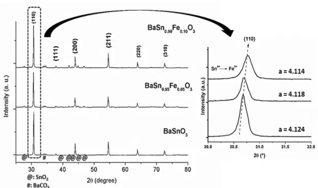
The XRD patterns of Fe-doped BaSnO3 are shown in
Figure 3. Highly crystalline cubic BaSnO3 was observed
while no peaks assigned to Fe2O3 (ICDD 03-065-3107) were
found. A slight shift in the diffraction peaks towards higher 2θ values was observed after doping indicating that Fe3+ got
into BaSnO3 lattice. The lattice parameters, a, of BaSnO3 were calculated and indicated that a small lattice decrease took place, which is assigned to the smaller ionic radius of Fe3+ (0.64 Å) compared to Sn4+ (0.69 Å)34.
Figure 4 shows the Raman spectra of the Fe3+-doped
BaSnO3 (0; 0.05 and 0.1 in mol). The group theory predicts the absence of active modes in the Raman spectra for a perfect
Pm3m perovskite structure. In spite of this, Cerda et. al.35
reported bands at 238, 408, 543 and 724 cm-1, attributed to
distortions of the cubic structure of BaSnO3 due to defects, which modify the internal symmetry of the perovskite phase,
leading to unexpected modes in Raman spectra. These modes
were assigned to the six fundamental vibrations of SnO6 with Oh symmetry. Similar studies on various perovskite compounds show that distortions of these materials are due to the presence of defects (VOx, V•
O, V ••
O,Sn 2+)36-38.
Balamuragan et al.39,40 evaluated the optical and electromagnetic properties of Fe-doped BaSnO3. According to the authors, when iron is added into the perovskite lattice, a center of extrinsic defects is formed with the formation of oxygen vacancies for charge compensation, as showed
in Equation (1).
(1)
Fe O
BaSnO2
Fe
SnV
3
O
X
2 3 0 0
3
+
r+
Figure 1. a) TG and b) DTG curve of the precursor after heat treatment in the O2 atmosphere at 300°C.
Figure 2. XRD patterns of the BaSnO3 heat treated at different temperatures.
In the present work, the mode at 150 cm-1 was attributed to
the vibration of carbonate groups. Undoped BaSnO3 showed
bands at similar regions to those reported by Cerda et al.35) indicating that distortions are present in the structure. After
doping, dislocation of the bands to 252, 413, 535 and 663
cm-1 took place. A higher definition was observed for the
bands at 252 and 413 cm-1, which may be correlated to the
oxygen vacancies, which change the symmetry.
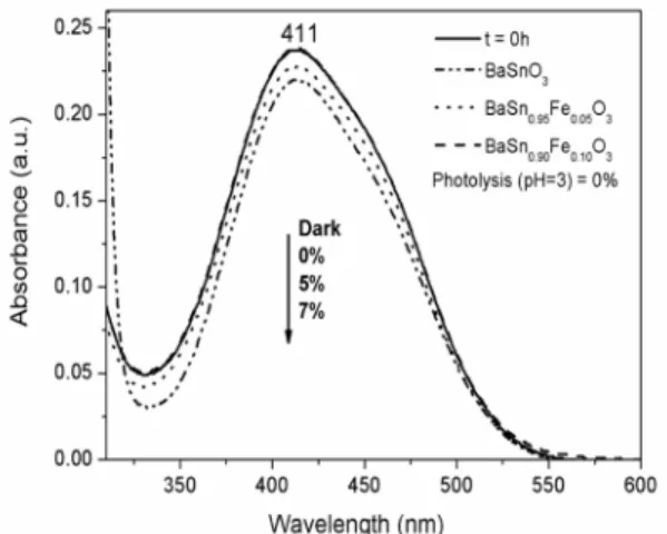
Figure 5 shows the absorption spectra of Fe3+- doped
BaSnO3, with a strong absorption in the visible region. The optical absorption edge of BaSnO3 was observed around
477 nm, with a red shift as doping concentration increases. The band gap values of the as-synthesized samples were estimated from diffuse reflectance spectra using the Wood-Tauc method41. Incorporation of Fe3+ into the lattice resulted
in a band gap decrease, indicating that intermediate levels were formed inside the band gap.
3.2 Photocatalytic properties
The photocatalytic decomposition of RNL by Fe-doped
BaSnO3 is presented in Figure 6. The highest degradation
efficiency occurred at pH = 3 and the lowest degradation occurred at pH = 6 (aqueous solution of the dye).
Photocatalysis may occur by two different mechanisms:
direct or indirect one. For the direct mechanism, dye is adsorbed on the photocatalyst surface and electron transfer takes place without the formation of intermediate compounds. During the indirect mechanism, hydroxyl radicals are formed due to
electron/hole transfer between the surface and compounds
as O2, H2O and OH-. Then, hydroxyl radicals in solution
react with the substrate.
The possibility of a discoloration by a direct mechanism
Moura et al.
320 Materials Research
Figure 3. XRD patterns of the BaSn1-xFexO3 (0; 0.05 and 0.1 in mol) samples. Detail of the (110) peak in the XRD patterns as a function of the iron concentration.
Figure 4. Raman spectra of BaSn1-xFexO3 (0; 0.05 and 0.1 in mol)
samples.
Figure 5. UV-Vis spectra of samples BaSn1-xFexO3 (0; 0.05 and 0.1 in mol).
the dye on the material surface is a requested prerequisite step for direct charge transfer42. Results displayed in Figure 7 indicate that the maximum discoloration due to adsorption
process was 7% for the BaSn0.9Fe0.1O3 sample, much smaller
than the discoloration percentage under UVC irradiation (93 %). This small adsorption indicates that the indirect
mechanism prevails for this system.
Figure 6. Results of the photocatalytic decomposition of RNL as a function of pH for the photocatalysts BaSn1-xFexO3 (x = 0, 0.05 at 0.1 in mol): a) pH = 6; b) pH = 3 and c) Percentage of photodegradation of the RNL.
Figure 7. Evaluation of RNL adsorption at pH = 3 after 4 h for the
samples BaSn1-xFexO3 (x = 0, 0.05 and 0.1 in mol).
mechanism at low pH when TiO2 is used as photocatalyst. On
the other hand, according to Guo et al.44, hydrogen radicals
also take part in the photodegradation of phenol using TiO2 as
photocatalyst. These •H radicals may be produced from H 2O
molecules and also from H3O
+ ions especially in acid media,
and may react with O2 forming HO2• which finally convert to •OH. According to Texeira et. al.45) the RNL azo dye has three
pKa values: the sulphonic group is deprotonated at pH = 3, the
sulphate group is deprotonated at pH = 3.5 and the amide group is deprotonated at pH = 6, which results in a large negative charge. Therefore, an attractive force between the positive
surface charge of the perovskite and the negative charge of
the azo dye occurs at pH 3 favoring the dye attraction and
Moura et al.
322 Materials Research
In the present work, the highest efficiency at acid media cannot
be assigned to a direct mechanism, as only a small adsorption was detected. It seems clear that RNL photodegradation is promoted by •OH radicals whose formation is favored at acidic
conditions, probably due to the highest amount of H3O + ions.
Figure 6c shows that the color was halved after 1 h of photocatalysis with BaSn1-xFexO3 (x = 0.05 and 0.10), 2.5x higher than undoped BaSnO3. For longer times, doped samples also presented a higher photoactivity than pure one, increasing
with Fe content. After 4 h, a photocatalytic efficiency of about 93% was attained for the sample BaSn0.90Fe0.10O3.
Several papers using Fe-doped TiO2 nanoparticles 17-22
assumed that a higher photoactivity for Fe-doped samples is possible in comparison with the undoped material, especially because Fe3+ can act as both hole and electron traps to enhance
lifetimes of electrons and holes.
Fe-doped BaSnO3 has been studied for magneticelectronics
applications, classified as oxide-diluted magnetic semiconductor, displaying ferromagnetism even with small doping amounts. This
property is enhanced due to a F-center exchange mechanism,
which enables Fe-ions to order ferromagnetically. This F-center
is characterized by a Fen+-V O-Fe
n+ configuration which is able
to trap electrons39,40,47.
In the present work, XRD patterns and Raman spectra indicated that Fe3+ was added into the BaSnO
3 lattice leading
to a shift of the absorption onset to the visible region due to the formation of intermediate levels inside the band gap.
These intermediate levels may trap electrons preventing
the electron-hole recombination. As a consequence, higher
photocatalytic efficiency is obtained.
4. Conclusions
BaSn1-xFexO3 (x = 0, 0.05 and 0.10) was successfully
synthesized by the modified Pechini method, with crystallization around 600°C. XRD patterns and Raman
spectra indicated that Fe3+ got into the perovskite lattice
leading to a decrease of the band gap. The samples showed
high potential for photodegradation of the RNL azo-dye at
pH = 3 with prevalence of indirect mechanism. Efficiency
was improved by Fe3+ doping probably due to the formation
of intermediate levels inside the band gap, which may trap electrons avoiding electron-hole recombination.
5. Acknowledgements
This work was supported by Brazilian Funding Agencies CT-INFRA/FINEP/MCTIC and CAPES.
6. References
1. Omeiri S, Hadjarab B, Bouguelia A, Trari M. Electrical, optical
and photoelectrochemical properties of BaSnO3-δ: Applications to hydrogen evolution. Journal of Alloys and Compounds.
2010;50(2):592-597.
2. Lee CW, Kim DW, Cho IS, Park S, Shin SS, Seo SW, et al.
Simple synthesis and characterization of SrSnO3 nanoparticles with enhanced photocatalytic activity. International Journal of Hydrogen Energy. 2012;37(14):10557-10563.
3. Wang W, Bi J, Wu L, Li Z, Fu X. Hydrothermal synthesis and
catalytic performances of a new photocalalyst CaSnO3 with
microcube morphology. Scripta Materialia.
2009;60(3):186-189.
4. Junploy P, Thongtem S, Thongtem T. Photoabsorption and
photocatalysis of SrSnO3 produced by a cyclic microwave radiation. Superlattices and Microstructures. 2013;57:1-10.
5. Lobo TM, Lebullenger R, Bouquet V, Guilloux-Viry M, Santos
IMG, Weber IT. SrSnO3:N - Nitridation and evaluation of
photocatalytic activity. Journal of Alloys and Compounds.
2015;649:491-494.
6. Sales HB, Bouquet V, Députier S, Ollivier S, Gouttefangeas F, Guilloux-Viry M, et al. Sr1-xBaxSnO3 system applied in the
photocatalytic discoloration of an azo-dye. Solid State Sciences.
2014;28:67-73.
7. Mizoguchi H, Woodward PM, Park CH, Keszler DA. Strong
near-infrared luminescence in BaSnO3. Journal of the American Chemical Society. 2004;126(31):9796-9800.
8. Moshtaghi S, Zinatloo-Ajabshir S, Salavati-Niasari M.
Nanocrystalline barium stannate: facile morphology-controlled preparation, characterization and investigation of optical and photocatalytic properties. Journal of Materials Science: Materials in Electronics. 2016; 27(1):834-842.
9. Moshtaghi S, Zinatloo-Ajabshir S, Salavati-Niasari M. Preparation
and characterization of BaSnO3 nanostructures via a new simple surfactant-free route. Journal of Materials Science: Materials in Electronics. 2016;27(1):425-435.
10. Borse PH, Joshi UA, Ji SM, Jang JS, Lee JS, Jeong ED, et al.
Band gap tuning of lead-substituted BaSnO3 for visible light photocatalysis. Applied Physics Letters. 2007;90(3):034103.
11. Borse PH, Lee JS, Kim HG. Theoretical band energetics of
Ba(M0.5Sn0.5)O3 for solar photoactive applications. Journal of Applied Physics. 2006;100(12):124915.
12. Yuan Y, Lv J, Jiang X, Li Z, Yu T, Zou Z, et al. Large impact of
strontium substitution on photocatalytic water splitting activity of BaSnO3. Applied Physics Letters. 2007;91(9):094-107.
13. Weber AS, Grady AM, Koodali RT. Lanthanide modified
semiconductor photocatalysts. Catalysis Science & Technology.
2012;2(4):683-693.
14. Mostaghni F, Abed Y. Structural, Optical and Photocatalytic Properties of Co-TiO2 Prepared by Sol-Gel Technique. Materials
Research. 2016;19(4):741-745.
15. Surendar T, Kumar S, Shanker V. Influence of La-doping on
phase transformation and photocatalytic properties of ZnTiO3
nanoparticles synthesized via modified sol-gel method. Physical Chemistry Chemical Physics. 2014;16(2):728-735.
16. Banić ND, Abramović BF, Šojić DV, Krstić JB, Finčur NL, Bočković IP. Efficiency of neonicotinoids photocatalytic
17. Pham TD, Lee BK, Lee CH. The advanced removal of benzene
from aerosols by photocatalytic oxidation and adsorption of
Cu-TiO2/PU under visible light irradiation. Applied Catalysis
B: Environmental. 2016;182:172-183.
18. Sood S, Umar A, Mehta SK, Kansal SK. Highly effective
Fe-doped TiO2 nanoparticles photocatalysts for visible light
driven photocatalytic degradation of toxic organic compounds.
Journal of Colloid and Interface Science. 2015;450:213-223.
19. Garza-Arévalo JI, García-Montes I, Hinojosa Reyes M, Guzmán-Mar JL, Rodríguez-González V, Hinojosa Reyes L.
Fe doped TiO2 photocatalyst for the removal of As(III) under
visible radiation and its potential application on the treatment of As-contaminated groundwater. Materials Research Bulletin.
2016;73:145-152.
20. Ma J, He H, Liu F. Effect of Fe on the photocatalytic removal of NOx over visible light responsive Fe/TiO2 catalysts. Applied
Catalysis B: Environmental. 2015;179:21-28.
21. Tian F, Wu Z, Tong Y, Wu Z, Cravotto G. Microwave-Assisted
Synthesis of Carbon-Based (N, Fe)-Codoped TiO2 for the
Photocatalytic Degradation of Formaldehyde. Nanoscale
Research Letters. 2015;10:360.
22. Li Z, Shen W, He W, Zu X. Effect of Fe-doped TiO2 nanoparticle derived from modified hydrothermal process on the photocatalytic
degradation performance on methylene blue. Journal of Hazardous Materials. 2008;155(3):590-594.
23. Hemmati Borji S, Nasseri S, Mahvi AH, Nabizadeh R, Javadi AH. Investigation of photocatalytic degradation of phenol
by Fe(III)-doped TiO2 and TiO2 nanoparticles. Journal of
Environmental Health Science & Engineering. 2014;12:101.
24. Vieira FTG, Oliveira ALM, Melo DS, Lima SJG, Longo E, Maia AS, et al. Crystallization study of SrSnO3:Fe. Journal of
Thermal Analysis and Calorimetry. 2011;106:507-512.
25. Alves MCF, Souza SC, Lima HHS, Nascimento MR, Silva MRS, Espinosa JWM, et al. Influence of the modifier on the
short and long-range disorder of stannate perovskites. Journal of Alloys and Compounds. 2009;476(1-2):507-512.
26. Lucena GL, Maia AS, Souza AG, Santos IM. Structural changes in Fe-doped SrSnO3 perovskites during thermal analysis. Journal of Thermal Analysis and Calorimetry. 2014;115(1):137-144.
27. Lucena GL, Souza JJN, Maia AS, Soledade LEB, Longo E, Souza AG, et al. New methodology for a faster synthesis of SrSnO3
by the modified Pechini method. Cerâmica.
2013;59(350):249-253.
28. Udawatte CP, Kakihana M, Yoshimura M. Preparation of pure
perovskite-type BaSnO3 powders by the polymerized complex method at reduced temperature. Solid State Ionics.
1998;108(1-4):23-30.
29. Li B, Tang Y, Luo L, Xiao T, Li D, Hu X, et al. Fabrication
of porous BaSnO3 hollow architectures using BaCO3@SnO2
core-shell nanorods as precursors. Applied Surface Science.
2010;257(1):197-202.
30. Huang C, Wang X, Liu X, Tian M, Zhang T. Extensive analysis
of the formation mechanism of BaSnO3 by solid-state reaction
between BaCO3 and SnO2. Journal of the European Ceramic
Society. 2016;36(3):583-592.
31. Manju MR, Kumar VP, Dayal V. Investigation of ferromagnetic
properties in Fe/Co substituted BaSnO3 perovskite stannates.
Physica B: Condensed Matter. 2016;500:14-19.
32. Ochoa YH, Schipani F, Aldao CM, Ponce MA, Savu R, Rodríguez-Páez JE. Electrical behavior of BaSnO3 bulk samples formed by slip casting: Effect of synthesis methods used for
obtaining the ceramic powders. Materials Research Bulletin.
2016;78:172-178.
33. Kumar AA, Kumar A, Quamara JK, Dillip GR, Joo SW, Kumar J. Fe (III) induced structural, optical, and dielectric behavior
of cetyltrimethyl ammonium bromide stabilized strontium stannate nanoparticles synthesized by a facile wet chemistry route. RSC Advances. 2015;5:17202-17209.
34. Balamurugan K, Kumar ES, Ramachandran B, Venkatesh S, Kumar NH, Rao MS, et al. Dielectric resonance and magnetic
properties of Fe-3% doped BaSnO3 thin films grown by pulsed
laser deposition. Journal of Applied Physics. 2012;11(7)074107.
35. Cerdà J, Arbiol J, Diaz R, Dezanneau G, Morante JR. Synthesis
of perovskite-type BaSnO3 particles obtained by a new simple wet chemical route based on a sol-gel process. Materials Letters.
2002;56(3):131-136.
36. Stanislavchuk TN, Sirenko AA, Litvinchuk AP, Luo X, Cheong
SW. Electronic band structure and optical phonons of BaSnO3 and Ba0.97La0.03SnO3 single crystals: Theory and experiment.
Journal of Applied Physics. 2012;112(4):044108.
37. Zhang W, Tang J, Ye J. Structural, photocatalytic, and
photophysical properties of perovskite MSnO3 (M = Ca,
Sr, and Ba) photocatalysts. Journal of Materials Research.
2007;22(7):1859-1871.
38. Zheng H, de Györgyfalva GDCC, Quimby R, Bagshaw H, Ubic R, Reaney IM, et al. Raman spectroscopy of B-site
order-disorder in CaTiO3-based microwave ceramics. Journal of the
European Ceramic Society. 2003;23(14):2653-2659.
39. Balamurugan K, Harishn Kumar N, Arout Chelvane J, Santhosh
PN. Room temperature ferromagnetism in Fe-doped BaSnO3.
Journal of Alloys and Compounds. 2009;472(1-2):9-12.
40. Balamurugan K, Harish Kumar N, Arout Chelvane J, Santhosh PN. Effect of W co-doping on the optical, magnetic and electrical
properties of Fe-doped BaSnO3. Physica B: Condensed Matter.
2012;407(13):2519-2523.
41. Wood DL, Tauc J. Weak Absorption Tails in Amorphous
Semiconductors. Physical Review B. 1972;5(8):3144-3151.
42. Tang WZ, Huang CP. Photocatalyzed oxidation pathways of 2,4-dichlorophenol by CdS in basic and acidic aqueous solutions.
Water Research. 1995;29(2):745-756.
43. Akpan UG, Hameed BH. Parameters affecting the photocatalytic degradation of dyes using TiO2-based photocatalysts: A review.
Journal of Hazardous Materials. 2009;170(2-3):520-529.
44. Guo Z, Ma R, Li G. Degradation of phenol by nanomaterial TiO2
in wastewater. Chemical Engineering Journal. 2006;119(1):55-59.
45. Teixeira TPF. Avaliação da eficiência do uso de hidrotalcitas
calcinadas na remoção de azo corantes aniônicos presentes
Moura et al.
324 Materials Research
46. Teixeira TPF, Pereira SI, Aquino SF, Dias A. Use of Calcined Layered Double Hydroxides for Decolorization of Azo Dye Solutions: Equilibrium, Kinetics, and Recycling Studies.
Environmental Engineering Science. 2012;29(7):685-692.
47. Swatsitang E, Karaphun A, Phokha S, Putjuso T. Characterization
and magnetic properties of BaSn1-xFexO3 nanoparticles prepared
by a modified sol-gel method. Journal of Sol-Gel Science and