QUÍMICA NOVA, 23(6) (2000) 749 INCLUSION COMPLEX OF THE ANTIVIRAL DRUG ACYCLOVIR WITH CYCLODEXTRIN IN AQUEOUS
SOLUTION AND IN SOLID PHASE
Carlos von Plessing Rossel*
Facultad de Farmacia - Departamento de Farmacia - Casilla, 237 - Universidad de Concepción - Concepción - Chile Jacqueline Sepúlveda Carreño
Facultad de Ciencias Biológicas - Departamento de Farmacología - Casilla, 160-C - Universidad de Concepción - Concepción - Chile Mario Rodríguez-Baeza
Facultad de Ciencias Químicas - Departamento de Polímeros - Casilla, 3-C - Universidad de Concepción - Concepción - Chile Joel Bernabé Alderete
Facultad de Ciencias Químicas - Departamento de Orgánica - Casilla, 3-C - Universidad de Concepción - Concepción - Chile
Recebido em 29/10/99; aceito em 18/4/00
Complexation between acyclovir (ACV), an antiviral drug used for the treatment of herpes sim-plex virus infection, and βββββ-cyclodextrin (βββββ-CD) was studied in solution and in solid states. Com-plexation in solution was evaluated using solubility studies and nuclear magnetic resonance spec-troscopy (1H-NMR). In the solid state, X-ray diffraction, differential scanning calorimetry (DSC), thermal gravimetric analysis (TGA) and dissolution studies were used. Solubility studies suggested the existence of a 1:1 complex between ACV and βββββ-CD. 1H-NMR spectroscopy studies showed that the complex formed occurs with a stoichiometry ratio of 1:1. Powder X-ray diffraction indicated that ACV exists in a semicrystalline state in the complexed form with βββββ-CD. DSC studies showed the existence of a complex of ACV with βββββ-CD. The TGA studies confirmed the DSC results of the complex. Solubility of ACV in solid complexes was studied by the dissolution method and it was found to be much more soluble than the uncomplexed drug.
Keywords: cyclodextrin; acyclovir; inclusion complex. ARTIGO
INTRODUCTION
Acyclovir (9-((2-hydroxyethoxy)-methyl)-guanine, is an acyclic analogue of the natural nuleoside 2'-deoxyguanine with antiviral activity ”in vitro” against herpes simplex viruses (HSV), varicella zoster virus (VZV), Epstein-Barr virus (EBV), cytomegalovirus (CMV) and human herpes virus 6 (HHV-6)1.
The fundamental pharmacokinetic properties of acyclovir are well established2,3. The intravenous administration of acyclovir is described as a two-compartment open model and the absorption of orally administered acyclovir is slow, variable and incomplete with an oral bioavailability of 15-30%4,5. The effects of dosage size on the extent of oral absorption are not well understood. Some reports suggest that absorption from the gastrointestinal tract may be a saturable, dose-dependent process6,7. In contrast, another study reported a relative constancy in the urinary recovery of unchanged drug and in the bioavailability calculated from urinary excretion data, concluding that the net absorption of acyclovir is nearly proportional to the dose8.
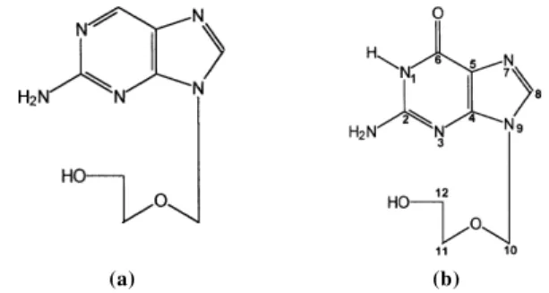
The limited absorption of ACV can be improved with a sustained release formulation9, by using a prodrug of ACV (Descyclovir)10 (Fig.1), or with ACV modified by acetylation and coupling obtaining a drug-polymer conjugate11. Another possible way to overcome this problem is by modifying the solubility of the drug by using cyclodextrins and their derivatives. Cyclodextrins are able to form inclusion complexes with a wide variety of drugs and the interaction can affect many of the observed physicochemical properties, such as aqueous solubility and stability12-14. Formation of inclusion
complexes with β-CD has been used extensively to improve the solubility of poorly soluble drugs2-4,12-15 and their dissolu-tion rate5. Loftsson et al.16 were able to double the ACV
solubility by incorporating it in a 50% w/w aqueous solution of 2-hydroxypropyl-β-cyclodextrin.
*e-mail: cvonples@udec.cl
Figure 1. Structural formulae of Descyclovir (A) and Acyclovir (B).
(a) (b)
The purpose of this investigation is to study the complexa-tion of ACV with CDs and their effect on ACV solubility by using techniques obtained from studying other type of complexes (e.g. X-ray diffraction, solubility studies, dissoluti-on rate, differential scanning calorimetry, thermal gravimetry and nuclear magnetic resonance spectroscopy).
MATERIALS AND METHODS Materials
750 QUÍMICA NOVA, 23(6) (2000) France). All other materials were of analytical grade. Deionized
and double-distilled water was used throughout the study.
Methods
Solubility studies
Solubility studies were carried out according to the Higuchi and Connors method17. β-CD solutions of different concentra-tions (13.2 - 2,81 * 10-3 M) were added to a supersaturated solution of ACV and shaken at room temperature (22 ± 1oC) for 36 hours. After reaching equilibrium, the solutions were filtered. The concentration of ACV in the filtrate was determi-ned spectrophotometrically at 290 nm with reference to a suitably constructed standard curve.
Preparation of the solid complex
A solid complex of β-CD with ACV was prepared at a 1:1 molar ratio. The physical mixture (Ph.M) was prepared by simply mixing the powders with a spatula. A freeze-dried complex (C) was prepared by drying an aqueous solution containing ACV/ β-CD in a 1:1 molar ratio in a Virtis“ Freeze-Dryer.
Differential Scanning Calorimetry (DSC) and Thermal Gravimetric Analysis (TG)
Thermal analyses were performed on solid and freeze-dried samples obtained directly from the preparation medium. DSC and TGA data were obtained using a Polymer Laboratories (STA-625) Thermal analyzer. Samples (2-6 mg) were heated in sealed aluminum pans under nitrogen flow (50 cm3 min-1) at
a heating rate of 10oC min-1, from 50 to 400oC.
X-ray diffraction studies
Powder X-ray diffraction patterns were recorded using a Siemens Kristalloflex D-501 with the following conditions: Ni-filtered CuK radiation, voltage 36kV, current 26 mA, at a scanning rate of 2o/min.
Dissolution studies
Accurately weighed amounts of the powdered ACV, Ph.M. and C, each containing 100 mg ACV, were used to carry out the studies in a Dissolution Test 6 device (Hanson Research Corp. Northridge, California, USA) using the USP XXIII paddle method. The dissolution medium consisted of 900 mL of distilled water at 37oC ± 0.5oC and a stirring speed = 50 rpm. The
progress of the dissolution was followed by circulating the dis-solution medium through the cell of the spectrophotometer for continuous recording over 30 min, at 290 nm.
Nuclear magnetic resonance studies
The inclusion complexes of ACV-β-CD were characterized in deutered water by 1H-NMR. The spectra were obtained in a
Brucker AC250 (250.13 MHz for 1H) spectrophotometer. The chemical displacements are reported as relative to the solvent residual signal, HDO (δ = 4.800 ppm). The conditions employed were: 16 K data points, weep width 5 khz, pulse width 4µs and numbers of scan 600.
RESULTS AND DISCUSSION
The solubility of ACV in water is very low, 1.3 mg.mL-1
at 25oC, as previously reported18. Figure 2 shows the
solubility curve obtained for ACV with β-CD. The resulting linear curve can be classified, in general, as type AL17,
indicating that complexes with a 1:1 stoichiometry will be present in the solution.
Figure 2. Higuchi phase solubility diagram of βCD and acyclovir.
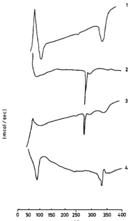
Figure 3. DSC thermograms of (1) βCD, (2) acyclovir, (3) physical mixture, (4) inclusion complex (1:1).
UV absorbance of ACV is not affected in the presence of CD. The apparent solubility constant K1:1 was calculated from the curve according to Eq. 1 and was found to be 22 M-1
K1:1 = slope/So (1-slope) (1)
where So is the solubility of ACV in the absence of β-CD. DSC measurements (Fig. 3) show an endothermic peak at the ACV melting point (249oC). However, with the physical mixture, this peak was absent in the diagram of the inclusion compound. Because of its molecularly dispersed distribution and the protection that the complexation affords, the peak was not detected due to its displacement to the decomposition temperature of the β-CD.
QUÍMICA NOVA, 23(6) (2000) 751 Figure 4. TG thermograms of (1) acyclovir, (2) physical mixture, (3)
βCD, (4) inclusion complex (1:1).
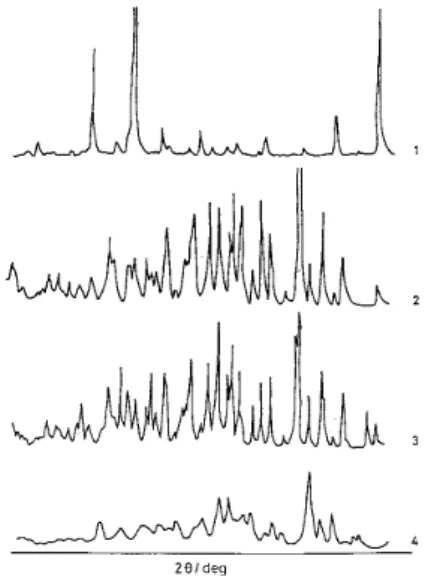
Figure 5. X-Ray diffractograms of (1) acyclovir, (2) βCD, (3) physical mixture, (4) inclusion complex (1:1).
With the physical mixture, a small increase in dissolution rate was obtained which can be explained due to the formation of a minimum quantity of the complex. After 3 minutes, the quantity of ACV dissolved from the β-CD complex was 1.3 times greater than that of the physical mixture. During the same period, the quantity of dissolved ACV alone was 2.0 times lower than that dissolved from the complex.
Although solubility studies indicated the existence of comple-xation between ACV and β-CD in solution, a deeper insight into the complexation mechanism was obtained from 1H-NMR. 1
H-NMR spectra for acyclovir in DMSO-d6 and D2O for the ACV/β
-CD complex in D2O are shown in Table 1. Due to the interchange
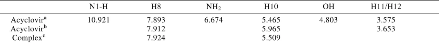
with deuterium, in deutered water only signals corresponding to protons H8, H10 and H11/H12 of the ACV were detected. In the case of the complex, a small displacement of the signals corresponding to H8 (0.012 ppm) and H10 (0.014 ppm) of the ACV was observed. The slight displacement at low field of these signals indicates a low efficiency in the acyclovir complexation. In spite of this fact it was possible to determine the stoichiometry of the complex using the continuous variation method29. For this study equimolar solutions of ACV and β-CD were prepared and mixed at a standard volume (1 mL) in such proportions that the sum of the concentrations of ACV and β-CD remained constant and equal to 10 mM. Afterwards, the change in the chemical displacement of signal H8 of ACV as a function of r, where r is the ratio of the initial concentrations defined by R= (ACV)/ ((ACV) + (β-CD)), was determined. The results of these experiments are shown in Figure 7. As can be seen in this figure, the curve achieves a maximum at R= 0.5, indicating the formation of a 1:1 stoichiometric complex. However, due to the low com-plexation efficiency, it is very difficult to compare the magnitude of the signals displacement of 1H of β-CD making it impossible to obtain information about the complex structure.
Figure 6. Dissolution studies of (1) acyclovir, (2) physical mixture, (3) inclusion complex (1:1).
is observed in the degradation area of the drug indicating that it has acquired a state different to that of the drug and that of the physical mixture, that is, an inclusion complex has been formed.
The X-ray diffraction pattern of the inclusion compound differed considerably from that of the drug or the β-CD alone (Fig. 5). In contrast the diffraction pattern of the physical mixture showed that in addition to the presence of the pure drug, the complex formation is also obtained, a fact shown by appearance of new peaks. The diffractogram of the complex indicated a new solid phase, the inclusion compound; whose diffractogram shows a different crystalline state which is simi-lar to that of other complexes described in literature19.
The dissolution rate (Fig. 6) of the drug in the inclusion compound was evidently higher than that of the drug alone. This can be attributed to the increase in solubility. Another factor that could have been involved in the improved solubility is the decrease in crystallinity of the inclusion complex which was confirmed by the X-ray diffraction results.
752 QUÍMICA NOVA, 23(6) (2000) CONCLUSION
Acyclovir forms an inclusion compound (1:1) with β-CD in the solid and the solution state. This was confirmed by DSC, TGA, X-ray diffraction and NMR spectroscopy. Com-plexation by inclusion increases ACV solubility and disso-lution. Dissolution increase is due to the decreased activity of the complexed ACV in solution and the low crystallinity of the formed complex as confirmed by the X-ray diffraction pattern. The solubility of the physical mixture, when compared to that of the complex was improved to a lesser degree.
ACKNOWLEDGEMENT
This research was supported by the Dirección de Investigación, Universidad de Concepción (Grant No 96.074.022 - 1.1D).
REFERENCES
1. Wagstaff Antona, J.; Faulds, D.; Goa Karen, L.; Drugs
1994, 47, 153.
2. Duchene, D.; Vaultion, C.; Glomot, F.; Drug Dev. Ind. Pharm. 1986, 12, 2193.
3. Erden, N.; Celebi, N.; Int. J. Pharm. 1988, 48, 83. 4. Hamada, Y.; Nambu, N.; Nagai, T.; Chem. Pharm. Bull.
1975, 23, 1205.
5. Corrigan, O. I.; Stanly, C. T.; J. Pharm. Pharmacol. 1982,
34, 621.
6. Bridgen, D.; Fowle, A.; Rosling, A.; Developments in antiviral therapy (Eds. Collier LH & Oxford I.) Academic Press, 1980; p. 53-62.
7. Vergin, H.; Kikuta, C.; Mascher, H.; Metz, R.;
Arzneimittel-Forschung/Drug Research 1995, 45, 508. 8. de Miranda, P.; Krasny, H. C.; Page, D. A.; Elion, C. B.;
Am. J. Med. 1982, 73, 31.
9. Lewis, L. D.; Fowle, A. S. E.; Bittner, S. B.; Bye A. and Isaacs, P. E. T.; Br. J. Clin. Pharmacol. 1986, 21, 459. 10. Richards, D. M.; Carmine, A. A.; Brodgen, R. N.; Heel,
R. C.; Speight, T. M.; Drugs 1983, 26, 378.
11. Fiamona, G.; Puglisi, G.; Cavallaro, G.; Spadaro A.; and Pitanesi, G.; J. Controlled Release 1995, 32, 261. 12. Saenger, W.; Angew. Chem. Int. Ed. Engl. 1980, 19, 344. 13. Szejtli, J.; Controlled Drug Bioavailability (Ed. W. F. Smolen; L. A. Ball), Vol. 3 Wiley, N.Y. 1985; p. 365. 14. Szejtli, J.; Medicinal Applications of Cyclodextrins, in
Me-dicinal Research Reviews, 1994; Vol. 14, Nª 3, p. 364 15. Frömming, K. H.; Prc. Ist Int. Symp. Cyclodextrins (Ed.
J. Szejtli) Reidel, Drodrecht, 1982; p. 367
16. Loftsson, Th.; Bodor, N.; Acta Pharm. Nord. 1989, 1, 185. 17. Higuchi, T.; Connors, K. A.; Adv. Anal. Chem. Instrum.
1965, 4, 117.
18. Drug Information 95, American Hospital Formulary Service, Ed. Gerald, K. McEvoy, USA, 1995; p.2388. 19. Fridrich, R.; Mehnert, W.; Frömming, K. H.; Minutes of
the 5th Int. Symp. on Cyclodextrins, (Ed. D. Duchene), Editions de Santé Publishers, Paris, 1990; p. 299. 20. Loukas, Y. L.; J. Pharm. Pharmacol. 1997, 49, 944. Table 1. Chemical displacements of ACV and ACV-βCD complex.
N1-H H8 NH2 H10 OH H11/H12
Acyclovira 10.921 7.893 6.674 5.465 4.803 3.575
Acyclovirb 7.912 5.965 3.653
Complexc 7.924 5.509
a In DMSO-d
6. Solvent signal used as reference (2.600 ppm) b In D
2O. In this case due to the interchange with deuterium the signals of interchangeable 1H are not detected. c In D