Article
Printed in Brazil - ©2017 Sociedade Brasileira de Química0103 - 5053 $6.00+0.00*e-mail: [email protected]
Differentiation of Toxic and Non-Toxic Leaves of
Jatropha curcas
L. Genotypes by
Leaf Spray Mass Spectrometry
Igor Pereira,a Thays C. de Carvalho,a Wanderson Romão,b Paulo R. Filgueiras,b
Bruno G. Laviola,c Clenilson M. Rodrigues,c Patrícia V. Abdelnurc and Boniek G. Vaz*,a
aInstituto de Química, Universidade Federal do Goiás, 74690-970 Goiânia-GO, Brazil
bDepartamento de Química, Universidade Federal do Espírito Santo, 29075-910 Vitória-ES, Brazil
cEmbrapa Agroenergia, 70770-901 Brasília-DF, Brazil
Jatropha curcas L. is an oil crop that has been studied as a potential source of biodiesel. A high protein pie is produced as a byproduct of the biodiesel production, which could be used as animal feed. However, the pie has toxic compounds, as phorbol esters and other toxins, which prevents the use as animal feed.For this reason, Embrapa (Brazilian Agricultural Research Corporation) has been working in genetic breeding to develop non-toxic J. curcas genotypes. To evaluate this process, a simple and fast analytical technique was employed to obtain responses in a short time. Leaf spray (LS) is a recent ambient ionization mass spectrometry technique in which the sample itself serves as support and ion source. Here, toxic and non-toxic J. curcas leaves were differentiated by LS using a linear ion trap mass spectrometer and partial least squares discriminant analysis (PLS-DA) model chemometrics. It was possible to differentiate toxic and non-toxic leaves and to identify the m/z valuesthat contribute to discrimination between the groups.
Keywords: ambient mass spectrometry, PLS-DA, discrimination analysis
Introduction
Jatropha curcas L. is a Euphorbiaceae of great interest to biofuels production due to its low production cost, hardiness, adaptability, drought resistance and high oil content in its seeds.1 To add value to the productive chain of biodiesel, the pie resulting from the extraction of oil from seeds, is being studied as a potential agent of animal feed due to its high protein value.2 However, because some toxins and anti-nutritional factors, pie has not been used yet as animal feed. The major toxic compounds are the phorbol esters,3 tetracyclic diterpenes with biological activities such as tumor promotion, platelet aggregation, apoptosis, cell differentiation and other adverse metabolic effects.4 Additionally, other toxic compounds are reported in leaves, seeds and pie of J. curcas, such as trypsin inhibitors, tannins, saponins, phytates, lectins and curcin.3
Researchers are working on J. curcas to produce non-toxic crops. In Brazil, researches at Embrapa (Brazilian Agricultural Research Corporation) are using genetic breeding approaches to obtain a J. curcas genotype fully
well-adapted in the country’s growing conditions, with good productivity, resistant to pests and diseases and not presenting toxic compounds. To monitor the efficiency of the detoxification process based on genetic breeding carried out by Embrapa, it is necessary to employ analytical techniques that provide precise and accurate information in a short time. Although HPLC-UV (high performance liquid chromatography ultraviolet) is often used in the routine analysis of toxic compounds in J. curcas,5-7 there are few studies using liquid chromatography mass spectrometry (LC-MS).8-10 However, these methods require sample preparation and separation steps, resulting in a time-consuming and laborious method. So, there is a lack of studies to determine these compounds quickly and comprehensively.
has emerged, a large number of techniques with the same purpose was emerging.11,14
An ambient ionization technique was recently developed, the leaf spray (LS), a technical variant of paper spray ionization.15 In the LS-MS technique, ions are generated directly from plant tissues,16 and no support is needed to deposit the sample because the plant acts simultaneously as sample and support for ions generation. The plant leaf is wetted with solvent, and a high voltage is applied in the leaf, producing ions to the mass spectrometer. Ions can be generated without adding solvent in plant tissue due to the natural juice present in fruit and vegetables, although spectra with more intense signals and better S/N ratio can be obtained when the solvent is added.17 LS-MS has been used in many applications, such as detection of ursolic and oleanolic acids and their oxidation products in medicinal plant Ocimum sanctum,18 glycosides analysis from Stevia leaves,19 profile analysis of saccharides, glycoalkaloids, organic acids, and glucosinolates in living plants and fresh fruits,20 and quantification of pesticide in fruits and vegetables.17 LS-MS provides simple and fast analysis, with little or no sample preparation.
In this paper, we describe the combination of an easily and simple ionization method, leaf spray mass spectrometry, with PLS-DA (partial least squares discriminant analysis) to create accurate and robust model to differentiate toxic and non-toxic J. curcas genotypes based on low-resolution mass spectra profile.
Experimental
Material and samples
HPLC-grade methanol was purchased from Sigma-Aldrich (Steinheim, Germany). The leaves of J. curcas
genotypes were provided by Embrapa Agroenergia (Brasilia, DF, Brazil). Leaves were selected from 40 genotypes, of which 16 were toxic and 24 non-toxic.21
Leaf spray-MS
J. curcas leaves were cut in the equilateral triangle shape with 1 cm sides. A high voltage (3.5 kV) was applied to the base of each triangular leaf previously wetted with 10 µL of solvent (0.1% formic acid in methanol). A charged microdroplets spray goes directly into mass spectrometer. Leaf was positioned about 4 mm from the MS input. Experiments were performed in triplicate. Mass spectra were recorded in the positive mode, LS(+). Other parameters used were: tube lens: 109 V; capillary voltage: 43 V; capillary temperature: 275 °C; scan range: 100-1200 m/z. The
experiments were performed using the mass spectrometer LTQ XL™ Ion Trap Linear (Thermo Scientific, San Jose, USA). To identify the major ions in the toxic and non-toxic leaves of J. curcas genotypes, a high-resolution MS was employed, the Q Exactive™ Hybrid Quadrupole-Orbitrap (Thermo Scientific, San Jose, USA). The parameters used were: resolution: 140,000; spray voltage: 3.5 kV; average of 3 micro-scans for each spectrum; capillary temperature: 275 °C; S-lens RF Level: 50%. The spectra were processed by the Xcalibur Analysis software package (version 2.0, Service Release 2, Thermo Electron Corporation). Molecular formula was acceptable only when the average differences between theoretical and experimental masses were less than 1.0 ppm. Molecular formula search was done using 12C, 1H, 16O, 14N, 23Na and 39K isotopologue ions.
Chemometrics
The chemometrics analysis was performed using PLS-DA model. To construct the model, 40 samples, 16 toxic and 24 non-toxic leaves, were divided into training (28 samples) and test (12 samples) sets using the duplex algorithm.22 The mass spectra dataset were autoscaled and the model was construct with five latent variables determined by leave-one-out cross-validation method. The percentage ratio of the ion intensities in the mass spectra was employed in the model construction. The threshold of discrimination between classes of samples was defined according to Bayes method.23 The forecasting results of classification models were evaluated for sensitivity, specificity, accuracy and prevalence.24 The sensitivity indicates the probability model to detect a toxic sample, since the sample is actually toxic; specificity reflects the probability model to identify correctly a non-toxic sample, when it really is non-toxic; accuracy is the model’s ability to classify correctly a toxic and non-toxic sample; the prevalence is split between the positive predictive value, which reflects the probability that the model is wrong when it identifies a sample as toxic, and negative predictive value, which reflects the probability that the model is wrong when it identifies a sample as non-toxic. From regression coefficients of the PLS-DA, the most important variables, m/z, were selected and a new model was built. All calculations were performed in Matlab® 7.8.0 software using the classification toolbox 3.0.25
Results and Discussion
preparation. Figure 1 illustrates the scheme of LS-MS homemade source developed.
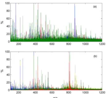
Figure 2 shows the mass spectra of all toxic (Figure 2a) and non-toxic (Figure 2b) leaves. Most of the ions were detected in both set, however, mass spectra of toxic leaves have shown more compounds. Some low-resolution mass spectra of toxic (Figure S1) and non-toxic (Figure S2) leaves are shown in the Supplementary Information. Mass spectra for toxic and non-toxic exhibited various common ions.
To identify the most intense ions, positive mass spectra of J. curcas leaves were recorded with high-resolution mass spectrometer Q-Exactive Hybrid Quadrupole-Orbitrap. Figure 3a shows a LS(+)-Orbitrap mass spectrum of a toxic leaf and Figure 3b shows a mass spectrum of a non-toxic leaf. Similarly to the low-resolution mass spectra, the high-resolution mass spectra for toxic and non-toxic also exhibited various ions in common. The ions of m/z 219,
Figure 1. Schematic of leaf spray-MS homemade source.
Figure 2. Positive leaf spray mass spectra of (a) all toxic leaves; (b) all non-toxic leaves.
391, 429, 480 and 819 were some of the most intense in the mass spectra of all 40 leaves analyzed.
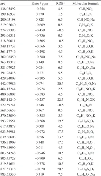
Table 1 shows the LS(+)-Orbitrap MS molecular formula assignments of various ions detected in the toxic and non-toxic leaves of J. curcas genotypes. The most intense ions at m/z 391, 429 and 819 were detected as phthalate, that are contaminant compounds. Phthalates are common plasticizers widely used in commercial products, including laboratory equipment parts.26 The low-intensity ions at m/z 522 and 803 were also detected as contaminant compounds, being stearyl-palmityl dimethyl ammonium and diisooctyl phthalate, respectively.
Due to the complexity of the data, excess of variables and similarity of mass spectra, PLS-DA chemometric model was applied to assist in the differentiation of ions
related to toxic and non-toxic leaves. Figure 4 displays the regression coefficients of the PLS-DA model constructed with five latent variables. The higher coefficient intensity, the greater the importance of this variable in the model construction and consequently to discriminate toxic and non-toxic leaves samples. The horizontal line at 1.03 × 10⁻³ represents a threshold for identification of the m/z values most important for class of toxic samples, whereas horizontal line at −1.03 × 10⁻³ is the same for the non-toxic samples. These m/z values are shown in Table S1 (Supplementary Information).
The average value of the percentage intensities of selected toxic and non-toxic samples are shown in Figure 5. Between the variables 7 to 43, the average m/z value for toxic samples is superior to non-toxic samples, while between the variables 53 to 70, the average intensity of non-toxic samples is greater than the toxic samples.
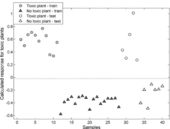
From regression coefficients of the PLS-DA, the most important variables, m/z, were selected to build a new model. Figure 6shows the results from PLS-DA model calculated with five latent variables for identification of toxic leaves samples. The y-axis represents the calculated
Table 1. Molecular formula assigned to ions detected in J. curcas leaves by LS(+)-Orbitrap MS
m/z Error / ppm RDBa Molecular formula
138.05492 −0.254 4.5 C7H8NO2
199.16937 0.570 1.5 C12H23O2
200.03198 0.828 6.5 C9H7NO3Na
219.02640 −0.669 0.5 C6H12O6K
274.27393 −0.459 −0.5 C16H36NO2
293.06311 −0.736 0.5 C9H18O8K
318.30011 −0.505 −0.5 C18H40NO3
349.17737 −0.566 3.5 C18H30O4K
361.17746 −0.298 4.5 C19H30O4K
362.08450 −0.380 7.5 C15H17NO8Na
363.19312 0.149 8.5 C22H28O3Na
381.07925 0.084 6.5 C15H18O10Na
391.28418 −0.271 5.5 C24H39O4
429.24008 −0.205 5.5 C24H38O4K
442.07986 0.074 20.5 C25H13N3O4Na
464.15244 −0.924 2.5 C17H31NO11K
480.36807 −0.583 4.5 C28H50NO5
505.14240 −0.237 22.5 C31H22N4OK
522.59741 0.348 −0.5 C36H76N
568.56653 0.367 0.5 C36H74NO3
570.23090 −0.385 5.5 C25H41NO11K
593.27551 −0.568 19.5 C35H37N4O5
617.43878 0.008 4.5 C35H62O7Na
623.36981 −0.972 17.5 C37H47N6O3
639.36603 0.656 13.5 C39H52O6Na
736.31909 0.348 17.5 C37H46N5O11
779.48999 0.011 4.5 C38H71N2O14
803.54248 −0.946 10.5 C48H76O8Na
805.45728 −0.909 6.5 C40H69O16
819.51654 −0.778 10.5 C48H76O8K
871.57318 −0.020 20.5 C55H75N4O5
983.55530 0.319 7.5 C49H84O18Na
aRDB: ring/double bond equivalent.
Figure 4. Regression coefficients of the PLS-DA model for the toxic and non-toxic leaves with five latent variables.
response to identify toxic leave samples. A horizontal line was determined from training samples using the Bayes method for maximum discrimination between toxic and non-toxic leaves samples. Samples located above the horizontal line are identified as belonging to the toxic leaf group. Note that all training and testing samples were correctly classified by the PLS-DA model built with only the most important ions to discrimination between the leaves of plants toxic and non-toxic.
All the training set samples were correctly classified. Thus, their statistical parameters showed 100% efficiency (Table 2). The model with all variables applied to the test set showed 80% of sensitivity, 88% of negative prevalence and 100% of positive prevalence. However, the model with variable selection presented 100% for all parameters showing the efficiency from model built with only the most important variables.
A spatial representation of the samples is shown in Figure 7, where 76.7% of the total variability of the spectra is represented by scores of the first three latent variables. It is observed that the non-toxic samples are less scattered in three-dimensional subspace and form a defined group. This
spatial distribution, combined with the high accuracy of the model, provides the identification of the most important variables for discrimination of class samples by model regression coefficients. Samples of toxic leaves show large dispersion indicating greater variability identified in these samples.
Conclusions
Leaf spray mass spectrometry is a simple and fast method for analysis and screening of compounds in Jatropha curcas
leaves without any sample preparation. Leaves of J. curcas
genotypes can be differentiated between toxic and non-toxic using LS-MS associated with chemometric model PLS-DA. The scores plot discriminate the two classes of samples. Based on the model coefficients, it was possible to identify the m/z values that contribute to discrimination between toxic and non-toxic leaves. Discrimination can be done using a routine and low resolution mass spectrometer. Thus, this paper shows a simple and efficient method for the quality control of the detoxification process in leaves of J. curcas genotypes.
Table 2. Results of the statistical parameters for classification of leaves samples according to the PLS-DA model with 5 latent variables
Parameter All variables After variable selection
Training / % Test / % Training / % Test / %
Sensibility 100 80 100 100
Specificity 100 100 100 100
Accuracy 100 92 100 100
Positive predictive value 100 100 100 100
Negative predictive value 100 88 100 100
Figure 6. Calculated response of the PLS-DA model with variable selection with five latent variables for identification of toxic leaves samples. The horizontal line represents a limit to discrimination of samples.
Supplementary Information
Supplementary information (Figure S1, Figure S2 and Table S1) is available free of charge at http://jbcs.sbq.org. br as PDF file.
Acknowledgments
The authors thank CNPq, CAPES, FAPES, FAPEG and EMBRAPA for institutional and financial support.
References
1. Makkar, H. P. S.; Becker, K.; Eur. J. Lipid Sci. Technol. 2009,
111, 773.
2. Prasad, L.; Pradhan, S.; Das, L. M.; Naik, S. N.; Appl. Energy
2012, 93, 245.
3. Makkar, H. P. S.; Becker, K.; Sporer, F.; Wink, M.; J. Agric. Food Chem.1997, 45, 3152.
4. Goel, G.; Makkar, H. P.; Francis, G.; Becker, K.; Int. J. Toxicol.
2007, 26, 279.
5. Devappa, R. K.; Bingham, J. P.; Khanal, S. K.; Ind. Crops Prod.
2013, 49, 211.
6. Baldini, M.; Ferfuia, C.; Bortolomeazzi, R.; Verardo, G.; Pascali, J.; Piasentier, E.; Franceschi, L.; Ind. Crops Prod.2014, 59, 268. 7. Makkar, H. P. S.; Maes, J.; Greyt, W. D.; Becker, K.; J. Am. Oil
Chem. Soc.2009, 86, 173.
8. Ichihashi, K.; Yuki, D.; Kurokawa, H.; Igarashi, A.; Yajima, T.; Fujiwara, M.; Maeno, K.; Sekiguchi, S.; Iwata, M.; Nishino, H.; J. Am. Oil Chem. Soc.2011, 88, 851.
9. Liu, X.; Li, L.; Li, W.; Lu, D.; Chen, F.; Li, J.; Ind. Crops Prod.
2013, 47, 29.
10. Punsuvon, V.; Nokkaew, R.; Karnasuta, S.; ScienceAsia2012,
38, 223.
11. Alberici, R. M.; Simas, R. C.; Sanvido, G. B.; Romão, W.; Lalli, P. M.; Benassi, M.; Cunha, I. B. S.; Eberlin, M. N.; Anal. Bioanal. Chem.2010, 398, 265.
12. Takáts, Z.; Wiseman, J. M.; Cooks, R. G.; Anal. Chem.2011,
83, 4508.
13. Cody, R. B.; Laramée, J. A.; Durst, H. D.; Anal. Chem.2005,
77, 2297.
14. Harris, G. A.; Galhena, A. S.; Fernández, F. M.; Anal. Chem.
2011, 83, 4508.
15. Wang, H.; Liu, J.; Cooks, R. G.; Ouyang, Z.; Angew. Chem., Int. Ed. 2010,49, 877.
16. Liu, J.; Wang, H.; Cooks, R. G.; Ouyang, Z.; Anal. Chem. 2011,
83, 7608.
17. Malaj, N.; Ouyang, Z.; Sindona, G.; Cooks, R. G.; Anal. Methods2012, 4, 1913.
18. Sarkar, D.; Srimany, A.; Pradeep, T.; Analyst2012, 137, 4559. 19. Zhang, J. I.; Li, X.; Ouyang, Z.; Cooks, R. G.; Analyst2012,
137, 3091.
20. Peng, Y. E.; Zhang, S.; Wen, F.; Ma, X.; Yang, C.; Zhang, X.;
Anal. Chem.2012, 84, 3058.
21. Laviola, B. G.; Bhering, L. L.; Mendonça, S.; Rosando, T. B.; Albrecht, J. C.; Biosci. J.2011, 27, 371.
22. Snee, R. D.; Technometrics1997, 19, 415.
23. Pérez, N. F.; Ferré, J.; Boqué, R.; Chemom. Intell. Lab. Syst.
2009, 95, 122.
24. Neto, A. C.; Oliveira, E. C. S.; Lacerda Jr., V.; Castro, E. V. R.; Romão, W.; Silva, R. C.; Pereira, R. G.; Sten, T.; Filgueiras, P. R.; Poppi, R. J.; Fuel2014, 135, 387.
25. http://michem.disat.unimib.it/chm/, accessed in December 2016.
26. Qi, S. H.; Xu, Y.; Xiong, H. R.; Qian, P. Y.; Zhang, S.; World J. Microbiol. Biotechnol.2009, 25, 399.