5
REV. HOSP. CLÍN. FAC. MED. S. PAULO 58(1):5-8, 2003
ORIGINAL ARTICLE
From the Genetics Clinic Unit of the Children’s Institute 1 and the Department
of Hematology 2, Hospital das Clínicas,
Faculty of Medicine, University of São Paulo.
Received for publication on Sepetember 28, 2001.
HEMATOLOGICAL FINDINGS IN NOONAN
SYNDROME
Débora R. Bertola1, Jorge David A. Carneiro2, Élbio Antônio D’Amico2, Chong A. Kim1, Lilian Maria José Albano1, Sofia M.M. Sugayama1 and Claudette H. Gonzalez1
BERTOLA DR et al. - Hematological findings in Noonan syndrome. Rev. Hosp. Clín. Fac. Med. S. Paulo 58(1):5-8, 2003.
OBJECTIVE: Noonan syndrome is a multiple congenital anomaly syndrome, and bleeding diathesis is considered part of the clinical findings. The purpose of this study was to determine the frequency of hemostatic abnormalities in a group of Noonan syndrome patients.
METHOD: We studied 30 patients with clinical diagnosis of Noonan syndrome regarding their hemostatic status consisting of bleeding time, prothrombin time, activated partial thromboplastin time and thrombin time tests, a platelet count, and a quantitative determination of factor XI.
RESULTS: An abnormal laboratory result was observed in 9 patients (30%). Although coagulation-factor deficiencies, especially factor XI deficiency, were the most common hematological findings, we also observed abnormalities of platelet count and function in our screening.
CONCLUSIONS: Hemostatic abnormalities are found with some frequency in Noonan syndrome patients (30% in our sample). Therefore, we emphasize the importance of a more extensive hematological investigation in these patients, especially prior to an invasive procedure, which is required with some frequency in this disorder.
DESCRIPTORS: Noonan syndrome. Hematological findings. Coagulation-factor XI deficiency.
INTRODUCTION
Noonan syndrome (NS) is an auto-somal dominant disorder comprising short stature, distinct craniofacial fea-tures, short or webbed neck, congeni-tal heart disease, cryptorchidism in males, skeletal anomalies, and bleed-ing diathesis1.
The gene for NS was recently iden-tified (PTPN11) in the long arm of chromosome 12. In a study of 22 pa-tients affected by NS, 50% of them had a mutation in this specific gene, in-dicative of genetic heterogeneity in the disorder2.
Hematological findings in NS have
been described as early as the first re-ports of this condition, some of them with a life-threatening hemorrhage. Noonan (1968)3 described an affected
woman who had persistent thrombocy-topenia. Several other descriptions of abnormal platelet count4,5 and
func-tion6 in this disorder followed.
Defi-ciency of coagulation factors, espe-cially factor XI7, is also considered part
of the syndrome. Moreover, the
asso-ciation of myeloproliferative disorder, mainly chronic myelomonocytic leukemia in childhood, and NS was not considered fortuitous8. The
preva-lence of reported hemostatic abnor-malities in NS varies widely, ranging from 20%9 to as high as 74%10. A
study with a large cohort of 72 patients with NS by Sharland et al.11 found a
frequency of bleeding diathesis in 50%. Another study12 found similar
re-sults (56%) in an evaluation of 18 pa-tients. The most common abnormality in both studies was a factor XI defi-ciency.
6
REV. HOSP. CLÍN. FAC. MED. S. PAULO 58(1):5-8, 2003 Hematological findings in Noonan syndrome
Bertola DR et al.
a previous knowledge of the hemos-tatic status of these patients is essen-tial for a better management of them.
We therefore screened 30 patients with a clinical diagnosis of NS for hemostatic abnormalities.
METHODS
Patients with a possible diagnosis of NS were selected from our clinic. Twenty-six of these probands fulfilled the clinical criteria described by van der Burgt et al.13 and were included in
this study. Their first-degree relatives were also examined for a possible di-agnosis of NS, and 5 of them were con-sidered affected by this disorder. One of these relatives had died prior to this study, resulting in a total of 30 partici-pant individuals. All the participating patients underwent a standardized questionnaire, including a detailed history of tendency to bleed, a com-plete physical examination, and a car-diac work-up, comprising an EKG and echocardiogram. A chromosome analy-sis was performed in all probands.
The hematological study consisted of a bleeding time (BT) test using the Ivy method, a prothrombin time (PT) test, an activated partial thromboplastin time (APTT) test, a thrombin time (TT) test, a platelet count, and a quantitative deter-mination of factor XI. If any of these tests yielded abnormal results, it was followed by a more extensive work-up. Ingestion of medications that could interfere in the accuracy of the BT was avoided 2 weeks prior to the tests.
In this study, the normal values for the hematological tests are as follows: BT: up to 7 minutes in children until 10 years of age and 10 minutes above that age;
Platelet count: 150000-400000/mm3 ;
PT (R): 1-1.20 (patient PT/pool of controls PT);
APTT (R): 0.76-1.16 (patient APTT/ pool of controls APTT);
Coagulation factors VIIIC and IX: 60%-160%;
Coagulation factors XI, XII, and ristocetin cofactor: 50%-150%; Von Willebrand factor: 60%-150%.
The patients were included in this study only after a written consent form was obtained.
RESULTS
Thirty patients (17 males and 13 females) were included in this study. Ages ranged form 3 months to 41 years (mean 12 years and 4 months). Twelve probands (46%) were white, 10 were mulatto (38%), 3 were black (12%), and 1 was oriental (4%).
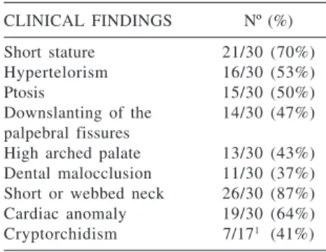
The main clinical findings of the affected NS patients are summarized in Table 1.
The most frequent cardiac anomaly observed in this group of patients was pulmonary stenosis (14 individuals), followed by hypertrophic cardiomy-opathy in 3 patients. One individual had aortic stenosis, and another one had ventricular septal defect (VSD).
All probands had a normal karyo-type.
Nine patients (30%) reported a ten-dency to bleed, characterized by easy bruising, mild nose bleeding, and pro-longed local bleeding after a simple cut and dental extraction. A surgical
procedure had been performed in 15 patients (50%); some of them had more than one surgical procedure. These included primarily heart surgery and/or cardiac catheterization (6 pa-tients), an orchidopexia (5) and an ENT surgery - tonsillectomy (3). The other surgical procedures included a herniorraphy and correction of the pto-sis of the palpebral fissures and a craniectomy in one patient due to an ophthalmic artery aneurysm. Only 2 of the patients who had an open-heart surgery bled profusely, requiring blood transfusion. In one of them, the bleeding was attributed more to the procedure itself, since re-operation stopped the bleeding. In the other pa-tient, we could not recover details of the procedure.
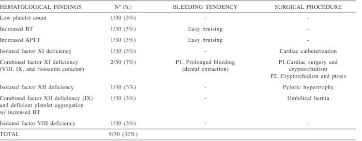
An abnormal hemostatic profile was obtained in 9 patients (30%) (Ta-ble 2). Coagulation-factor deficiencies were observed in 5 individuals (iso-lated factor XI deficiency in 1 patient and combined in another 2; isolated factor XII deficiency in 1 patient and combined in another patient, and fac-tor VIII deficiency in 1 patient). One individual with a combined deficiency of factor XII and IX also had a platelet aggregation defect. Isolated low plate-let count and an increasing BT were also found.
DISCUSSION
NS is a well recognized genetic disorder, and although its incidence is not accurately known, it is estimated at 1/1000 to 1/250014, making this
dis-order one of the most common syn-dromes associated with congenital heart disease.
Due to its broad phenotypic spec-trum, NS caught the attention of dif-ferent clinicians. Studies in these pa-tients have been performed in attempt to better understand this heterogene-ous disorder.
Table 1 - Clinical findings in 30
patients with Noonan syndrome.
CLINICAL FINDINGS Nº (%) Short stature 21/30 (70%) Hypertelorism 16/30 (53%)
Ptosis 15/30 (50%)
Downslanting of the 14/30 (47%) palpebral fissures
High arched palate 13/30 (43%) Dental malocclusion 11/30 (37%) Short or webbed neck 26/30 (87%) Cardiac anomaly 19/30 (64%) Cryptorchidism 7/171 (41%)
7
REV. HOSP. CLÍN. FAC. MED. S. PAULO 58(1):5-8, 2003 Hematological findings in Noonan syndrome Bertola DR et al.
Several reports have emphasized the importance of the hematological aspects in NS, which include abnormal platelet count and function, as well as deficiency of various coagulation fac-tors.
A bleeding diathesis has been con-sidered part of NS, but its prevalence varies widely. The largest study11
esti-mated its frequency in 50%. In our study, an abnormal laboratory screen was observed in 30% of our patients, which is a significant finding.
The most frequent hematological abnormality described in NS is a fac-tor XI deficiency11. It is interesting to
note that congenital deficiency of fac-tor XI was described originally in per-sons of Jewish ethnicity, where the ho-mozygous state could present a severe form of the disease7. In our study,
fac-tor XI deficiency was also the most common hematological finding (33%), and none of our patients were of Jew-ish ethnicity. The background of the patients studied had been emphasized in other papers, prompting the conclu-sion that there is an association be-tween this hematological abnormality and NS.
It is known that there is a poor cor-relation between the tendency to bleed and the level of circulating fac-tor XI12. In our study, only 1 patient
who showed a mild deficiency of fac-tor XI (41%), reported prolonged bleeding after a dental extraction, but had normal clotting in 2 surgical pro-cedures (correction of his heart defect and cryptorchidism). The other 2 pa-tients denied any bleeding tendency. One of them had a cardiac catheteri-zation, and the other had correction of the cryptorchidism and palpebral pto-sis without bleeding problems. Both also had a mild deficiency of factor XI (37% and 40%).
The factor XII deficiency (31% and 32%), observed in 2 of our patients, did not lead to a bleeding problem, as expected. One of these patients, who complained only of easy bruising, also had a factor IX (49%) deficiency, as well as a platelet function abnormal-ity (hypoaggregation to ADP and adrenalin). He underwent a correction of an umbilical hernia without abnor-mal bleeding.
Another patient, who also com-plained of easy bruising, had a
pro-longed bleeding time and a platelet hypoaggregation to ristocetin. The rest of the tests were repeatedly normal, and a diagnosis of von Willebrand dis-ease could not be established.
The multiplicity of unrelated types of bleeding abnormalities and the variability of their expression in NS are difficult to attribute to a single gene defect15. It is possible that the
gene responsible for NS somehow in-teracts with the regulation of other genes involved in the coagulation pathway. This could explain the great variety of the hematological abnor-malities seen in this disorder.
Although the hematological find-ings in NS are complex and the corre-lation between the clinical findings and the circulating levels of some co-agulation factors is not always precise, all clinicians should be aware of this problem and be prompted to conduct a more extensive hematological work-up in these patients. Prior knowledge of a hemostatic abnormality in case of an invasive procedure could be assur-ing of a better management if bleed-ing occurs.
Table 2 - Hematological findings in 30 patients with Noonan syndrome.
HEMATOLOGICAL FINDINGS Nº (%) BLEEDING TENDENCY SURGICAL PROCEDURE
Low platelet count 1/30 (3%) -
-Increased BT 1/30 (3%) Easy bruising
-Increased APTT 1/30 (3%) Easy bruising
-Isolated factor XI deficiency 1/30 (3%) - Cardiac catheterization
Combined factor XI deficiency 2/30 (7%) P1. Prolonged bleeding P1.Cardiac surgery and
(VIII, IX, and ristocetin cofactor) (dental extraction) cryptorchidism
P2. Cryptorchidism and ptosis
Isolated factor XII deficiency 1/30 (3%) - Pyloric hypertrophy
Combined factor XII deficiency (IX) 1/30 (3%) - Umbilical hernia
and deficient platelet aggregation w/ increased BT
Isolated factor VIII deficiency 1/30 (3%) -
-TOTAL 9/30 (30%)
8
REV. HOSP. CLÍN. FAC. MED. S. PAULO 58(1):5-8, 2003 Hematological findings in Noonan syndrome
Bertola DR et al.
RESUMO
to aos aspectos hematológicos que consistiu de tempo de sangramento, tempo de protrombina, tempo de tromboplastina parcial ativada, tempo de trombina, contagem de plaquetas e dosagem do fator de coagulação XI.
RESULTADOS: Um resultado
la-boratorial anormal foi observado em 9 desses pacientes (30%). Apesar dos achados mais comuns terem sido as deficiências dos fatores de coagulação, especialmente do fator XI, também ob-servamos anormalidades no número e na função plaquetária.
CONCLUSÕES: Anormalidades
BERTOLA DR e col. - Achados hema-tológicos na síndrome de Noonan.
Rev. Hosp. Clín. Fac. Med. S. Pau-lo 58(1):5-8, 2003.
OBJETIVO: A síndrome de
Noonan é uma patologia de múltiplas anomalias congênitas e, dentre os achados clínicos, a diátese hemor-rágica está incluída. O propósito des-te estudo é dedes-terminar a freqüência de anormalidades hemostáticas nos paci-entes afetados.
MÉTODO: Nós estudamos 30
pa-cientes afetados pela síndrome
quan-hemostáticas são observadas com cer-ta freqüência em pacientes com sín-drome de Noonan (30% em nossa amostra). Enfatizamos, portanto, a im-portância de uma investigação hema-tológica mais detalhada nesses pacien-tes, especialmente antes da realização de um procedimento invasivo, o qual é requerido com certa freqüência na síndrome.
DESCRITORES: Síndrome de
Noonan. Achados hematológicos. De-ficiência do fator XI de coagulação.
REFERENCES
1 . NOONAN JA – Noonan syndrome. An update and review for the primary pediatrician. Clin Pediatr 1994; 33: 548-555. 2 . TARTAGLIA M, MEHLER EL, GOLDBERG R, et al. - Mutations
in PTPN11, encoding the protein tyrosine phosphatase SHP-2, cause Noonan syndrome. Nat Genet 2001; 29: 465-468. 3 . NOONAN JA – Hypertelorism with Turner phenotype. A new
syndrome with associated congenital heart disease. Am J Dis Child 1968; 116: 373-380.
4 . CARALIS DG, CHAR F, GRABER JD, et al. – Delineation of multiple cardiac anomalies associated with the Noonan syndrome in an adult and review of the literature. Hopkins Med J 1974; 134: 346-355.
5 . PHILLIPS WG, DUNNILL MGS, KURWA AR et al. – Orbital oedema; an unusual presentation of Noonan’s syndrome. Br J Dermatol 1993; 129: 190-192.
6 . FESTEN C – Une complication particulière de l’orchidopexie dans le syndrome de Noonan. Chir Pédiatr 1980; 21: 393-395.
7 . KITCHENS CS, ALEXANDER JA – Partial deficiency of coagulation factor XI as a newly recognized feature of Noonan syndrome. J Pediatr 1983; 102: 224-227.
8 . BADER-MEUNIER B, TCHERNIA G, MIÉLOT F, et al. – Occurrence of myeloproliferative disorder in patients with Noonan syndrome. J Pediatr 1997; 130: 885-889.
9 . ALLANSON JE – Noonan syndrome. J Med Genet 1987; 24: 9-13.
10. SHARLAND M, PATTON M, CHITTOLIE A, et al. – Coagulation abnormalities in Noonan syndrome. J Med Genet 1990; 27: 646.
11. SHARLAND M, PATTON MA, TALBOT S, et al. – Coagulation factor deficiencies and abnormal bleeding in Noonan’s syndrome. Lancet 1992; 339: 19-21.
12. MASSARANO AA, WOOD A, TAIT, RC, et al. – Noonan syndrome: coagulation and clinical aspects. Acta Paediatr 1996; 85: 1181-1185.
13. VAN DER BURGT I, BERENDS, E, LOMMEN, E, et al. – Clinical and molecular studies in a large Dutch family with Noonan syndrome. Am J Med Genet 1994; 53: 187-191.
14. NORA JJ, NORA AH, SINHA AK, et al. – The Ullrich-Noonan syndrome (Turner phenotype). Am J Dis Child 1974; 127: 48-55.