Cop
yright
© ABE&M t
odos os dir
eit
os r
eser
vados
.
Comparison of body composition
methods in overweight and
obese Brazilian women
Comparação de métodos da composição corporal em mulheres brasileiras com sobrepeso e obesidade
Valeria Bender Braulio1, Valéria Cristina Soares Furtado2,
Maria das Graças Silveira1, Maria Helena Fonseca1, José Egídio Oliveira1
absTRacT
Objective: The purpose of this study was to compare skinfold thickness (SKF) and bioelectrical im-pedance analysis (BIA) of body composition using three different equations against dual-energy X-ray absorptiometry (DXA) in overweight and obese Brazilian women. Subjects and methods: Thirty-four women (age 43.8 ± 10.9 years; body mass index [BMI] 32.1 ± 4.3 kg/m2) had percentage body fat (BF%), fat mass (FM) and fat-free mass (FFM) estimated by DXA, SKF and BIA (BIA-man: manufacturer’s equation; and predictive obesity-speciic equations of Segal and of Gray). Regres-sion analysis, Bland-Altman plot analysis and intra-class correlation coeficient (ICC) were used to compare methods. Results: Absolute agreement between DXA and BIA-man was poor for all measures of body composition (BF% -6.8% ± 3.7%, FM -3.1 ± 3.6 kg, FFM 5.7 ± 2.8 kg). BIA-Segal equation showed good absolute agreement with DXA for BF% (1.5% ± 1.5%), FM (1.0 ± 3.2 kg) and FFM (1.5 ± 2.6 kg), albeit the limits of agreement were wide. BIA-Gray equation showed good absolute agreement with DXA for FM (2.3 ± 4.1 kg), and smaller biases for BF% (0.05% ± 4.4%) and FFM (0.2 ± 2.9 kg), although wide limits of agreement. BIA-Gray and DXA showed the highest ICC among the pairs of methods. A good absolute agreement was observed between DXA and SKF for BF% (-2.3% ± 5.8%), FM (0.09 ± 4.7 kg), and FFM (2.4 ± 4.4 kg), although limits of agreement were wider and ICC between DXA and SKF for BF% indicated poor degree of reproducibility. Con-clusion: These indings show that both BIA-Segal and BIA-Gray equations are suitable for BF%, FM and FFM estimations in overweight and obese women. Arq Bras Endocrinol Metab. 2010;54(4):398-405 Keywords
Absorptiometry, photon; electric impedance; skinfold thickness; body composition; obesity; overweight
Resumo
Objetivo: Veriicar a concordância dos métodos de impedância bioelétrica (BIA) usando três equa-ções diferentes, e medida das pregas cutâneas (PC) com absorciometria de raios-X de dupla energia (DEXA), para análise da composição corporal de mulheres com sobrepeso e obesidade. Sujeitos e métodos: Em 34 mulheres (43,8 ± 10.9 anos; índice de massa corporal [IMC] 32,1 ± 4,3 kg/m2) foram avaliados: percentual de gordura total (%GT), massa gorda (MG) e massa magra (MM) por DEXA, PC, BIA-Fab (equação do fabricante), BIA-Segal e BIA-Gray (equações para obesidade). Foram utiliza-dos: análise de regressão, método de Bland-Altman e coeiciente de correlação intraclasse (CCI). Re-sultados: A concordância absoluta entre DEXA e BIA-Fab foi fraca para todas as medidas (BF% -6,8% ± 3,7%, FM -3,1 ± 3,6 kg, FFM 5,7 ± 2,8 kg). BIA-Segal apresentou boa concordância absoluta com DEXA para %GT (1,5% ± 1,5%), MG (1,0 ± 3,2 kg) e MM (1,5 ± 2,6 kg), mas com amplos limites de concordância. BIA-Gray teve boa concordância absoluta com DEXA para MG (2,3 ± 4,1 kg) e peque-nos vieses para %GT (0,05% ± 4,4%) e MM (0,2 ± 2,9 kg), mas com amplos limites de concordância. BIA-Gray e DEXA tiveram maior CCI entre métodos. Houve boa concordância absoluta entre DEXA e PC para %GT (-2,3% ± 5,8%), MG (0,09 ± 4,7 kg) e MM (2,4 ± 4,4 kg), mas com amplos limites de con-cordância e pouca reprodutibilidade no CCI, entre DEXA e PC para %GT. Conclusão: A BIA, utilizando equação especíica para obesidade, foi o método que melhor concordou com DEXA, para estimar %GT, MG e MM em mulheres com sobrepeso e obesidade. Arq Bras Endocrinol Metab. 2010;54(4):398-405 Descritores
Absorciometria de fóton; impedância elétrica; pregas cutâneas; composição corporal; obesidade, sobrepeso 1 Divisãode Metabolismo
e Nutrição, Faculdade de Medicina, Universidade Federal do Rio de Janeiro (UFRJ), Rio de Janeiro, RJ, Brazil 2 Departamento de Nutrição Aplicada, Escola de Nutrição, Universidade Federal do Estado do Rio de Janeiro (Unirio), Rio de Janeiro, RJ, Brazil
Correspondence to: Valeria Bender Braulio Rua Professor Rodolpho Paulo Rocco, 255, 9° andar, sala 9E-14, Ilha do Fundão 21941-913 − Rio de Janeiro, RJ, Brazil
Cop
yright
© ABE&M t
odos os dir
eit
os r
eser
vados
.
inTRoDucTion
O
verweight and obesity are increasing at alarming rates throughout the world (1,2). Obesity is char-acterized by an excessive amount of body fat, which is the main cause for health hazards such as hypertension, diabetes, and heart disease (3,4). In addition, the pres-ervation of fat-free mass is a target in the treatment of this disease (5). Hence, in this context it is important to identify a simple, reliable, non-invasive and cost-effec-tive technique for body composition assessment.There is no direct method for measuring adiposi-ty, and the most frequently used indirect methods are dual-energy X-ray absorptiometry (DXA), bioelectrical impedance analysis (BIA) and anthropometry. DXA is considered to be a valid and reliable reference method for body composition assessment (6). DXA measures three body compartments: fat mass, lean body mass, and bone content. However, the use of DXA is limited in some situations due to the cost of the equipment, the need for training an operator, and lack of portabi-lity, all of which limit its application in diverse facilities, including clinic rooms and private health centers (7). Consequently, there is a need for simpler, more accura-te and reliable, as well as less expensive and less invasive body composition measuring than the DXA technique.
Bioelectrical impedance analysis and skinfold thick-ness (SKF) are non-invasive methods for body compo-sition assessment that can be readily performed by trai-ned operators in most facilities since these equipment are easily transported. They also have the advantage of being relatively quick to perform (8).
BIA is based on the conduction of an electrical cur-rent through the body (9). Using the assumption of a conductive material constant resistance, and estimating the length of the conductive path from an individual’s height, total body water can be calculated from impedan-ce measuring through the low of a small current. Body composition can then be estimated assuming that total body water constitutes a ixed percentage of lean mass (usually 73%). Speciic prediction equations have been developed to evaluate body composition from height, weight, and impedance (10). As BIA accuracy mainly depends on the equation used, several researchers have developed speciic equations to use with obese adult po-pulations (11-14). However, no irm conclusions can be drawn regarding the predictability of those equations.
Skinfold thickness is a long established method for determining body fat. The sum of four SKF provides an
estimation of the fat mass, and subtracting it from total body weight, fat-free mass is valuated (15). Although with altered fat distribution conditions the method has been questioned, and also due to inter-observer errors, in clinical facilities, it has been the most simple and inexpensive technique available, and its applicability is possible in a large number of subjects (16).
Validity of body composition measurements is scar-ce for Brazilian obese people data (17,18). Therefore, the current study worked with SKF and BIA for body composition assessment in overweight and obese Bra-zilian women. BIA was assessed using standard and obesity-speciic equations. Both methods were com-pared with DXA as a reference method to verify the degree of similarity among techniques.
subJecTs anD meTHoDs
subjects
Thirty-four overweight and obese Brazilian women with mean body mass index (BMI) 32.1 ± 4.3 kg/m2
(range 25.0-39.7 kg/m2), mean age 43.8 ± 10.9
ye-ars participated in this study. Subjects were outpatients beginning a hospital weight-loss program. Exclusion criteria included pregnancy, hypothyroidism, and body weight over 120 kg, anterior-posterior abdominal dia-meter exceeding 30 cm, and use of medication inluen-cing luid balance and metabolism.
Informed consent which was approved by the local research ethics committee of the Hospital of Federal University of Rio de Janeiro, was obtained from all par-ticipants before entering the study.
Protocol
Subjects were studied four hours after their last meal, and after they had emptied their bladders before their body weight and height were recorded. Height was measured to the nearest 0.1 cm using a Sanny®
stadio-meter, and subjects stood up erect without shoes. Wei-ght was measured to the nearest 0.1 kg using Welmy®
scale. The scale was calibrated prior to each test. BMI was calculated as body weight (in kg) divided by squa-red height (in meters).
Cop
yright
© ABE&M t
odos os dir
eit
os r
eser
vados
.
1. DXA scanning (Lunar DPX-IQ, version 4.7e, Lunar Radiation Corporation, Madison, WI, USA), ac-cording to standardized procedures recommended by the manufacturer. Calibration using the manufacturer’s phantom was performed on each scanning day. Subjects reclined on a lat couch, dressed in light clothing, and wore no metal objects. The whole body was scanned. Scan duration typically ranged from 10 to 30 min de-pending on the subject’s size, with radiation dose for a scan being approximately 0.5 µSv. The results were pre-sented in kg for FFM, FM, and in % for BF%. FFM was calculated using the sum of lean tissue mass and bone mineral content estimations for each subject. DXA was the reference method.
2. BIA was undertaken with a tetrapolar bioanalyzer device (Model 310, Biodynamics Corp., Seattle, WA − USA). Measurements were undertaken as previous-ly described (9) by a single trained observer. Subjects reclined on a lat couch, with limbs not touching each other. Electrodes were placed on the right side of the body between the distal prominences of the radius and ulna; the distal end of the third metacarpal; between the median and lateral malleoli at the ankle; and at the distal end of the third metatarsal. FFM, FM and BF% were calculated from the measurements of resistance made at 50 kHz using the formula provided by the instrument manufacturer (BIA-man). In addition, the resistance directly read from the impedance device was considered along with height, weight, and age in two obesity-speciic equations published by Segal and cols. (BIA-Segal) (13) and Gray and cols. (BIA-Gray) (11).
3. SKF measurements were performed by a single ob-server at four sites (triceps, biceps, subscapular and su-prailiac) according to standard techniques. Skinfold mea-surements were performed on the body’s right side with a Lange skinfold caliper (Cambridge Scientiic Instru-ments, Cambridge, MD, USA). Three sets of measure-ments were averaged for each site. Body density was cal-culated from the sum of the four skinfold measurements according to Durnin and Wormersley (15), and BF% was then calculated by Siri’s equation (19). FFM (in kg) was calculated subtracting body fat from total body weight.
statistical analysis
Values are reported in the text as mean ± standard de-viation (SD). Statistical analysis were performed using SPSS version 13.0 (SPSS, Inc., Chicago, IL), and Med-Calc version 9.6.4.0 (MedMed-Calc Software, Mariakerke, Belgium). One-way ANOVA was used to compare
di-fferences among body composition values determined using different techniques. When ANOVA showed a statistically signiicant mean effect, Bonferroni’s test was used to determine differences among means. Re-gression analysis was used to determine the level of relative agreement among different techniques. Bland-Altman analysis (20) was used to determine absolute limits of agreement between body composition varia-bles assessed by BIA and SKF against DXA. The in-tra-class correlation coeficient (ICC) was used to test the reproducibility of BF%, FM and FFM measured by BIA-man, BIA-Gray and SKF compared to DXA. (21) Values of ICC below 0.4 were considered as an indication of poor reproducibility, values between 0.4 and 0.7, medium-term reproducibility and values abo-ve 0.7, good reproducibility. Statistical signiicance was considered when p < 0.05.
ResuLTs
As can be seen in table 1, BIA-man results underestima-ted BF% and overestimaunderestima-ted FFM when compared with DXA, whereas no difference was observed between mean BF% and FFM assessed by BIA-Segal, BIA-Gray and SFK, and DXA. Regarding FM measurements, no differences were found between the four techniques compared with DXA.
Cop
yright
© ABE&M t
odos os dir
eit
os r
eser
vados
.
Table 2. Comparison of methods of body composition with DXA
comparison
Regression analysis bland and altman intra-class correlation coeficient
r2 p-Value mean bias ± sD of bias
95% limits of
agreement r p-Value
BIA-manufactory vs. DXA
% body fat Fat mass (kg) Free-fat mass (kg)
0.751 0.899 0.841
< 0.001 < 0.001 < 0.001
-6.8 ± 3.7 -3.1 ± 3.6 5.7 ± 2.8
-14.0 to 0.3 -10.3 to 3.8 0.2 to 11.3
0.179 0.791 0.340
0.149 < 0.001
0.022
BIA-Segal vs. DXA
% body fat Fat mass (kg) Free-fat mass (kg)
0.802 0.920 0.833
< 0.001 < 0.001 < 0.001
-1.5 ± 1.5 1.0 ± 3.2 1.5 ± 2.6
-8.7 to 5.8 -5.3 to 7.3 -3.6 to 6.6
0.471 0.897 0.784
0.002 < 0.001 < 0.001
BIA-Gray vs. DXA
% body fat Fat mass (kg) Free-fat mass (kg)
0.700 0.867 0.825
< 0.001 < 0.001 < 0.001
0.05 ± 4.4 2.3 ± 4.1 0.2 ± 2.9
-8.5 to 8.6 -5.7 to 10.3 -5.5 to 5.9
0.706 0.833 0.812
< 0.001 < 0.001 < 0.001
SKF vs. DXA
% body fat Fat mass (kg) Free-fat mass (kg)
0.248 0.819 0.746
0.158 < 0.001 < 0.001
-2.3 ± 5.8 0.09 ± 4.7 2.4 ± 4.4
-17.3 to 9.1 -9.2 to 9.4 -6.3 to 11.12
0.167 0.764 0.611
0.166 < 0.001 < 0.001
DXA: dual X-ray absorptiometry; BIA: bioelectrical impedance analysis; SKF: skinfold thickness.
Table 1. Percentage body fat, fat mass, and fat-free mass assessed by different body composition methods
method % body fat fat mass (kg) fat-free mass (kg)
DXA 44.6 ± 5.5 33.6 ± 8.0 43.6 ± 4.3
BIA-manufacturer 37.8 ± 4.4* 30.4 ± 6.4 49.3 ± 5.2*
BIA-Segal 43.2 ± 3.6 34.6 ± 6.6 45.1 ± 4.6
BIA-Gray 44.7 ± 5.8 35.9 ± 7.8 43.8 ± 5.2
SKF 42.3 ± 3.6 33.7 ± 5.4 45.9 ± 6.6
Values are mean ± standard deviation. DXA: dual X-ray absorptiometry; BIA: bioelectrical impedance analysis; SKF: skinfold thickness.
* P < 0.05.
SKF overestimated FFM by 2.4 kg, and underestimated BF% by 2.3%. There was, on average, good absolute agreement between SKF and DXA for all measures of body composition with wider limits of agreement for each variable (Table 2; Figure 4).
ICC results were indicative of good reproducibility between BIA-man and DXA for FM (0.791), Se-gal and DXA for FM (0.897) and FFM (0.784), BIA-Gray and DXA for BF% (0.706), FM (0.833) and FFM (0.812), and between SKF and DXA for FM (0.764). The ICC between BIA-Segal and DXA for BF% (0.471), and between SKF and DXA for FFM (0.611) represen-ted moderate reproducibility. The ICC obtained be-tween BIA-man and DXA for BF% (0.179) and FFM (0.340), and between SKF and DXA for BF% (0.167) was indicative of poor reproducibility (Table 2).
Discussion
The main inding of this study was that, in a group of overweight and obese Brazilian women, all body com-position assessments provided good relative agreement with DXA, as indicated by high correlation coeficients, except for BF% assessed by SKF. However, in absolute terms, all methods were associated with wide limits of agreement. Compared with DXA, BIA-man substan-tially underestimated BF% and overestimated FFM, whereas BIA-Segal and BIA-Gray as well as SKF me-thods provided good absolute agreement for all body components assessed. In addition, BF%, FM, and FFM measurements obtained by BIA-Gray provided higher ICC indicating good reproducibility between BIA-Gray and DXA methods.
Cop
yright
© ABE&M t
odos os dir
eit os r eser vados . A
10 20 30 40 50 60
20
10
0
-10
-20
BF (BIA-man + DXA) / 2 (%)
Diff. BF ( BI A-man - DXA) ( %) Mean -6.8 -1.96 SD -14.0 +1.96 SD 0.3 B
10 20 30 40 50 60
20
10
0
-10
-20
FM (BIA-man + DXA) / 2 (kg)
D iff . FM (B IA -m an D X A ) ( kg ) Mean -3.3 -1.96 SD -10.3 +1.96 SD 3.8 C
10 20 30 40 50 60
20
10
0
-10
-20
FFM (BIA-man + DXA) / 2 (kg)
D iff . FF M (B IA -m an D X A ) ( kg ) Mean 5.7 -1.96 SD 0.2 +1.96 SD 11.3
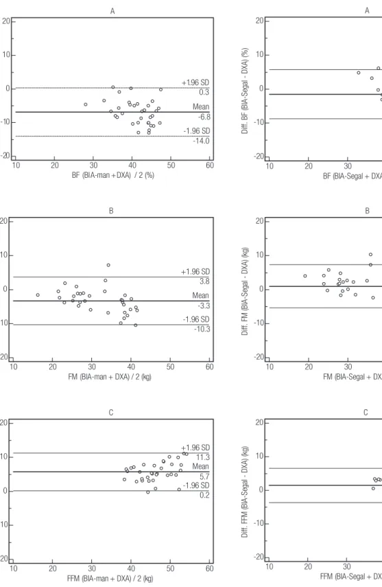
figure 1. Bland-Altman plots showing the limits of agreement between (a) percentage body fat (BF%), (b) fat mass (FM), and (c) fat-free mass (FFM) as determined by bioelectrical impedance analysis (BIA) using the equation provided by the manufacturer of the instrument (BIA-man) and dual energy X-ray absorptiometry (DXA). The center line represents the mean differences between the two methods, and the other two lines represent two SDs from the mean.
figure 2. Bland-Altman plots showing the limits of agreement between (a) percentage body fat (BF%), (b) fat mass (FM), and (c) fat-free mass (FFM) as determined by bioelectrical impedance analysis (BIA) using the obese-specific equation of Segal (BIA-Segal) and dual energy X-ray absorptiometry (DXA). The center line represents the mean differences between the two methods, and the other two lines represent two SDs from the mean.
A
10 20 30 40 50 60
20
10
0
-10
-20
BF (BIA-Segal + DXA ) / 2 (%)
D iff . B F (B IA -S eg al D X A ) ( % ) Mean -1.5 -1.96 SD -8.7 +1.96 SD 5.8 B
10 20 30 40 50 60
20
10
0
-10
-20
FM (BIA-Segal + DXA) / 2 (kg)
D iff . FM (B IA -S eg al D X A ) ( kg ) Mean 1.0 -1.96 SD -5.3 +1.96 SD 7.3 C
10 20 30 40 50 60
20
10
0
-10
-20
FFM (BIA-Segal + DXA) / 2 (kg)
Cop
yright
© ABE&M t
odos os dir
eit
os r
eser
vados
.
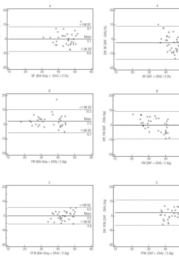
figure 4. Bland-Altman plots showing the limits of agreement between (a) percentage body fat (BF%), (b) fat mass (FM), and (c) fat-free mass (FFM) as determined by skinfold thickness (SKF) and dual energy X-ray absorptiometry (DXA). The center line represents the mean differences between the two methods, and the other two lines represent two SDs from the mean.
figure 3. Bland-Altman plots showing the limits of agreement between (a) percentage body fat (BF%), (b) fat mass (FM), and (c) fat-free mass (FFM) as determined by bioelectrical impedance analysis (BIA) using the obese-specific equation of Gray (BIA-Gray) and dual energy X-ray absorptiometry (DXA). The center line represents the mean differences between the two methods, and the other two lines represent two SDs from the mean.
A
10 20 30 40 50 60
20
10
0
-10
-20
BF (BIA-Gray + DX A ) / 2 (%)
Diff.
BF
(
BI
A-Gray - D
XA
) (%
)
Mean 0.1
-1.96 SD -8.6 +1.96 SD 8.7
B
10 20 30 40 50 60
20
10
0
-10
-20
FM (BIA-Gray + DXA) / 2 (kg)
D
iff
.
FM
(B
IA
-G
ra
y
-
D
X
A
) (
kg
)
Mean 2.3
-1.96 SD -5.7 +1.96 SD 10.3
C
10 20 30 40 50 60
20
10
0
-10
-20
FF M (B IA -Gray + DXA) / 2 (kg)
Diff.
FFM
(B
IA
-Gra
y
-
DXA)
(kg
)
Mean 0.2 -1.96 SD -5.6 +1.96 SD
6.0
B
10 20 30 40 50 60
20
10
0
-10
-20
FM (SKF + DXA) / 2 (kg)
Diff. FM (SKF - DXA) (kg)
Mean 0.1
-1.96 SD -9.2 +1.96 SD 9.4
A
10 20 30 40 50 60
20
10
0
-10
-20
BF (SKF+DXA) / 2 (%)
D
iff
.
B
F
(S
K
F
-
D
X
A
) (
%
)
Mean -2.3
-1.96 SD -13.8 +1.96 SD 9.1
C
10 20 30 40 50 60
20
10
0
-10
-20
FFM (S KF + DXA) / 2 (kg)
Diff.
FFM
(S
KF
- D
XA
) (k
g)
Mean 2.4
-1.96 SD -6.3 +1.96 SD
Cop
yright
© ABE&M t
odos os dir
eit
os r
eser
vados
.
Although BIA-man demonstrated a good relative agreement with DXA, it provided the poorest absolute levels of agreement with a large bias in underestima-ting body fat content (~7%) and overestimaunderestima-ting FFM (~6 kg). This inding is consistent with the results of previous studies (22,25-27) which indicated that BIA-man results signiicantly underestimated body fat con-tent in overweight and obese women (22,26,27), and non-obese subjects (25) when compared with DXA.
Results of the current study and of the three previous studies (22,26,27) indicate that BIA-man provides a poor level of absolute agreement with DXA, suggesting that BIA-man may not be suitable for the measurement of body composition in overweight and obese women in clinical research as well as clinical facilities.
Thus, in a population of obese people, equations developed to estimate lean mass in the healthy popu-lation through BIA are probably not valid to assess body fat by subtracting estimated lean mass from total body weight (26). In fact, whenever the manufacturer’s equations are used, BIA may overestimate lean tissue and underestimate fat in obese subjects (11,13,28,29).
The current study employed two obesity-speciic BIA analysis equations because studies have shown that the predictive accuracy for BIA method may be impro-ved using obesity-speciic equations to estimate an obe-se population’s body composition. The Gray obesity-speciic equation validated the Segal equation over a wide range of body fat values (11,13).
We found that the bias for the absolute differences between DXA and BIA-Segal or BIA-Gray were small. These indings of good absolute agreement between BIA-Segal or BIA-Gray and DXA, taken together with the good relative agreement among methods, indicate that BIA using either obesity-speciic equations such as Segal (13) or Gray equation (11) is a reliable method for assessing body fat and free-fat mass at the group level in overweight/obese populations. However, the wide limits of agreement conine the utility of this te-chnique to any accurate determination of an individual body composition, and its use, therefore, seems limi-ted in clinical facilities. Additionally, these wide limits of agreement are in accordance with previous reports using obesity-speciic equations (24).
The most widely applied method for calculating total body fat from measured SKF in current use was developed by Durnin and Womersley (15). Body den-sity obtained by Durnin and Womersley (15) is derived from a spectrum of lean to moderately obese subjects,
and are expected to lose precision in severely obese sub-jects with BF% far higher than their validation popula-tion means (30). The results of the current study show good relative agreement between SKF and DXA for the assessment of FM, as well as good absolute agreement and good reproducibility. Also, the mean difference be-tween these methods was not signiicant. Thus, for FM, it is reasonable to consider the SKF method very useful to assess overweight and obese women, and as the most simple and inexpensive method. In contrast, no signii-cant correlation was found between SKF and DXA for BF% assessment. Despite the good absolute agreement, there were large biases between SKF and DXA mea-surements, along with poor degree of reproducibility obtained by ICC for BF%.
Usually total body fat estimations are expressed as percentage body weight. However, a problem in this approach is that the relationship between total body fat and body weight has a nonzero intercept. The result is that a curvilinear relationship exists between total body fat, expressed as a percent of body weight and body weight or BMI (31). Indeed, the complex relationship between percentage of fat and body weight can result in some indeinite situations.
A severely obese patient could lose a relatively large amount of weight and yet have a relatively small change in percentage of fat. The SKF method cannot be recom-mended for overweight or obese individuals although it can be used to validate the estimation of BF% for di-verse healthy age and ethnic groups, because increasing levels of body fat leads to subcutaneous total body fat proportional changes, thereby affecting the relationship between the sum of skinfolds and relative body fat (14). Finally, according to our results, BIA-Segal as well as BIA-Gray equations provided the most accurate and precise estimations of BF%, FM, and FFM in compari-son to DXA in overweight and obese Brazilian women. However, due to broad limits of agreement, we can only recommend these equations to groups of popula-tions, not of individuals.
Acknowledgments: the authors gratefully acknowledge Mrs. Ro-sângela A. M. Noé for statistical assistance.
Disclosure: no potential conlict of interest relevant to this article was reported.
RefeRences
Cop
yright
© ABE&M t
odos os dir
eit
os r
eser
vados
.
2. Mokdad AH, Bowman BA, Ford ES, Vinicor F, Marks JS, Koplan JP. The continuing epidemics of obesity and diabetes in the United States. JAMA. 2001;286(10):1195-200.
3. Okosun IS, Chandra KM, Choi S, Christman J, Dever GE, Prewitt TE. Hypertension and type 2 diabetes comorbidity in adults in the United States: risk of overall and regional adiposity. Obes Res. 2001;9(1):1-9.
4. Rader DJ. Effect of insulin resistance, dyslipidemia, and intra-ab-dominal adiposity on the development of cardiovascular disease and diabetes mellitus. Am J Med. 2007;120(3 Suppl 1):S12-8. 5. Ritz P, Salle A, Audran M, Rohmer V. Comparison of different
me-thods to assess body composition of weight loss in obese and diabetic patients. Diabetes Res Clin Pract. 2007;77(3):405-11. 6. Prior BM, Cureton KJ, Modlesky CM, Evans EM, Sloniger MA,
Saunders M, et al. In vivo validation of whole body composition estimates from dual-energy X-ray absorptiometry. J Appl Phy-siol. 1997;83(2):623-30.
7. Thomson R, Brinkworth GD, Buckley JD, Noakes M, Clifton PM. Good agreement between bioelectrical impedance and dual-energy X-ray absorptiometry for estimating changes in body composition during weight loss in overweight young women. Clin Nutr. 2007;26(6):771-7.
8. King S, Wilson J, Kotsimbos T, Bailey M, Nyulasi I. Body compo-sition assessment in adults with cystic ibrosis: comparison of dual-energy X-ray absorptiometry with skinfolds and bioelectri-cal impedance analysis. Nutrition. 2005;21(11-12):1087-94. 9. Lukaski HC. Methods for the assessment of human body
compo-sition: traditional and new. Am J Clin Nutr. 1987;46(4):537-56. 10. Kyle UG, Bosaeus I, De Lorenzo AD, Deurenberg P, Elia M, Gomez
JM, et al. Bioelectrical impedance analysis − part I: review of prin-ciples and methods. Clin Nutr. 2004;23(5):1226-43.
11. Gray DS, Bray GA, Gemayel N, Kaplan K. Effect of obesity on bioelectrical impedance. Am J Clin Nutr. 1989;50(2):255-60. 12. Jakicic JM, Wing RR, Lang W. Bioelectrical impedance analysis
to assess body composition in obese adult women: the effect of ethnicity. Int J Obes Relat Metab Disord. 1998;22(3):243-9. 13. Segal KR, Van Loan M, Fitzgerald PI, Hodgdon JA, Van Itallie TB.
Lean body mass estimation by bioelectrical impedance analysis: a four-site cross-validation study. Am J Clin Nutr. 1988;47(1):7-14. 14. Stolarczyk LM, Heyward VH. Assessing body composition of
adults with diabetes. Diabetes Technol Ther. 1999 Fall;1(3):289-96. 15. Durnin JV, Womersley J. Body fat assessed from total body den-sity and its estimation from skinfold thickness: measurements on 481 men and women aged from 16 to 72 years. Br J Nutr. 1974;32(1):77-97.
16. Kamimura MA, Avesani CM, Cendoroglo M, Canziani ME, Draibe SA, Cuppari L. Comparison of skinfold thicknesses and bioelec-trical impedance analysis with dual-energy X-ray absorptiometry for the assessment of body fat in patients on long-term haemo-dialysis therapy. Nephrol Dial Transplant. 2003;18(1):101-5.
17. Bottaro MM HV, Lindolfo JB. Cross-validation of bioimpe-dance (BI) equations for Brazilian women using dual-energy x-ray absorptiometry (DXA) [abstract]. Med Sci Sports Exerc. 1999;31:S200.
18. Oliveira FL, Taddei JA, Escrivao MA, Cobayashi F, Barros ME, Vi-tolo MR, et al. Accuracy of obesity diagnosis in Brazilian adoles-cents: comparison of Cole et al and Must et al criteria with DXA percentage of fat mass. Nutr Hosp. 2006;21(4):484-90.
19. Siri WE. Body composition from luid spaces and density: analy-sis of methods. 1961. Nutrition. 1993;9(5):480-91; discussion, 92. 20. Bland JM, Altman DG. Statistical methods for assessing
agre-ement between two methods of clinical measuragre-ement. Lancet. 1986;1(8476):307-10.
21. Bartko JJ, Carpenter WT Jr. On the methods and theory of reliabi-lity. J Nerv Ment Dis. 1976;163(5):307-17.
22. Andreoli A, Melchiorri G, De Lorenzo A, Caruso I, Sinibaldi Sa-limei P, Guerrisi M. Bioelectrical impedance measures in diffe-rent position and vs dual-energy X-ray absorptiometry (DXA). J Sports Med Phys Fitness. 2002;42(2):186-9.
23. Frisard MI, Greenway FL, Delany JP. Comparison of methods to assess body composition changes during a period of weight loss. Obes Res. 2005;13(5):845-54.
24. Pateyjohns IR, Brinkworth GD, Buckley JD, Noakes M, Clifton PM. Comparison of three bioelectrical impedance methods with DXA in overweight and obese men. Obesity (Silver Spring). 2006;14(11):2064-70.
25. Bolanowski M, Nilsson BE. Assessment of human body compo-sition using dual-energy x-ray absorptiometry and bioelectrical impedance analysis. Med Sci Monit. 2001;7(5):1029-33.
26. Panotopoulos G, Ruiz JC, Guy-Grand B, Basdevant A. Dual x-ray absorptiometry, bioelectrical impedance, and near infrared interac-tance in obese women. Med Sci Sports Exerc. 2001;33(4):665-70. 27. Sun G, French CR, Martin GR, Younghusband B, Green RC, Xie
YG, et al. Comparison of multifrequency bioelectrical impedance analysis with dual-energy X-ray absorptiometry for assessment of percentage body fat in a large, healthy population. Am J Clin Nutr. 2005;81(1):74-8.
28. Deurenberg P, van der Kooy K, Leenen R, Weststrate JA, Seidell JC. Sex and age speciic prediction formulas for estimating body composition from bioelectrical impedance: a cross-validation study. Int J Obes. 1991;15(1):17-25.
29. Ellis KJ. Human body composition: in vivo methods. Physiol Rev. 2000;80(2):649-80.
30. Teran JC, Sparks KE, Quinn LM, Fernandez BS, Krey SH, Steffee WP. Percent body fat in obese white females predicted by anthro-pometric measurements. Am J Clin Nutr. 1991;53(1):7-13. 31. Webster JD, Hesp R, Garrow JS. The composition of excess