*e-mail: [email protected]
1. Introduction
Titanium is widely-used as a biomaterial because it is biocompatible, chemically inert, and possesses good mechanical properties. It is not an ideal material from a medical standpoint as it does not present good bioactivity, i.e. bone does not grow spontaneously around it when implanted in the human body1-3. We are conducting studies to improve the bioactivity of this material.
Currently, there are two main lines of research focused on improving the bioactivity of titanium through changes in it is surface properties. These are based on the growth of a layer of hydroxyapatite, Ca10(PO4)6(OH)2, the principal mineral component of bone, on the metal surface. Hydroxyapatite (HA) has superior biocompatibility to any other known material4. Many studies propose the modiication of the titanium surface to accelerate the spontaneous precipitation of apatite when in contact with the body; others propose the deposition of an HA layer.
The layer deposition techniques used to produce calcium phosphate include “plasma spray”5-8, laser ablation, “magnetron sputtering”9,10, sol-gel11-14, electrophoresis15,16, and biomimetic methods17-19. In the latter technique, the hydroxyapatite is precipitated from a saturated calcium phosphate solution, similar to that of blood. Among the techniques mentioned above, the only one commercially available is plasma spray, but the layers produced present problems. The connection between the hydroxyapatite and metal is purely mechanical, without chemical interaction between the two materials. Consequently, coatings behave as a brittle ceramic, presenting a low resistance to fatigue,
delamination and degradation when used in implants for prolonged periods20,21. The particles released into body can
cause inlammatory reactions22.
An alternative for the production of highly adherent layers using a cheap and simple methodology is Plasma Electrolytic Oxidation (PEO). Using PEO coatings can be deposited onto samples from an aqueous electrolyte, by applying a potential of a few hundred volts, which generates localized discharges on the sample surface23. The electrolytes dissolved in water dissociate to form anions and cations. When a voltage is applied between the electrodes, anions and cations are attracted to the anode and cathode, respectively. Thus, at the anode, the oxidation process begins. Initially, an insulating layer formed on the anode causes a signiicant drop in system current. With the increase of the applied voltage, the current is forced to pass through the coating. Intense electric ields around the samples generated localized electrical discharges, called micro-arcs. The coating is formed by oxidation of the samples and by deposition of elements from the electrolytic solution. As the micro-arcs produce high temperatures the oxide layer can be sintered and incorporate elements into the coating. The coating is deposited across the whole surface immersed in the solution. An improvement in adhesion of these layers is observed compared to that of hydroxyapatite layers deposited by plasma spray24,25. In addition, the layers are porous and rough, and resistant to wear and corrosion26,27, which are interesting features for good bioactivity.
Recent studies have demonstrated the possibility of the deposition of HA films by PEO28-32. The different concentrations of calcium and phosphorus and the different
Hydroxyapatite Coating Deposited on Grade 4 Titanium by Plasma Electrolytic Oxidation
César Augusto Antônioa*, Nilson Cristino Cruza, Elidiane Cipriano Rangela,
Rita de Cássia Cipriano Rangela, Tamires do Espirito Santo Araujoa, Steven Frederick Durranta,
Bruna Antunes Másb, Eliana Aparecida Rezende Duekc
aTechnological Plasmas Laboratory, Paulista State University – UNESP,
Experimental Campus of Sorocaba, Sorocaba, SP, Brazil
bDepartment of Material Engineering, Faculty of Mechanical Engineering,
Campinas State University – UNICAMP, Campinas, SP, Brazil
cLaboratory of Biomaterials, Pontifícia Universidade Católica de São Paulo – PUC-SP,
Sorocaba, SP, Brazil
Received: April 7, 2014; Revised: December 18, 2014
The present study reports the deposition of coating using Plasma Electrolytic Oxidation (PEO) onto grade 4 titanium to produce novel surface features. Samples were treated in an electrolytic solution of calcium acetate and sodium glycerolphosphate. The temporal evolution of hydroxyapatite coatings with high Ra roughness and a maximum thickness of 120 µm was obtained. X-ray spectra revealed the presence of hydroxyapatite, rutile and calcium phosphate. Cell growth measurement by MTT assay showed that the coatings were not toxic because cells grew on all samples.
Antônio et al.
1428 Materials Research
structures of these ilms are easily controlled by varying the concentration of the reagents used and the pH of the electrolyte solution28. Reagents commonly used for the electrolyte solution are tripolyphosphate, calcium acetate, calcium glycerophosphate, monosodium phosphate, and trisodium phosphate29,31. Some results show that it is possible to deposit various phases of calcium phosphate, including hydroxyapatite30,32.
Currently, the formation of HA in coatings produced by PEO is possible using long treatment times together with high polarization frequencies. As conirmed by XRS, however, the amount of HA produced is small. Thus, the objective of this work is to obtain a coating containing a signiicant amount of HA using short depositions times.
2. Experimental Description
Grade 4 titanium disks of 8 mm diameter and 2 mm thickness were produced. After machining and polishing, the samples were sterilized and stored in suitable containers. The samples treatment was carried out in a stainless steel tank of capacity 2 l. The tank was enclosed in a cooling system that maintains the temperature of the solution at approximately 50 °C. The sample was attached at the anode and the tank served as the cathode itself. The electrolytic solution used for the treatment was 0.2 M calcium acetate ((CH3COO)2Ca) and 0.02 M sodium glycerophosphate (C3H5(OH2)PO4Na) diluted in 1 liter of deionized water. The samples were treated with a current density that varied between 1.7 and 0.6 A cm–2 (both ± 0.2 A cm–2) for times of 120, 300 and 600 s. The voltage, frequency and duty cycle were 480 V, 100 Hz and 60%, respectively. Treatment of the samples was carried out in the potentiostatic mode, during which the voltage is maintained constant and the current varies as a function of time. After PEO treatment, the samples were cleaned with deionized water and dried.
In this study, we investigated the structure, thickness, roughness, morphology, chemical composition and cytocompatibility of the coating. The coating thickness was measured by scanning electron microscopy (SEM) after preparation of a metallographic sample section. Sample surface roughness was veriied by proilometry. SEM was used to analyze the morphology of the coating. X-ray diffraction using Cu-Kα radiation (λ = 1.54 Å) with angles between 20 and 60° and a step-size of 0.1°/ min, was used to examine the phase of the coating.
The cytocompatibility, i.e. the ability of the samples to support adhesion and cell growth of primary osteoblasts after 24 hours and 5 days of culture, was assessed using the colorimetric MTT metabolism assay (3 - (4,5-dimethylthiazol-2-yl) -2,5 diphenyl tetrazolium bromide). Osteoblasts were obtained by extraction from calvariae of three 20-day-old Wistar rats according to Declercq et al.33. Osteoblasts were cultured in Dulbecco’s
modiied essential medium (DMEM) supplemented with 10% (v/v) heat-inactivated fetal bovine serum (FBS), gentamicin (50 μg/mL) and amphotericin B (5 μg/mL). The cells were then harvested using trypsin/EDTA and seeded onto 48-well culture plates containing the samples. The culture medium was changed every 2 days. For the MTT assay, 1x10³ cells/mL were seeded in wells containing the
samples. After 24 hours and 5 days of culture, the culture medium was removed and the wells washed twice with 0.1 M PBS, pH 7.4, at 37 °C, followed by the addition of 200 μL of DMEM and MTT reagent (20 μL/well, containing 5 mg of MTT reagent/mL). After incubation for 4 h at 37 °C, the cells were lysed by adding 200 μL of dimethylsulphoxide (DMSO). Subsequently, 100 μL aliquots of the solution in each well were transferred to a new plate and the absorbance then measured at 570 nm using a microplate reader. The data for the cytocompatibility assay was expressed as the mean ± standard deviation (SD) for n = 4 experiments. Analysis of variance (One-Way ANOVA) followed by the Tukey test (with p < 0.05) was used to assess the signiicance of the data. For the data analyses the BioEstat® 5.0 statistical software was used.
3. Results and Discussion
3.1. Current density characteristics
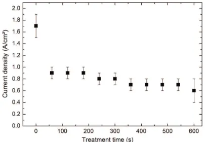
When the supply is turned on, the voltage between the electrodes rises automatically to 480 V in 10 s. At this treatment voltage, the sample surface is entirely covered by micro-arcs and the formation of the coating begins. The current, however, varies as function of treatment time since the coating growing on the titanium is electrically isolated, which changes the electrical conductivity of the system. Consequently, as shown in Figure 1, the current density varied from 1.7 to 0.6 Acm–2 (both ± 0.2 A cm–2). Up to 50s of treatment time there is a rapid fall in the current density; subsequently, it falls at a lower rate.
3.2. Thickness and roughness
The coating growth process occurs both by sample oxidation and by incorporation of the species present in the electrolytic solution. In both of these processes recombination of species occurs, forming new materials on the treated sample surface. Figure 2 shows micrographs obtained by scanning electron microscopy (SEM) of transverse sections of samples treated using PEO. From these, the average coating thickness was 22, 135 and 120 µm, respectively. Observe that the thickness of the sample treated for 600 s is less than treated to 300 s. This may be due to ejection of material from the coating caused by high density micro-arcs.
Observation of the transverse section of the coating reveals that it is formed by a heterogeneous and porous layer. The large thickness shows that the coating completely covers the titanium.
An analysis of Figure 1 reveals that the current density initially falls rapidly, but at greater treatment times it falls at lesser rate. As the coating thickness increases, the electrical resistance of the system increases. Consequently, as the electric ield is high near the samples, the more intense electrical discharge leads to the formation of high intensity micro-arcs. Thus, deposited material is ejected from the growing coating.
micro-arcs. The sample treated for 600 s has a greater roughness. Long treatment times generate more intense micro-arcs. The action of these micro-arcs increases the roughness of the samples by ejection of already-deposited material. Inspection of the standard deviation of the roughness of the sample treated for 600 s shows that the surface roughness is not uniform in the sample.
3.3. Morphology of the coating
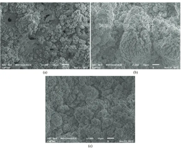
The morphology of the samples treated by PEO may be observed in Figure 4. Inspection of Figure 4(a) shows that a new structure forms on the porous matrix. As the treatment time increases this structure grows and covers the porous matrix. At a treatment time of 300 s, the structure grows to form of clusters with tridimensional columns. It may also
Figure 1. Current density as a function of treatment time.
Figure 2. SEM micrograph, of the section of the titanium samples treated by PEO for different periods of time: (a) 120 s with magniication
Antônio et al.
1430 Materials Research
be observed that the morphology of the layer is irregular, which explains the observed increase in roughness.
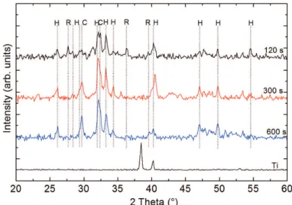
3.4. X-ray diffraction
The coatings produced by PEO, at a voltage of 480 V, on the titanium substrate are composed of HA, rutile and calcium phosphate (Ca2P2O7). The spectrum of the samples treated for 120 s, presented in Figure 5, has well-deined
peaks due to rutile and HA. Calcium phosphate is also detected, but the peaks have low intensities.
As the treatment time is increased the rutile phase disappears and the coating is composed only of HA and calcium phosphate. The peaks due to HA and calcium phosphate of the sample treated for 300 s have increased intensities compared to these of the spectrum of the sample treated for 120 s. Figure 4(a) shows that on the porous matrix a new cluster-like structure grows and as time passes the matrix is completely covered. Probably, the matrix bound to the titanium substrate is composed of rutile and the phase that covers the matrix is composed of HA and calcium phosphate. This interpretation is based on the observation that as the treatment time is increased rutile is no longer observed in the X-ray spectra. The irst layers formed are more susceptible to the presence of rutile, since rutile is formed by the oxidation of titanium from the substrate.
As the coatings grow and the supply of titanium for production of rutile is interrupted HA and calcium phosphate predominate. Hence the clusters formed in the porous matrix (Figure 4) are made of HA.
Note that there are no signiicant differences between the spectra of samples treated for 300 and 600 s, except for a slight reduction in the intensity of peaks due to HA. To obtain HA in the coating, therefore, a time of 300 s is suficient.
Figure 4. SEM micrograph, with magniication of 1000x, of titanium treated by PEO for different periods of time: (a) 120 s, (b) is 300
(c) 600 s.
Figure 3. Sample roughness of titanium treated by PEO in different
No peak due to titanium metal is observed in the spectrum of the coating, which demonstrates the complete coverage of the samples surface.
3.5. Cell viability
The cytocompatibility of the HA coated-titanium samples after 120s, 300s and 600s of coating time was assessed by MTT assay, which is based on the ability of viable cells to reduce yellow 3-(4, 5-dimethythiazol-2-yl)-2, 5-diphenyl tetrazolium bromide (MTT) by mitochondrial succinate dehydrogenase. Since osteoblasts are anchorage-dependent cells and only metabolically active osteoblasts can attach to a substrate, the MTT results were interpreted as a measure of cellular adhesion and proliferation.
Figure 6 shows that samples treated by PEO, regardless of coating time, allowed cells to adhere to the sample surface
after 24h of culture (p> 0.05). No statistical differences were found among the samples in comparison with tissue culture on untreated Ti, which were used as a control (p>0.05).
After 5 days of culture, it is observed in Figure 6 that all samples significantly stimulated cellular growth in relation to 24h of culture time (p<0.05). Among the HA coated samples, no statistically signiicant differences in cell densities were found (p>0.05); however, the amount of viable cells found in the samples coated for 600s was lower than those found in uncoated Ti samples (p<0.05).
It has been shown that coating of HA onto implantable surfaces enhances osteoblast affinity, improving cell adhesion and proliferation34,35. In the present study, however, these results were not confirmed. On the other hand, although cytocompatibility of the coated samples was not
Figure 5. XRD patterns of coating produced by plasma electrolytic oxidation for different times, where H – Hydroxyapatite (Ca5(PO4)3
(OH); C – Calcium Phosphate (Ca2P2O7) and R – Rutile (TiO2).
Figure 6. Cellular viability assay on specimens grown for growth times of 24 hours and 5 days. (The mean absorbance values and the
Antônio et al.
1432 Materials Research
impaired for cell adhesion and proliferation, it was not signiicantly improved.
Similar results were reported by Yeung et al.36, who assessed the cytocompatibility of PEO coated TiO2 with human osteosarcoma cells (MG-63s) and found that PEO coated samples had fewer viable cells in comparison with plasma-sprayed HA samples and TCPS. The same authors believe that cellular behavior may be inhibited by the speciic morphology or chemical composition of the PEO coating36.
For 5 days of cultivation, the sample treated for 600 s was the only that presented cellular growth signiicantly less than the untreated sample, which was used as a control to compare with the treated samples (p <0.01). The sample treated for 600 s presented detachment of the deposited material, due to smaller thickness than the sample treated for 300 s. The standard deviation of the roughness shows that the surface was strongly affected by exposure of the sample to a long treatment time. The chemical composition and surface topography may have reduced the coating stability, which promoted less cell growth.
In general we can say that the treated samples are cytocompatible and stimulate signiicant cellular growth.
4. Conclusion
In this study, coatings composed of HA, rutile and calcium phosphate were produced from a solution containing a source of Ca and P. The main results are given below.
Using PEO it was possible to produce a porous coating composed of HA, rutile and calcium phosphate, possessing a high surface roughness; the latter property is useful for biomedical applications.
In one step, with a treatment time of 120 s, it was possible to obtain a coating containing HA. The HA phases depend on the treatment time, but samples treated for 120 s already present interesting characteristics for biomedical applications.
The coatings produced at all treatment times studied are cytocompatible.
Acknowledgments
The authors thank the Brazilian agencies FAPESP and CNPq for inancial support of this study, and the company Conexão for supplying the samples.
References
1. Hanawa T and Ota M. Calcium phosphate naturally formed on titanium in electrolyte solution. Biomaterials. 1991; 12(8):767-774. http://dx.doi.org/10.1016/0142-9612(91)90028-9. PMid:1799652
2. Liu X, Poon RWY, Kwok SCH, Chu PK and Ding C. Structure and properties of Ca-plasma-implanted titanium. Surface and Coatings Technology. 2005; 191(1):43-48. http://dx.doi. org/10.1016/j.surfcoat.2004.08.118.
3. Hanawa T. In vivo metallic biomaterials and surface modification. Materials Science and Engineering A. 1999; 267(2):260-266. http://dx.doi.org/10.1016/S0921-5093(99)00101-X.
4. Ghomi H, Fathi MH and Edris H. Preparation of nanostructure hydroxyapatite scaffold for tissue engineering applications.
Journal of Sol-Gel Science and Technology. 2011; 58(3):642-650. http://dx.doi.org/10.1007/s10971-011-2439-2.
5. Yang CY, Wang BC, Lee TM, Chang E and Chang GL. Intramedullary implant of plasma-sprayed hydroxyapatite coating: an interface study. Journal of Biomedical Materials Research. 1997; 36(1):39-48. http://dx.doi.org/10.1002/ (SICI)1097-4636(199707)36:1<39::AID-JBM5>3.0.CO;2-M. PMid:9212387
6. Ong JL, Carnes DL and Bessho K. Evaluation of titanium plasma-sprayed and plasma-sprayed hydroxyapatite implants in vivo. Biomaterials. 2004; 25(19):4601-4606. http://dx.doi. org/10.1016/j.biomaterials.2003.11.053. PMid:15120505 7. Garcia-Alonso D, Parco M, Stokes J and Looney L.
Low-Energy Plasma Spray (LEPS) Deposition of Hydroxyapatite/ Poly-ε-Caprolactone Biocomposite Coatings. Journal of Thermal Spray Technology. 2012; 21(1):132-143. http://dx.doi. org/10.1007/s11666-011-9695-0.
8. Singh G, Singh S and Prakash S. Surface characterization of plasma sprayed pure and reinforced hydroxyapatite coating on Ti6Al4V alloy. Surface and Coatings Technology.
2011; 205(20):4814-4820. http://dx.doi.org/10.1016/j. surfcoat.2011.04.064.
9. Wang C, Chen Z, Guan L, Liu Z, Wang P, Zheng S, et al. Structural characterization of ion beam sputter deposited calcium phosphate coatings. Surface and Coatings Technology. 2000; 130(1):39-45. http://dx.doi.org/10.1016/S0257-8972(00)00705-2.
10. Bagratashvili VN, Antonov EN, Sobol EN, Popov VK and Howdle SM. Macroparticle distribution and chemical composition of laser deposited apatite coatings. Applied Physics Letters. 1995; 66(19):2451. http://dx.doi.org/10.1063/1.113992. 11. Lee J and Aoki H. Hydroxyapatite coating on Ti plate by a
dipping method. Bio-Medical Materials and Engineering. 1995; 5(2):49-58. PMid:7655318.
12. Li P, De Groot K and Kokubo T. Bioactive Ca10(PO4)6(OH)2− TiO2 composite coating prepared by sol-gel process. Journal of Sol-Gel Science and Technology. 1996; 7(1-2):27-34. http:// dx.doi.org/10.1007/BF00401880.
13. Choudhury P and Agrawal DC. Sol–gel derived hydroxyapatite coatings on titanium substrates. Surface and Coatings Technology. 2011; 206(2-3):360-365. http://dx.doi. org/10.1016/j.surfcoat.2011.07.031.
14. Büyükşaǧis A, Çiftçi N, Ergün Y and Kayali Y. The examination of corrosion behaviors of hap coated Ti implant materials and 316L SS by sol-gel method. Protection of Metals and Physical Chemistry of Surfaces. 2011; 47(5):670-679. http://dx.doi. org/10.1134/S2070205111050054.
15. Abdeltawab AA, Shoeib MA and Mohamed SG. Electrophoretic deposition of hydroxyapatite coatings on titanium from dimethylformamide suspensions. Surface and Coatings Technology. 2011; 206(1):43-50. http://dx.doi.org/10.1016/j. surfcoat.2011.06.034.
17. Kokubo T, Hata K, Nakamura T and Yamamuro T. Apatite formation on ceramics, metals, and polymers induced by a CaO, SiO2 based glass in a simulated body fluid. In: Bonfield W, Hastings GW and Tanner KE, editors. Bioceramics: Proceedings of the 4th International Symposium on Ceramics in Medicine; 1991; Oxford, London. Oxford: Butterworth-Heinemann; 1991. p. 113-120. v. 4.
18. Paz A, Martín Y, Pazos LM, Parodi MB, Ybarra GO and González JE. Obtención de recubrimientos de hidroxiapatita sobre titanio mediante el método biomimético. Revista de Metalurgia. 2011; 47(2):138-145. http://dx.doi.org/10.3989/ revmetalmadrid.1009.
19. Thair L, Ismaeel T, Ahmed B and Swadi AK. Development of apatite coatings on Ti–6Al–7Nb dental implants by biomimetic process and EPD: in vivo studies. Surface Engineering. 2011; 27(1):11-18. http://dx.doi.org/10.1179/174329409X439023. 20. García-Sanz FJ, Mayor MB, Arias JL, Pou J, León B and
Pérez-Amor M. Hydroxyapatite coatings: a comparative study between plasma-spray and pulsed laser deposition techniques.
Journal of Materials Science. Materials in Medicine. 1997; 8(12):861-865. http://dx.doi.org/10.1023/A:1018549720873. PMid:15348805
21. Sergo V, Sbaizero O and Clarke DR. Mechanical and chemical consequences of the residual stresses in plasma sprayed hydroxyapatite coatings. Biomaterial. 1997; 18(6):477-482. http://dx.doi.org/10.1016/S0142-9612(96)00147-0.
22. Nagase M, Nishiya H and Abe Y. The effect of crystallinity on hydroxyapatite-induced production of reactive oxygen metabolites by polymorphonuclear leukocytes. FEBS Letters. 1993; 325(3):247-250. http://dx.doi.org/10.1016/0014-5793(93)81082-B. PMid:8391480
23. Dunleavy CS, Golosnoy IO, Curran JA and Clyne TW. Characterisation of discharge events during plasma electrolytic oxidation. Surface and Coatings Technology. 2009; 203(22):3410-3419. http://dx.doi.org/10.1016/j. surfcoat.2009.05.004.
24. Tsui YC, Doyle C and Clyne TW. Plasma sprayed hydroxyapatite coatings on titanium substrates. Part 1: Mechanical properties and residual stress levels. Biomaterials. 1998; 19(22):2015-2029. http://dx.doi.org/10.1016/S0142-9612(98)00103-3. PMid:9870753
25. Tsui YC, Doyle C and Clyne TW. Plasma sprayed hydroxyapatite coatings on titanium substrates. Part 2: optimisation of coating properties. Biomaterials. 1998; 19(22):2031-2043. http:// dx.doi.org/10.1016/S0142-9612(98)00104-5. PMid:9870754 26. Curran JA and Clyne TW. Porosity in plasma electrolytic oxide
coatings. Acta Materialia. 2006; 54(7):1985-1993. http:// dx.doi.org/10.1016/j.actamat.2005.12.029.
27. Montazeri M, Dehghanian C, Shokouhfar M and Baradaran A. Investigation of the voltage and time effects on the formation
of hydroxyapatite-containing titania prepared by plasma electrolytic oxidation on Ti–6Al–4V alloy and its corrosion behavior. Applied Surface Science. 2011; 257(16):7268-7275. http://dx.doi.org/10.1016/j.apsusc.2011.03.103.
28. Rudnev VS, Morozova VP, Lukiyanchuk IV and Adigamova MV. Calcium-containing biocompatible oxide-phosphate coatings on titanium. Russian Journal of Applied Chemistry. 2010; 83(4):671-679. http://dx.doi.org/10.1134/ S107042721004018X.
29. Rudnev VS, Medkov MA, Yarovaya TP and Nedozorov PM. Calcium and strontium phosphates coatings on titanium formed by the plasma electrolytic oxidation. Russian Journal of Applied Chemistry. 2012; 85(12):1856-1860. http://dx.doi. org/10.1134/S1070427212120117.
30. Durdu S, Deniz ÖF, Kutbay I and Usta M. Characterization and formation of hydroxyapatite on Ti6Al4V coated by plasma electrolytic oxidation. Journal of Alloys and Compounds. 2013; 551:422-429. http://dx.doi.org/10.1016/j.jallcom.2012.11.024.
31. Cheng T, Chen Y and Nie X. Surface morphology manipulation and wear property of bioceramic oxide coatings on titanium alloy. Surface and Coatings Technology. 2013; 215:253-259. http://dx.doi.org/10.1016/j.surfcoat.2012.07.093.
32. Sun J, Han Y and Huang X. Hydroxyapatite coatings prepared by micro-arc oxidation in Ca- and P-containing electrolyte.
Surface and Coatings Technology. 2007; 201(9-11):5655-5658. http://dx.doi.org/10.1016/j.surfcoat.2006.07.052.
33. Declercq H, Van den Vreken N, De Maeyer E, Verbeeck R, Schacht E, De Ridder L, et al. Isolation, proliferation and differentiation of osteoblastic cells to study cell/biomaterial interactions: comparison of different isolation techniques and source. Biomaterials. 2004; 25(5):757-768. http://dx.doi. org/10.1016/S0142-9612(03)00580-5. PMid:14609664
34. Whiteside P, Matykina E, Gough JE, Skeldon P and Thompson GE. In vitro evaluation of cell proliferation and collagen synthesis on titanium following plasma electrolytic oxidation. Journal of Biomedical Materials Research. Part A. 2010; 94(1):38-46. http://dx.doi.org/10.1002/jbm.a.32664. PMid:20091708
35. Robinson HJ, Markaki AE, Collier CA and Clyne TW. Cell adhesion to plasma electrolytic oxidation (PEO) titania coatings, assessed using a centrifuging technique.
Journal of the Mechanical Behavior of Biomedical Materials. 2011; 4(8):2103-2112. http://dx.doi.org/10.1016/j. jmbbm.2011.07.009. PMid:22098910
36. Yeung WK, Reilly GC, Matthews A and Yerokhin A. In vitro biological response of plasma electrolytically oxidized and plasma-sprayed hydroxyapatite coatings on Ti-6Al-4V alloy.