35 Ureteric jet spectral Doppler waveform
Radiol Bras. 2010 Jan/Fev;43(1):35–38 Original Article • Artigo Original
Ureteric jet Doppler waveform: interobserver agreement.
A prospective study with asymptomatic children*
Formato da onda do jato ureteral ao estudo Doppler espectral: comparação interobservadores. Estudo prospectivo em crianças assintomáticas
Carolina Freitas Lins1, Gabriela Ferreira Lima2, Adonis Born Muniz Filho2, João Vicente Ribeiro Neto1, Silvio Cavalcanti de Albuquerque2, Eduardo Just da Costa e Silva3
OBJECTIVE: Early diagnosis of voiding dysfunction can minimize social and psychological repercussions and avoid renal lesions. The ureteric jet can be evaluated by color Doppler, and a good correlation has been observed between this method and patients’ clinical data in the diagnosis of voiding dysfunction. This study was aimed at evaluating the interobserver agreement in the assessment of the ureteral jet in asymptomatic children. MATERIALS AND METHODS: Interobserver agreement was prospectively evaluated. A total of 41 patients were sequentially evaluated by two medical sonographers. For each patient, three consecutive Doppler waveforms were obtained from each of the two ureteral jets. The number of peaks on each wave was observed and classified. The maximum velocity of the highest peak of each waveform was also observed. Kappa coefficients (κκκκκ) were calculated. RESULTS: Moderate interobserver agreement was observed (κκκκκ = 0.48; 95% confidence interval: 0.36–0.60). Most frequently a plateau pattern was observed for the ureteric jet. Maximum velocities measured by the two observers were respectively 32.37 cm/s and 35.63 cm/s. CONCLUSION: Moderate interobserver agreement was observed in the evaluation of the ureteric jet by means of color Doppler.
Keywords: Ureter; Urination; Children.
OBJETIVO: Diagnóstico precoce de distúrbios miccionais pode diminuir as repercussões sociais e psicológi-cas e evitar lesões renais. O jato ureteral pode ser avaliado por estudo Doppler, método que apresenta boa associação com dados clínicos dos pacientes no que diz respeito ao diagnóstico de disfunção miccional. O objetivo deste estudo é avaliar a concordância interobservadores entre os tipos de jato ureteral. MATERIAIS E MÉTODOS: Estudo prospectivo de concordância interobservadores. Um total de 41 pacientes foi examinado sequencialmente por dois médicos ultrassonografistas. Para cada paciente, três curvas dopplerfluxométricas foram obtidas de jatos consecutivos de cada ureter. O número de picos em cada curva foi observado e clas-sificado. A velocidade máxima do maior pico de cada onda foi observada. Coeficientes kappa (κκκκκ) foram
calcu-lados. RESULTADOS: A concordância interobservadores foi moderada (κκκκκ = 0,48; intervalo de confiança 95%:
0,36–0,60). O padrão platô foi o mais frequente. As velocidades máximas dos ureteres, medidas pelos dois observadores, foram de 32,37 cm/s e 35,63 cm/s, respectivamente. CONCLUSÃO: O exame das curvas dopplerfluxométricas do jato ureteral é método que demonstrou moderada concordância interobservadores.
Unitermos: Ureter; Micção; Crianças. Abstract
Resumo
* Study developed at Instituto de Medicina Integral Prof. Fer-nando Figueira, Recife, PE, Brazil.
1. MDs, Radiologists at Instituto de Medicina Integral Prof. Fernando Figueira, Recife, PE, Brazil.
2. Graduate students of Medicine, Faculdade Pernambucana de Saúde (FPS), Recife, PE, Brazil.
3. Master, Fellow PhD degree of Children’s and Teenagers’ Health, Universidade Federal de Pernambuco (UFPE), MD, Ra-diologist at Instituto de Medicina Integral Prof. Fernando Figueira, Preceptor of Imaging Anatomy at Faculdade Pernambucana de Saúde (FPS), Recife, PE, Brazil.
minimize the social and psychological re-percussions of urinary incontinence and avoiding renal injury with development of scarring and function loss(1).
The urodynamic study allows the con-firmation of the diagnosis of lower urinary tract dysfunction(1). By means of such study it is possible to evaluate the functions of bladder storage and emptying and the ac-tivity of the ureteral sphincter complex. However, the urodynamic assessment is not always available, besides the fact of being considered as an invasive procedure be-cause of the need of using urethral cathe-terism(1–3).
Lins CF, Lima GF, Born AMF, Ribeiro Neto JV, Albuquerque SC, Costa e Silva EJ. Ureteric jet Doppler waveform: interob-server agreement. A prospective study with asymptomatic children. Radiol Bras. 2010;43(1):35–38.
ner. The alterations in the function of the lower urinary tract may be divided into neu-rological (neurogenic bladder), most fre-quently resulting from spinal dysraphism and cerebral palsy; and functional, caused by disorders in children with no evidence of neurological diseases(1,2). The relevance of early diagnosis is related to the possibil-ity of establishing the treatment in order to
0100-3984 © Colégio Brasileiro de Radiologia e Diagnóstico por Imagem
Mailing address: Dr. Eduardo Just da Costa e Silva. Instituto de Medicina Integral Prof. Fernando Figueira – Radiologia. Rua dos Coelhos, 300, Boa Vista. Recife, PE, Brazil, 50070-550. E-mail: [email protected]
Received June 10, 2009. Accepted after revision November 9, 2009.
INTRODUCTION
man-36
Lins CF et al.
Radiol Bras. 2010 Jan/Fev;43(1):35–38 The ureteric jet is a phenomenon caused
by urine ejection into the vesical lumen by ureteral peristalsis(1,4,5). This phenomenon can be visualized with the use of color Doppler(6). Studies evaluating the ureteric jet by means of spectral Doppler demon-strate the presence of six waveform pat-terns as follows: monophasic, biphasic, triphasic, polyphasic, square and
continu-ous(2–4,6–9). As far as the ureteric jet is
con-cerned, the diagnosis of urinary diseases, including vesical dysfunctions, is highly associated with clinical data of the patients and those of Doppler
ultrasonogra-phy(2,4,5,8).
However, as the evaluation of the ure-teric jet is based on the observation of a spectral curve, difficulties in its interpreta-tion may be raised because of individual variations among investigators with re-gards to patterns classification caused by different understanding of what character-izes the different patterns.
The present study is aimed at evaluat-ing the interobserver agreement on differ-ent ureteric jet patterns in children without urinary complaints.
MATERIALS AND METHODS
The present prospective study was pre-viously approved by the Committee for Ethics in Research of the Institution. Free and informed consent was obtained from parents or guardians of the children.
The sample of the present study in-cluded children in the age range between 6 and 12 years referred to the unit of imag-ing diagnosis service of a pediatric school hospital to be submitted to ultrasonography because of complaints not related to the urinary tract. The sampling method was non casual and by convenience.
Patients whose examination was sched-uled for a certain time during the week were selected. Such sampling method was chosen for allowing the inclusion of pa-tients that were examined when the radi-ologists involved in the present study were present in the unit. The patients followed the standard recommendations for abdomi-nal ultrasonography. In the authors’ insti-tution, US studies are scheduled for spe-cific days in the week by order of arrival, with no preferential scheduling for any
specific time slot, so that, in principle, the sampling is not affected by any special characteristic that might contribute to a selection bias.
Patients with a previous history of uri-nary surgery or anatomic abnormalities likely to be detected at ultrasonography were excluded.
The sample size was arbitrarily defined, considering that data on the appropriate sample size for studies involving kappa statistics are scarce in the literature.
Variables
The waveform patterns were classified into six types according to Leung et al.(4,6). A monophasic jet is characterized by one peak. Biphasic and triphasic jets are char-acterized by two and three peaks, respec-tively. Four or more peaks characterize a polyphasic jet. A wave with a plateau for-mat with a duration of up to 20 seconds characterizes a square pattern, while a du-ration above 20 seconds characterizes a continuous jet(2).
Operationalization
The patients were sequentially exam-ined by two sonographers, one of them a specialist in clinical pediatrics and radiol-ogy, with a five-year experience in pediat-ric radiology. The other investigator had a three-year experience in general and pedi-atric ultrasonography. The patients were not stimulated to drink a larger than usual amount of liquids, being instructed to in-form when the voiding desire started, in order to initiate the study, avoiding the examination with a strong voiding desire. All the patients were evaluated in the supine position, with the bladder being ob-served in the transverse plane and identifi-cation of the ureteric jets with the aid of color Doppler. After obtaining the signal with the color Doppler, samples of the spectral curves were obtained for each ure-teric jet. The Doppler sample volume was sufficiently wide to comprise the whole ureteric jet and was positioned on the cen-ter of the jet which corresponded to the point of largest flow. The angle was limited to remain between 30° and 60°.
For each patient, three successive waves of the ureteric jets from each side were obtained. The number of peaks on each
wave was observed and classified, with the most frequent one being selected whenever a variation occurred among the three samples. The maximum peak velocity of the highest peak was also observed.
Statistical analysis
The kappa coefficient (κ) was calcu-lated to evaluate the interobserver agree-ment in relation to the results regarding wave types(10). Velocities were compared by means of the Students t-test. A signifi-cance level of 95% was adopted.
RESULTS
A total of 41 children (82 ureteric jets) were evaluated. In the cases of two chil-dren, the evaluation of one ureter was com-promised as these children became unco-operative before the end of the examina-tion. Thus, 80 ureteric jets were evaluated by one observer and 82 by the other, there-fore corresponding to 486 waveforms.
The mean examination time was not recorded; however extremely long or dif-ficult examinations were scarce.
The interobserver agreement was mod-erate (κ = 0.48; 95% confidence interval: 0.36–0.60).
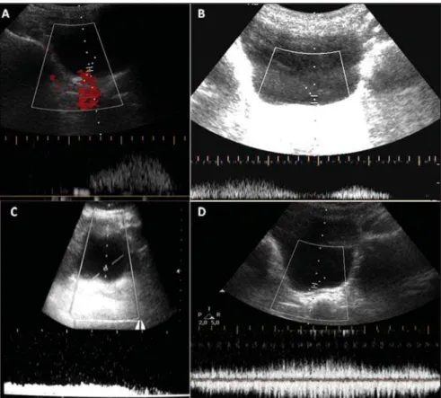
The most frequent jet pattern was the square type, visualized in 186 jets (38.2%). The other types were detected as follows: biphasic, 119 (24.4%); monophasic, 101 (20.7%); triphasic, 64 (13.1%); continuous, 10 (2%) and polyphasic, 6 (1.2%). No sta-tistically significant difference was ob-served between the two observers with re-spect to the patterns frequency (p = 0.41). Figure 1 shows the most common pat-terns in the study. One difficulty observed by the investigators was the differentiation between small oscillations on square jets and true peaks (Figure 1D).
The maximum jet velocities observed by the investigators were 32.37 cm/s and 35.63 cm/s, respectively, with no statisti-cally significant difference (p = 0.25). Also, statistically significant differences were not observed between maximum ve-locities observed in the right and left ure-ters either.
37 Ureteric jet spectral Doppler waveform
Radiol Bras. 2010 Jan/Fev;43(1):35–38
The relevance of the study of such pat-terns is the demonstration of a possible relation between the occurrence of the im-mature pattern and the presence of urinary tract diseases, such as vesicoureteral reflux, urinary infection and nocturnal enuresis(4,8), with the possibility that further studies bring a significant contribution to the un-derstanding of the physiopathology and diagnosis of these common conditions in children, which are still in need of a defined investigation strategy(13).
The introduction of new methods or patterns, either in the daily clinical practice or in research models, must always take into consideration the reproducibility of proposed findings, i.e., the capacity of ob-taining similar results when the examina-tion in a same patient, under the same physiological conditions is performed by different investigators or equipment. A common way of testing the imaging stud-ies performance is the evaluation of the interobserver agreement.
The moderate interobserver agreement in the evaluation of ureteric jet patterns ob-served in the present study must be cau-tiously evaluated, as there is no other study approaching this matter in the literature. In the author’s institution, such type of evalu-ation is not yet a part of the daily routine. A square like pattern may be easily con-fused with a polyphasic or triphasic pattern, considering that minimum variations in the wave’s height may be interpreted as peaks or not. This difficulty was experienced by the authors as the present study was devel-oped.
A great number of studies evaluating the patterns approached in the present study have been developed by a single group with a wide experience in this type of imaging study, which contributes to a better stan-dardization of findings interpretation. More strict parameters for the definition of a peak may be instrumental in reducing this varia-tion in a clinical or research practice. For example, a peak might be differentiated from a non significant variation in veloc-ity either by the variation or by its relation with the mean velocity. A longer learning curve would be another form of improving the interobserver agreement, contributing to the method application. Other possible explanation for the moderate interobserver
Figure 1. Common patterns in the study. On A, a well defined peak is noticed, characterizing a monophasic ureteric jet. The two peaks identified on B indicate a biphasic ureteric jet. On C, the ureteric jet corre-sponding to a flat waveform (plateau type). A difficulty encountered on waveforms such as the one shown on D. The variation in velocity may be interpreted as a simple oscillation, leading to a classification as a plateau type, or as distinct peaks, characterizing the ureteric jet as biphasic or triphasic.
DISCUSSION
The present results demonstrate a mod-erate interobserver agreement regarding the type of ureteric jet observed at the color Doppler study in children. No statis-tically significant difference was observed in the evaluation of the maximum jet ve-locity. The square pattern was the most fre-quently observed, followed by the biphasic pattern.
The identification of ureteric jets on im-aging studies such as gray scale ultrasonog-raphy and excretory urogultrasonog-raphy is common. The utilization of color Doppler facilitates such visualization, even in individuals un-der normal hydration conditions(11). Early studies have identified curve patterns at the spectral Doppler study, particularly charac-terized by the presence of peaks in variable numbers, or by continuous tracing without peaks(3,11). The classification proposed by Leung et al. comprises six different types of wave patterns, and has been the subject
38
Lins CF et al.
Radiol Bras. 2010 Jan/Fev;43(1):35–38 agreement, which is coherent with the
cur-rent level of knowledge about ureteric jets behavior, lies on the fact that the imaging studies were sequentially evaluated.
It is already known that the ureteric jets morphology vary as the bladder becomes full. So, the ureteric jets may have pre-sented variations over time, which may have contributed to the level of interob-server agreement. An effort was made to maintain the closest possible temporal proximity between the two investigators evaluations in order to minimize such an effect. The instruction to patients about not drinking more liquids than usual was an attempt to avoid rapid variations in the vesical volume. However this bias certainly could not be completely eliminated with this measure. Again, the absence of simi-lar studies evaluating the interobserver agreement on the patterns does not allow comparisons to be made. A proposal for a study that could eliminate this variable would be obtaining hard copies of the waveforms which would then be separately evaluated by two different investigators.
The predominance of the square pattern observed by the present study group is dif-ferent from reports in the literature(2). Some factors may have contributed for such a discrepancy. As previously described, the difficulty in characterizing a waveform may have easily biased many patterns of the triphasic or polyphasic types to a square type classification. Moreover, the patients were evaluated under physiological condi-tions, without being instructed to drink a larger than usual amount of liquids. In the previously mentioned studies, the patients were always instructed to drink large vol-umes of liquids. Considering that the pat-terns are known to be influenced by physi-ological conditions, this would be a plau-sible further explanation for the difference in patterns. Curiously, the square pattern is usually related to situations of forced diure-sis, which was not the case with the patients in the present study(6). However, the evalu-ation of pattern predominance was not the objective of the present study, so that fol-lowing the same method employed in other studies in the literature would not be strictly necessary, provided the two observers evalu-ated the patients under identical conditions.
The maximum velocities of the evalu-ated peaks did not present a significant interobserver variation, and are similar to those described in the literature(2).
The fact that 57% of the evaluations demonstrated variations of wave type in the three samples may indicate a limitation of the method, as more samples per ureter might be necessary to define the predomi-nant pattern. Such finding is similar to the one reported by Leung et al.(4). A proposed explanation for the variation would be the increase in urine volume in the bladder during the examination, affecting the cor-respondent waveform shape(6). This fact is particularly relevant, as a child presenting two patterns of the biphasic type and one pattern of the monophasic type would be considered as presenting a mature pattern. A new sample in this same ureter present-ing a monophasic pattern would be enough to change the classification to immature. It is possible that in future researches a higher number of samples may be defined in or-der to characterize the predominant pattern, making the evaluation less susceptible to ureteric jets occasionally observed out of the pattern. Another practical measure would be establishing maximum vesical volumes, above which the ureteric jets should not be evaluated.
The present study faced several limita-tions. Although they have previously been mentioned, it is convenient to review them in this topic. The fact that the ureteric jet evaluation by means of color Doppler is not routinely performed in the authors’ institu-tion was certainly one of them, since a greater experience with the method could improve the interobserver agreement. The sample size is another factor that that may have reduced eventual differences from a statistical point of view. There was no stan-dardization of the hydration level of the children, which may have contributed to the occurrence of patterns that are less fre-quently found in children. Further studies should evaluate the average time spent in the performance of the examinations, to better define its feasibility in the daily prac-tice.
In conclusion, the study of wave pat-terns of ureteric jets by means of color Dop-pler is a method that demonstrated a
mod-erate interobserver agreement. A better definition of the parameters for differentia-tion between waveform types and a longer learning curve may be useful for the appli-cation of the method. The observed varia-tion between wave patterns obtained from the same ureter in different samples, indi-cates a possible necessity of a higher num-ber of waveforms per ureter for the defini-tion of the predominant type. Further stud-ies including the evaluation of hard copstud-ies of the waveforms by different observers may prove to be useful.
REFERENCES
1. Fonseca EM, Monteiro LM. Clinical diagnosis of bladder dysfunction in enuretic children and ado-lescents. J Pediatr (Rio J). 2004;80:147–53.
2. Leung VY, Chu WC, Yeung CK, et al. Ureteric jet Doppler waveform and bladder wall thickness in children with nocturnal enuresis. Pediatr Res. 2006;60:582–6.
3. Cox IH, Erickson SJ, Foley WD, et al. Ureteric jets: evaluation of normal flow dynamics with color Doppler sonography. AJR Am J Roentgenol. 1992;158:1051–5.
4. Leung VY, Metreweli C, Yeung CK. The ureteric jet doppler waveform as an indicator of vesico-ureteric sphincter function in adults and children. An observational study. Ultrasound Med Biol. 2002;28:865–72.
5. Sakate M, Teixeira AS, Sakate ATY, et al. Study of the ureterovesical jet by means of color Dop-pler in patients with and without vesicoureteral reflux. Radiol Bras. 2006;39:425–8.
6. Leung VY, Chu WC, Yeung CK, et al. Doppler waveforms of the ureteric jet: an overview and implications for the presence of a functional sphincter at the vesicoureteric junction. Pediatr Radiol. 2007;37:417–25.
7. Leung VY, Metreweli C. Doppler waveform of the ureteric jet in pregnancy. Ultrasound Med Biol. 2002;28:879–84.
8. Leung VY, Metreweli C, Yeung CK. Immature ureteric jet doppler patterns and urinary tract in-fection and vesicoureteric reflux in children. Ul-trasound Med Biol. 2002;28:873–8.
9. Leung VY, Metreweli C, Yeung CK, et al. Ure-teric jet in the anaesthetised child. Ultrasound Med Biol. 2003;29:1237–40.
10. Kundel HL, Polansky M. Measurement of ob-server agreement. Radiology. 2003;228:303–8. 11. Jequier S, Paltiel H, Lafortune M. Ureterovesical
jets in infants and children: duplex and color Doppler US studies. Radiology. 1990;175:349– 53.
12. Leung VY, Chu WC, Yeung CK, et al. Gender difference in achieving rate of maturity of the vesicoureteric junction. Pediatr Radiol. 2007;37: 189–93.