Case 11325
Uncommon pelvic mass: GIST mimicking adnexal tumour
Mariana Horta , Teresa Margarida Cunha , Joana Ferreira , Paula Chaves 1 2 3 3
Genital (Female) Imaging Section:
2013, Nov. 22 Published:
66 year(s), female Patient:
Authors' Institution
[1] Serviço de Radiologia, Centro Hospitalar Lisboa Ocidental, EPE, Lisboa; Email: mariana_horta@hotmail.com
[2] Serviço de Radiologia, Instituto Português de Oncologia de Lisboa Francisco Gentil, EPE, Lisboa, Portugal
[3] Serviço de Anatomia Patológica, Instituto Português de Oncologia de Lisboa Francisco Gentil, EPE, Lisboa, Portugal
Clinical History
A 66-year-old woman was referred to our Institute to define a sonographic finding of a pelvic mass. She presented with a history of pelvic pain, asthenia and weight loss. Gynaecological examination : Upper deflection of the cervix by a compressive lesion in the posterior cul-de-sac. CA125= normal.
Imaging Findings
The patient underwent a pelvic MR that showed a large, lobulated tumour localized in the pouch of Douglas, measuring 15, 4x10, 6x11, 7cm.
It did not invade the uterus or the sigmoid colon and it was in close relation with the intestinal loops. The ovaries were not visualized.
T1W1 were hyperintense on T2WI and did not enhance, which suggests hemorrhagic and necrotic nature.
The patient underwent a small bowel and rectal partial resection with total hysterectomy and bilateral anexectomy. The resected specimen contained, in a paraintestinal location, a 12cm partially cystic and necrotic nodular tumour.
The histopathologic diagnosis was small bowel malignant GIST.
Discussion
Gastrointestinal stromal tumours (GISTs) are mesenchymal tumours which account for less than 1% of all gastrointestinal tumours [1, 2]. They can occur throughout the GI tract, mesentery, omentum and retroperitonuem, with 20-30% arising in the small bowel [3, 4].
They range from benign tumours to aggressive tumours (10-30%) [3].
The majority of GISTs presents at ages above 40 years. Gastrointestinal hemorrhage due to mucosal ulceration is a frequent symptom. Abdominal pain, vomiting, weight loss and intestinal obstruction can also occur [4].
Extraluminal and eccentric growth is frequently encountered in malignant small intestine GISTs, which can encase non contiguous segments [2, 4].
The solid component of GISTs is typically hypointense on T1WI and shows intermediate/high signal intensity on T2WI, enhancing avidly following the administration of gadolinium. Signal intensity of focal hemorrhagic areas varies according to the age of hemorrhage [1, 2, 5]. Small bowel GIST's 5-year survival rate is 40-50% [1].
Female pelvic masses commonly arise from reproductive organs, however non gynaecological lesions originating from urinary and gastrointestinal systems, from the peritoneum, retroperitoneum and from adjacent soft tissues are important pitfalls that radiologists and gynaecologists must take into account [1].
Identification of the location of the mass (intra or extraperitoneal) is based on anatomic landmarks and should also take into account the patient's age, clinical and gynaecological history [1].
MRI is the modality of choice to evaluate US and CT indeterminate pelvic masses and has capability to differentiate benign from malignant lesions [1, 6].
In our case MR was performed to characterize an indeterminate adnexal mass that revealed to be of gastrointestinal origin.
Despite being an uncommon pelvic mass, GISTs must be considered in the differential diagnosis of large adnexal masses, since they may have similar imaging characteristics to ovarian cancer and its therapy and prognosis are very different.
Final Diagnosis
Small bowel malignant GIST
Differential Diagnosis List
Figures
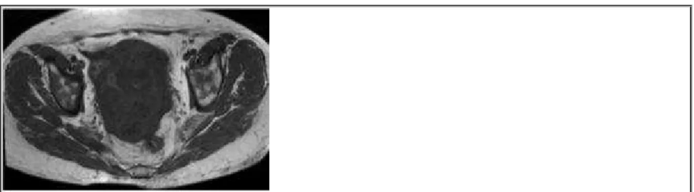
Figure 1 Axial non-enhanced T1-weighted MR image
Axial non-enhanced T1-weighted MR image demonstrates a heterogeneous lobulated pelvic
mass in the pouch of Douglas predominantly hypointense with focal areas of intermediate
signal and of remarkably hypointense signal.
© Caldeira JP, Department of Radiology,Instituto Português de Oncologia de Lisboa Francisco Gentil, Lisboa, Portugal
Area of Interest: Genital / Reproductive system female;
Imaging Technique: MR;
Procedure: Diagnostic procedure;
Special Focus: Neoplasia;
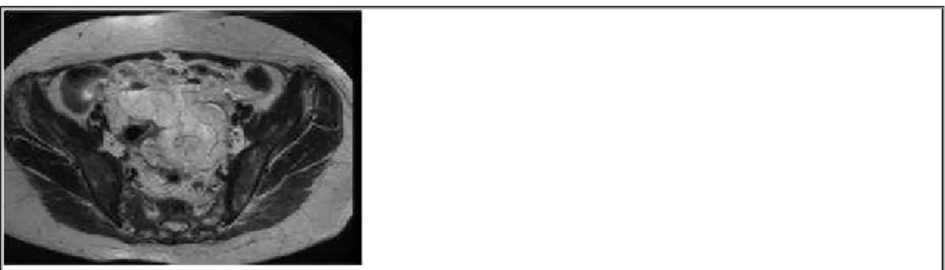
Figure 2 Axial T2- weighted MR image
Axial T2- weighted MR image shows a hyperintense mass with areas of strong hyperintense
signal.
© Caldeira JP, Department of Radiology,Instituto Português de Oncologia de Lisboa Francisco Gentil, Lisbon, Portugal
Area of Interest: Genital / Reproductive system female;
Imaging Technique: MR;
Procedure: Diagnostic procedure;
Special Focus: Neoplasia;
Tumour areas of hypointensity on T1W1 correspond to areas of hyperintensity on T2WI.
Tumour areas of intermediate signal on T1W1 correspond to areas of strong hypersignal on
T2WI.
© Caldeira JP, Department of Radiology,Instituto Português de Oncologia de Lisboa Francisco Gentil, Lisbon, Portugal
Area of Interest: Genital / Reproductive system female;
Imaging Technique: MR;
Procedure: Diagnostic procedure;
Special Focus: Neoplasia;
Figure 4 Axial non-enhanced T2-weighted MR image
Tumour areas of hypointensity on T1W1 correspond to areas of hyperintensity on T2WI.
Tumour areas of intermediate signal on T1W1 correspond to areas of strong hypersignal on
T2WI.
© Caldeira JP, Department of Radiology,Instituto Português de Oncologia de Lisboa Francisco Gentil, Lisbon, Portugal
Area of Interest: Genital / Reproductive system female;
Imaging Technique: MR;
Procedure: Diagnostic procedure;
Special Focus: Neoplasia;
Figure 5 Sagittal T2-weighted image
sigmoid colon posteriorly.
© Caldeira JP, Department of Radiology,Instituto Português de Oncologia de Lisboa Francisco Gentil, Lisbon, Portugal
Area of Interest: Genital / Reproductive system female;
Imaging Technique: MR;
Procedure: Diagnostic procedure;
Special Focus: Neoplasia;
Figure 6 Sagittal fat-supressed gadolinium-enhanced T1-weighted image
Sagittal fat-supressed gadolinium-enhanced T1-weighted image show avid contrast uptake
by the solid component of the tumour.
© Caldeira JP, Department of Radiology,Instituto Português de Oncologia de Lisboa Francisco Gentil, Lisbon, Portugal
Area of Interest: Genital / Reproductive system female;
Imaging Technique: MR;
Procedure: Diagnostic procedure;
Special Focus: Neoplasia;
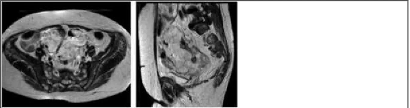
Figure 7 a) Axial T2-weighted image. b) Sagittal T2-weighted image.
Axial T2-weighted image. b) Sagittal T2-weighted image. The tumour was in close relation
with intestinal loops (arrow) , that crossed the tumour .
© Caldeira JP, Department of Radiology,Instituto Português de Oncologia de Lisboa Francisco Gentil, Lisbon, Portugal
Area of Interest: Genital / Reproductive system female;
Imaging Technique: MR;
Procedure: Diagnostic procedure;
Special Focus: Neoplasia;
The tumour shows restricted diffusion.
© Caldeira JP, Department of Radiology,Instituto Português de Oncologia de Lisboa Francisco Gentil, Lisbon, Portugal
Area of Interest: Genital / Reproductive system female;
Imaging Technique: MR;
Procedure: Diagnostic procedure;
Special Focus: Neoplasia;
Figure 9 Tumour
The tumor showed short intersecting fascicles of fusiform, pleomorphic cells with frequent
mitotic figures (>10 mitosis/50HPF) (islet, H-E, 400x). (H-E, 200x).
© Chaves P, Department of Pathology, Instituto Português de Oncologia de Lisboa Francisco Gentil, Lisbon, Portugal
Area of Interest: Genital / Reproductive system female;
Imaging Technique: Experimental;
Procedure: Diagnostic procedure;
Special Focus: Neoplasia;
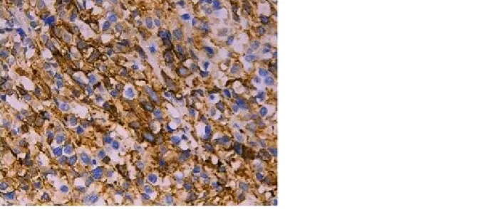
The neoplastic cells were positive for caldesmon
© Chaves P,Department of Patholgy, Instituto Português de Oncologia de Lisboa Francisco Gentil, Lisbon, Portugal
Area of Interest: Genital / Reproductive system female;
Imaging Technique: Experimental;
Procedure: Diagnostic procedure;
Special Focus: Neoplasia;
Figure 11 Neoplastic cells
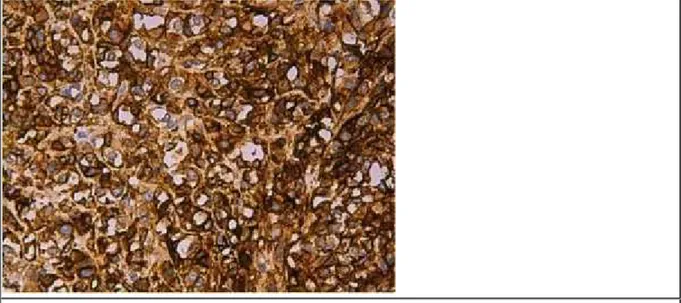
The neoplastic cells were positive for CD117
© Chaves P, Department of Pathology, Instituto Português de Oncologia de Lisboa Francisco Gentil, Lisbon, Portugal
Area of Interest: Genital / Reproductive system female;
Imaging Technique: Experimental;
Procedure: Diagnostic procedure;
Special Focus: Neoplasia;
The neoplastic cells were positive for DOG1
© Chaves P. Department of Pathology. Instituto Português de Oncologia de Lisboa Francisco Gentil, Lisbon, Portugal
Area of Interest: Genital / Reproductive system female;
Imaging Technique: Experimental;
Procedure: Diagnostic procedure;
Special Focus: Neoplasia;
Figure 13 Neoplastic cells
The neoplastic cells were negative for muscle markers namely actin and desmin
© Chaves P. Department of Pathology.Instituto Português de Oncologia de Lisboa Francisco Gentil, Lisbon, Portugal
Area of Interest: Genital / Reproductive system female;
Imaging Technique: Experimental;
Procedure: Diagnostic procedure;
Special Focus: Neoplasia;
References
Large Pelvic Masses Radiographics 23:403-424
[2] Mass Elli G, Colaiacomo MC, Marcelli G, Bertini L, Casciani E, d'Amici P, Caprasecca S, Polettini E, Gualdi G. (2012) MRI of small bowel: how to differentiate primary neoplasms and mimickers Br J Radiol 85(1014):824-37
[3] Webb WR, Brant W, Major N. (2006) Fundamentals of body CT Elsevier Health Sciences
[4] Levy A, Remotti H, Thompson W, Sobin L, Miettinen M (2003) Gastrointestinal Stromal Tumours: Radiologic Features with Pathologic Correlation Radiographics 23:283-304
[5] Caramella T, Schmidt S, Chevallier P, Saint Paul M, Bernard JL, Bidoli R, Bruneton JN (2005) MR Features of gastrointestinal stromal tumours Journal of Clinical Imaging 251-254
[6] Spencer J, Forstner R, Cunha TM (2010) ESUR guidelines for MR imaging of the sonographically indeterminate adnexal mass: an algorithmic approach. Eur Radiol 20: 25-35
Citation
Mariana Horta , Teresa Margarida Cunha , Joana Ferreira , Paula Chaves (2013, Nov. 22) 1 2 3 3 Uncommon pelvic mass: GIST mimicking adnexal tumour {Online}