IX
Radiol Bras 2007;40(3):IX–X
WHICH IS YOUR DIAGNOSIS?
Rafael Burgomeister Lourenço1, Marcelo Bordalo Rodrigues2
1. MD, Resident at Institute of Radiology, Hospital das Clínicas da Faculdade de Medicina da Universidade de São Paulo (InRad/HC-FMUSP), São Paulo, SP, Brazil. 2. Director for the Service of Radiology at Institute of Orthopedics and Traumatology, Hospital das Clínicas da Faculdade de Medicina da Universidade de São Paulo (IOT/ HC-FMUSP), São Paulo, SP, Brazil. Mailing address: Dr. Marcelo Bordalo Rodrigues. Avenida Doutor Enéas de Carvalho Aguiar, 255. São Paulo, SP, Brazil, 05403-001. E-mail: [email protected]
A female, 45-year-old patient complaining of pain on the anterior aspect of the left knee for about five years. At presentation, the patient reported a history of direct local trauma eight months ago, with worsening of the pain, swelling, and restricted movement amplitude. She denied previous surgical interventions or other relevant antecedents. At clinical examination she presented swelling in the infrapatellar region, tender to palpation and incomplete flexo-extension.
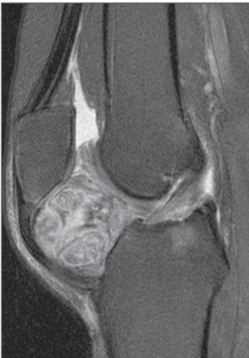
Figure 1. Lateral view plain radiograph. Figure 2. Sagittal proton density magnetic resonance image.
Figure 3. Sagittal T2-weighted magnetic resonance image with fat saturation.
X Radiol Bras 2007;40(3):IX–X
Imaging findings
Figure 1: Calcified mass with bone ma-trix on the infrapatellar fat pad.
Figure 2: Heterogeneous, solid expan-sive formation in the Hoffa’s fat pad.
Figure 3: Areas of high signal inten-sity in the middle of the lesion suggest-ing a chondroid component. Also, joint effusion and patellar tendinopathy can be observed. Free intra-articular bodies were not identified.
Figure 4: Lesion with heterogeneous impregnation and signs of synovitis.
Diagnosis: Chondroma of the Hoffa’s fat pad as an end-stage Hoffa’s disease.
COMMENTS
Hoffa’s disease (or infrapatellar fat pad syndrome) was first described by Albert Hoffa in 1904(1), and is character-ized by the development of chronic in-flammatory alterations associated with the impingement of the infrapatellar fat pad between the femorotibial and femoropatellar spaces. The process may be caused by a major acute (direct or in-direct) trauma or by chronic repetitive traumas, resulting in hemorrhage and edema in the infrapatellar fat pad. The resulting swelling predisposes to im-pingement and impact on the infrapatel-lar fat pad, accentuating local inflamma-tory alterations, fat pad hypertrophy and further injury and inflammation. The progress of chronic inflammation results in adipocytes fibrosis and fibroblast pro-liferation with marked fibrosis(2). Chon-droid metaplasia may eventually occur, and endochondral ossification is rare, resulting in the development of true os-teochondromas just like in the present case. Primary mechanical changes, such as genu recurvatum, rotational micro-instability or other conditions affecting the femorotibial and femoropatellar spaces contribute to the process and may result in primary impact, progressing to chronic entrapment with a similar appear-ance, in the absence of a triggering, trau-matic factor, this entity being character-ized as Hoffa’s syndrome(3).
In the literature, there are some case reports and series about patients
pre-senting with history, symptoms, radio-logical and anatomopathoradio-logical find-ings similar to those described for Hoffa’s disease, and characterized as “para-articu-lar” or “intracapsu“para-articu-lar” chondromas(4–7). Because of the similarity between these processes, some authors(8), have consid-ered that both entities are related, and that these chondromas, in truth, are more than simple areas of osteocartilaginous change or heterotopic ossification ran-domly developing within the infrapatel-lar fat pad, as a matter of fact represent-ing an end-stage Hoffa’s disease.
Main clinical symptoms are pain on the anterior aspect of the knee, and re-duced movement amplitude. The typical clinical finding is hypertrophied, hard-ened and painful fat tissue (Hoffa’s sign – pain at knee extension with digital com-pression over the infrapatellar fat pad).
The radiographic diagnosis is based on the direct identification of osteochon-droma and only can be achieved late in time, after ossification. Likewise, scintig-raphy is effective only during the active phase of the ossifying process(7). Mag-netic resonance imaging allows evaluat-ing the presence of inflammatory alter-ations both at acute and chronic phases of the disease. At the acute phase, ede-mas can be identified as areas of low-in-tensity signal on T1-weighted images and increased signal intensity on T2-weighted images. Also, areas with ex-tremely low signal intensity may be dem-onstrated in both sequences as a result of hemorrhage with hemosiderin depo-sition. At the chronic phase of the dis-ease, fibrous scarring tissues may be identified as irregular areas with low sig-nal intensity on T1- and T2-weighted images. Chondroid matrix may be identi-fied as nodular areas with increased sig-nal intensity on T2-weighted images. In case of endochondral ossification, areas with increased signal intensity may be identified within the lesion on T1-weighted images, representing prolifera-tion of medullary adipocytes. Regardless the phase of the disease, occurrence of contrast impregnation is frequent as a result of local inflammatory alterations.
Differential diagnosis at early phases
of the disease include infrapatellar plica syndrome. When signal abnormalities follow the course of the infrapatellar plica (a line extending from the inter-condylean sulcus to the retropatellar fat tissue, par-allel to the anterior cruciate ligament), most probably the diagnosis is plica syn-drome. More diffuse abnormalities affect-ing the whole posterior aspect of the infrapatellar fat pad indicate the diagno-sis of Hoffa’s disease(9).
Other differential diagnoses to be considered are: synovial chondromato-sis, pigmented villonodular synovitis, calcinosis, osteochondrosis, calcareous tendinopathy, femur, tibia or even patella tumors with infrapatellar fat tissue inva-sion, and, even though extremely rare, primary chondrosarcomas of the Hoffa’s fat pad.
The primary therapeutic approach should be clinical. Refractory cases re-quire surgical approach for lesion resec-tion, both the open(4–7) and the arthro-scopic(2,8,10) approaches being feasible. In most of cases, resection results in symp-toms resolution, without recurrence of the lesion.
REFERENCES
1. Hoffa A. Influence of adipose tissue with regard to the pathology of the knee joint. JAMA 1904;43: 795–796.
2. Magi M, Branca A, Bucca C, Langerame V. Hoffa disease. Ital J Orthop Traumatol 1991;17:211–216. 3. Jacobson JA, Lenchik L, Ruhoy MK, Schweitzer ME, Resnick D. MR imaging of the infrapatellar fat pad of Hoffa. RadioGraphics 1997;17:675–691. 4. Mosher JF, Kettelkamp DB, Campbell CJ. Intra-capsular or para-articular chondroma: a report of three cases. J Bone Joint Surg Am 1966;48:1561– 1569.
5. Milgram JW, Dunn EJ. Para-articular chondro-mas and osteochondrochondro-mas: a report of three ca-ses. Clin Orthop Relat Res 1980;148:147–151. 6. Smillie IS. Lesions of the infrapatellar fat pad and synovial fringes: Hoffa’s disease. Acta Orthop Scand 1963;33:371–377.
7. Böstman O, Karaharju E, Heikkonen L, Holms-tröm T. Extraskeletal ossifying chondroma in the knee: a case report. Acta Orthop Scand 1985;56: 87–89.
8. Krebs VE, Parker RD. Arthroscopic resection of an extrasynovial ossifying chondroma of the in-frapatellar fat pad: end-stage Hoffa’s disease? Ar-throscopy 1994;10:301–304.