OR
IGI
N
A
L
R
E
S
E
A
R
C
H
Corresponding address: Adriana Clemente Mendonça – Universidade Federal do Triângulo Mineiro – Rua da Constituição, 1009, sala 10, Abadia – Uberaba (MG), Brasil – Zip Code: 38026-280 – Phone: +55 (34) 3318-5524 – Email: [email protected] – Funding Source: Nothing to declare – Conlict of interest: Nothing to declare – Presentation: Sept. 2016 – Accepted for publication: Feb. 2017 – This study was approved by the Research Ethics Committee of Universidade Federal do Triângulo Mineiro under Opinion No. 785382. 1Post graduate in Dermato-Functional Physical Therapy by Universidade Federal do Triângulo Mineiro (UFTM) – Uberaba (MG), Brazil. 2Graduate in Physical Therapy by Universidade Federal do Triângulo Mineiro (UFTM) – Uberaba (MG), Brazil.
3Phd, Professor of the Physical Therapy course and Coordinator of the Post graduate course in Dermato-Functional Physical Therapy
of Universidade Federal do Triângulo Mineiro (UFTM) – Uberaba (MG), Brazil.
ABSTRACT | The cellulitis is a disorder of dermal and subcutaneous tissues. It afects 90% of women after puberty. Several predisposing factors interfere with its occurrence such as postural impairments. Photogrammetry has been considered reliable, easy to use, reproducible and with low cost to assess posture and cellulitis. It was evaluated if there is correlation between pelvic alignment and cellulitis in the gluteal region by photogrammetry. This is an observational cross-sectional study with 46 women (18-29 years old, mean age 23 years ± 4.2). Posture was evaluated by the software SAPO and the angle between the right and left anterior superior iliac spineswas calculated. Cellulitis was classiied according to Hexsel’s photonumeric scale. Analyses were performed by two independent and blinded evaluators and Pearson correlation tests investigated possible relationships between posture and cellulitis. The average value of the pelvic disalignment angle was 1.70 (+1.46), the mean value of the right gluteus cellulitis was 6.30 (+4.01) and left 6.76 (+3.61). There was no correlation between pelvic alignment and the level of cellulitis in the gluteal region (p = 0.38).
Keywords | Posture; Cellulitis.
RESUMO | O ibroedema geloide (FEG) é uma desordem dos tecidos dérmico e subcutâneo. Acomete 90% das mulheres após a puberdade. Vários fatores predisponentes interferem na sua ocorrência, entre os quais as alterações posturais. Para avaliação da postura e do FEG, a fotogrametria tem se mostrado um recurso coniável, de fácil aplicação, reprodução e de baixo custo. Avaliou-se Avaliou-se há correlação entre alinhamento pélvico e FEG na
40
região glútea por meio da fotogrametria. Estudo do tipo transversal observacional, com 46 mulheres de 18-29 anos (média 23 anos±4,2 anos). Foi realizada avaliação postural pelo software de avaliação postural (SAPO) e calculado o ângulo entre as espinhas ilíacasanterossuperiores direita e esquerda. O FEG foi classiicado de acordo com a escala fotonumérica de Hexsel. As análises foram realizadas por dois avaliadores independentes e cegos, e foi utilizado o teste de correlação de Pearson. Obteve-se como resultado o valor médio do ângulo de desalinhamento pélvico de 1,70 (±1,46), o valor médio do FEG no glúteo direito de6,30 (±4,01) e no esquerdo de 6,76 (±3,61). Não foi observada correlação entre o alinhamento pélvico e o grau de FEG na região glútea (p=0,38).
Descritores | Postura; Celulite.
RESUMEN | La dermopaniculosis vasculopática (DV) es un trastorno inlamatorio de los tejidos adiposo y subcutáneo. Un 90% de la población femenina presenta este trastorno tras la pubertad. Hay diversos factores que pueden desencadenar su aparición, entre ellos se destacan las alteraciones posturales. Un recurso iable, de fácil aplicación, reproducción y bajo costo para evaluar la postura y la DV es la fotogrametría. En este estudio se pretende evaluar la presencia de correlación entre la alienación de la pelvis y la DV en la región glútea a través de la fotogrametría. Se trata de estudio de tipo transversal observacional, del cual participaron 46 mujeres de 18 a 29 años (promedio de 23 años ± 4,2 años). Se realizó la evaluación de la postura por intermedio del software de evaluación postural (Sapo) y se calculó el ángulo entre las
Correlation between pelvic alignment and cellulitis
Correlação entre alinhamento pélvico e ibroedema geloide
Correlación entre la alienación de la pelvis y la dermopaniculosis vasculopática
Juliane Moreira Naves1, Camila Soares1, Vanessa de Almeida Svezzia2, Franciele Daiane Cussolim2,
espinas ilíacas anterosuperiores derecha e izquierda. La DV se clasiicó según la escala fotonumérica Hexsel. Los análisis fueron realizados por dos evaluadores independientes y aleatorios, en los cuales se utilizó de la prueba de correlación de Pearson. Como resultado el promedio del valor del ángulo de desalienación de
la pelvis fue de 1,70 (±1,46), el promedio del valor de la DV en el glúteo derecho fue de 6,30 (±4,01) y en el izquierdo 6,76 (±3,61). No se observó correlación entre la alienación de la pelvis y el grado de la DV en la región glútea (p=0,38).
Palabras clave | Postura; Celulitis.
INTRODUCTION
Cellulitis is a dermal and subcutaneous tissue disease that causes lipodystrophic changes with sclerosing response, which result in an unesthetic aspect. Its prevalence is estimated at around 80-90%; it may cause emotional distress and is a frequent complaint of patients1.
his process culminates in the accumulation of edema and later ibrotic nodules, which will favor the formation of cutaneous reliefs and possible painful sensation, mainly in the area of the buttocks, thighs and abdomen2,3.
he emergence of cellulite is characterized by wavy and irregular appearance of the skin, due to the protrusion of fat in the dermo-hypodermic interface4.
he female gender presents vertical fascial bands, and it is believed that cellulitis is a result of stretching of these bands, which are genetically determined5. he
stretching weakens and thins out the base of the dermal connective tissue, allowing the protrusion of fat in the dermo-hypodermic interface, causing the appearance of wavy and irregular skin6. he herniations of fat in
the dermis are a characteristic of female anatomy7.
Subcutaneous tissue in men, on the other hand, is characterized by horizontal and diagonal fascial bands, preventing herniation of fat8.
Cellulitis modiies the histological structure of the skin, altering the connective tissue, which results in increased retention of water, sodium and potassium, leading to increased interstitial pressure, compression of veins, lymphatic vessels and nerves, which generates a vicious cycle9.
Among the predisposing or aggravating factors of cellulitis, we can cite: female gender, race (more common in Caucasian), increase of the fat tissue, age, genetic and familial predisposition, psychosomatic factors, pregnancy, intestinal dysfunctions, external compressions, smoking, medications, postural changes, among others5,8-10.
he classiication criteria are variables, according to histopathological changes, macroscopic appearance of
the skin and its clinical form. All criteria are divided into levels according to the degree of commitment of the analyzed features11,12.
Several methods to evaluate cellulitis have been studied and described in the literature, but many of these do not apply due to high cost of the equipment. Amid these, photogrammetry is the most commonly used resource, because it is capable of reproduction, precision and low cost13. he photographic record has
long been used for documentation and comparison of plastic surgery results, repair surgeries, postural assessments, among others14-18.
he postural evaluation software (SAPO) is a computer program for quantitative analysis, developed by a team who attended clinical and methodological issues19. he program requires evaluators’ training and
voluntary acceptance to prolonged orthostasis. It is simple and free, generates linear measures, measurement of distances and body angles20.
Despite its easy reproducibility and repeatability, attested by several studies15,19,21, there is still a dearth of
literature in dermato-functional physical therapy. Some authors11,14 report diiculty in obtaining photographs
of architecture and tonicity of the skin, using resources such as gluteal contraction to assist in the assessment.
Although many authors cite postural changes as a predisposing factor of cellulitis, no studies in the literature have identiied and demonstrated such a relationship, thus the need to investigate it. he muscle fascia are diferent according to the region of the human body, presenting more slender in the abdomen and thicker and with greater amount of dense connective tissue in the glutei and hips22.
correlation between pelvic alignment and cellulitis in the gluteal region.
METHODOLOGY
A cross-sectional observational study with blinding of evaluators. Sample was composed of 46 female students of Universidade Federal do Triângulo Mineiro (UFTM) aged between 18 and 29 years old, 23 years old average (± 4.2 years). Inclusion criteria were: female gender and age (18-29 years). Pregnant women and volunteers who presented postural hypotension would be excluded from the study.
In the evaluation, the volunteers responded to a questionnaire in physical therapy with their personal data (name, age, gender, occupation, telephone), followed by a comprehensive assessment with SAPO. All of them signed an informed consent form. his study was approved by the Research Ethics Committee of UFTM with opinion no. 785382.
Photographic records were performed by a single examiner, previously trained in well-lit environment, with no relective background. he participants adopted the orthostatic position with bathing suit, tied hair, barefoot and without any kind of accessory. All anatomical points of SAPO protocol were previously palpated for the marking with a dermatograph pencil. Subsequently, styrofoam espheres were ixed with a double-sided adhesive tape on the skin, which was previously cleaned with rubbing alcohol to reduce skin impedance with the adhesive.
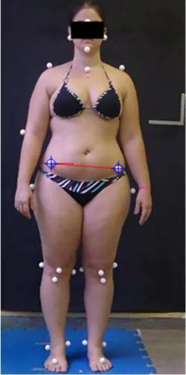
hirty-eight points were marked, with main focus on two: left anterior superior iliac spine and right anterior superior iliac spine, from where the pelvic tilt angle in the frontal plane was obtained (Figure 1). he participants were placed in a previously marked place, with a 3,5 m distance from the camera and 50 cm from the wall. To ensure and secure the distance, between the participant and the wall was used a blue-colored ethyl-vinyl-acetate (EVA) mat, 100 cm wide, 50 cm long and 5 cm thick.
On the back wall, a black-colored EVA with 1 cm thick, 120 cm wide and 250 cm long was ixed. he camera used was Sony Cybershot 14.1 megapixels with 1536x2048 megapixel resolution, positioned on a 100 cm high tripod and leveled in relation to horizontal and vertical.
In order to prevent compensation and imbalances, ten seconds were marked for taking the pictures
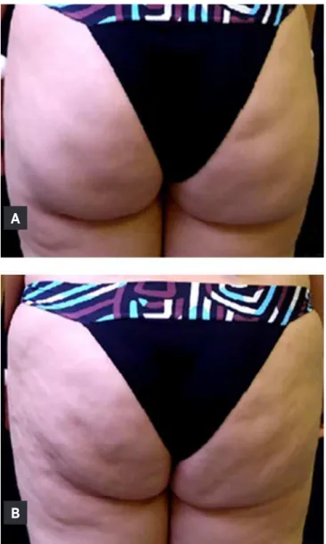
and 30 seconds of intervals for changing positions. Photographic records of the gluteal region were performed without and with gluteal contraction (Figure 2), with the machine positioned 100 cm high and 3 m away from the volunteer.
Figure 1. Postural record in anterior view
Evaluation of the cellulitis has been carried out according to the photonumeric scale proposed by Hexsel, Dal’Forno and Hexsel22, which evaluates the severity of
of Nürnberger and Müller4, only complements it,
subclassifying each grade. his evaluation was carried out by two blinded and independent examiners (experienced and previously trained for use of the scale), by means of photographic records. In case of divergence in the classiication, it was remade in the presence of both.
Figure 2. Gluteal region without contraction (A) and with contraction (B).
A
B
Descriptive analysis was performed from absolute and percent frequencies, and measures of centrality and dispersion. he correlation among variables was checked with the Pearson correlation test and the signiicance level adopted was 5%.
RESULTS
he classiication variables of cellulitis for the gluteus D and E and the pelvic angle in the previous view can be found in Table 1.
Among 46 volunteers, it is important to note that 20 presented grade I, 12 considered mild and eight moderate, corresponding to 43% of the sample; 25 of them presented grade II, ive mild, 11 moderate and nine severe, corresponding to 54% of the sample and one presented grade III, classiied as severe, corresponding to 3% of the sample.
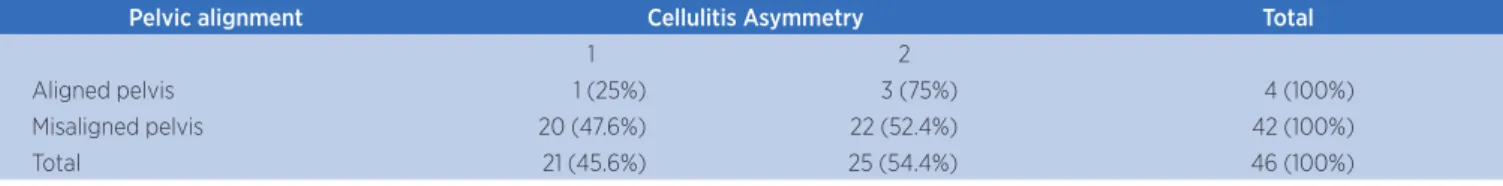
It was considered the correlation when there was pelvic misalignment associated with the asymmetry of the cellulitis in the gluteal region, or when there was pelvic alignment without asymmetry of the cellulitis in the gluteal region. It was not considered the correlation when there was pelvic misalignment without asymmetry of cellulitis in the gluteal region, or when there was pelvic alignment with asymmetry of cellulitis in the gluteal region.
he frequency of variables can be observed in Table 2: pelvic alignment and asymmetry of cellulitis.
Although there was no correlation between pelvic alignment and cellulitis, large asymmetries of cellulitis were observed in seven volunteers between gluteus D and gluteus E, higher than 3 points in the photonumeric scale. hese indings were found only in volunteers who presented pelvic misalignment and asymmetry of cellulitis.
Table 1. Variables evaluated
Variables Average Standard Deviation
Cellulitis right gluteus 6.3 points ±4.01
Cellulitis left gluteus 6.76 points ±3.61
For Sandoval24, postural changes as lat foot, lumbar
hyperlordosis and prolonged orthostatic can cause or aggravate cellulitis. Guirro and Guirro2 describe
the postural changes as associated with an important hemodynamic disorder, which may interfere in the emergence or worsening of the cellulitis. herefore, body postures that tend to compress the fatty tissue in speciic areas commonly exacerbate the ripples10.
Posture assessment is a method used in physical therapy to study the alignment of body parts; with a good evaluation we are able to draw a good treatment plan.
For Ribeiro et al.25, the low cost of photogrammetry,
the ease of interpretation, the high precision and reproducibility of the results, as well as the opportunity to archive and access records, are advantages that justify its extensive use. Photogrammetry is also a valuable record of the postural changes over time, since it is able to capture subtle transformations and to interrelate diferent parts of the body that are hard to measure26.
he photonumeric scale of Hexsel, Dal’Forno and Hexsel22 ofers a practical way to assess the severity
of cellulitis to distinguish and facilitate its diagnosis and direct the treatment according to the grade of involvement. It is important to note that the graduation shows important aspects, which are not considered in other forms of classiication such as the number and depth of depressions, the morphological aspects of skin and laccidity degree. here is a need for a more comprehensive and accurate evaluation, contributing to a better treatment plan and consequently a better result. It becomes so practical and efective that any professional of the area can apply it without major diiculties, therefore, it is an important tool for the evaluation of cellulitis in clinical practice.
CONCLUSION
No correlation was observed between pelvic alignment and severity and asymmetry of cellulitis. DISCUSSION
he hypothesis initially raised, that asymmetries of severity of cellulitis in the gluteal region could be related to pelvic misalignment, has not been conirmed in this study, which involved young volunteers (average of 23 years old) and the mild grade (grade I) of cellulitis, predominantly. Pelvic misalignment alters the request of muscle fascias of the gluteal region, either by high-side stretching or by low-high-side compression. However, it is likely that other factors interfere with the local microcirculation and may asymmetrically trigger/ aggravate the cellulitis in the region. Further studies are needed to investigate the asymmetry of cellulitis in the gluteal region. Other predisposing factors act similarly in both gluteus, and posture seems to be the only one of them that would act diferently bilaterally, which may explain the asymmetry.
Literature is scarce on this topic – we found a single scientiic article, which correlated posture with cellulitis. Milani, Natal Filho and João23 evaluated the possible
correlation between the angle of lumbar lordosis and the cellulitis grade. hey worked with the hypothesis that changes in the lumbar spine curvature could interfere in local blood low, inluencing on appearance of cellulitis. his hypothesis was raised because the severity of cellulitis is greater in the gluteal area and in posterior areas of the thigh. However, as in this study, they did not ind any correlation23. New studies should be conducted with a
larger sample number, with women with an average age greater than those used in this study and cellulitis grade greater than II by Nürnberger and Müller4.
In grade II, ibrosis is present, causing repercussions on the microcirculation described earlier, on the other hand it is not the case of grade I cellulitis, in which ibrosis is not yet installed. In this study, 20 volunteers (43%) had grade I cellulitis, therefore, it is likely that even with pelvic misalignment, little impact had occurred on the microcirculation of the subcutaneous and dermal tissue of these volunteers, directly interfering in the result. Table 2. Pelvic alignment x asymmetry of cellulitis
Pelvic alignment Cellulitis Asymmetry Total
1 2
Aligned pelvis 1 (25%) 3 (75%) 4 (100%)
Misaligned pelvis 20 (47.6%) 22 (52.4%) 42 (100%)
Total 21 (45.6%) 25 (54.4%) 46 (100%)
REFERENCES
1. Hexsel D, Soirefmann M, de Souza JS, Zafari D, David RB, Siega C. Avaliação do grau de celulite em mulheres em uso de três diferentes dietas. Surg Cosmet Dermatol. 2014;6(3):214-9. 2. Guirro ECO, Guirro RRJ. Fisioterapia dermato-funcional:
fundamentos, recursos e patologias. 3ª ed. Barueri: Manole; 2010.
3. Godoy JMP, Groggia MY, Laks LF, Godoy MFG. Intensive treatment of cellulite based on physiopathological principles. Dermatol Res Pract. 2012;2012:Article ID 834280. doi: 10.1155/2012/834280.
4. Nürnberger F, Müller G. So-called cellulite: an invented disease. J Dermatol Surg Oncol. 1978;4(3):221-9.
5. Piérard GE, Nizet JL, Piérard-Franchimont C. Cellulite: from standing fat herniation to hypodermal stretchmarks. Am J Dermatopathol. 2000;22(1):34-7.
6. Afonso JPJM, Tucunduva TCM, Pinheiro MVB, Bagatin E. Celulite: artigo de revisão. Surg Cosmet Dermatol. 2010;2(3):214-9.
7. Rosenbaum M, Prieto V, Hellmer J, Boschmann M, Krueger J, Leibel RL, et al. An exploratory investigation of the morphology and biochemistry of cellulite. Plast Reconstr Surg. 1998;101(7):1934-9.
8. Avram MM. Cellulite: a review of its physiology and treatment. J Cosmet Laser Ther. 2004;6(4):181-5. doi: 10.1080/14764170410003057.
9. Maio M. Etiologia e isiopatologia da celulite. In: Maio M, editor. Tratado de medicina estética. Vol. 3. São Paulo: Roca; 2004. p. 1481-6.
10. Quatresooz P, Xhaulaire-Uhoda E, Piérard-Franchimont C, Piérard GE. Cellulite histopathology and related mechanobiology. Int J Cosmet Sci. 2006;28(3):207-10. doi: 10.1111/j.1467-2494.2006.00331.x.
11. Rossi AB, Vergnanini AL. Cellulite: a review. J Eur Acad Dermatol Venereol. 2000;14(4):251-62.
12. Rona C, Carrera M, Berardesca E. Testing anticellulite products. Int J Cosmet Sci. 2006;28(3):169-73. doi: 10.1111/j.1467-2494.2006.00317.x.
13. Mendonça AMS, Pádua M, Ribeiro AP, Milani GB, João SMA, et al. Coniabilidade intra e interexaminadores da fotogrametria na classiicação do grau de lipodistroia ginóide em mulheres assintomáticas. Fisioter Pesqui. 2009;16(2):102-6.doi: 10.1590/S1809-29502009000200002.
14. Gherardini G, MatarassoA, Serure AS, Toledo LS, DiBernardo BE. Standardization in photography for body contour
surgery and suction-assisted lipectomy. Plast Reconstr Surg. 1997;100(1):227-37.
15. Penha PJ, João SMA, Casarotto RA, Amino CJ, Penteado DC. Postural assessment of girls between 7 and 10 years of age. Clinics. 2005;60(1):9-16.doi: 10.1590/ S1807-59322005000100004.
16. American Society for Photogrammetry and Remote Sensing. What is ASPRS? [Internet].Bethesda: American Society for Photogrammetry and Remote Sensing; 2000. [acesso em 2008out 2]. Disponível em:http://www.asprs.org/About-Us/ What-is-ASPRS.html
17. Nayler JR. Clinical photography: a guide for the clinician. J Postgrad Med. 2003;49(3):256-62.
18. Talamas I, Pando L. Speciic requirements for preoperative and postoperative photos used in publication. Aesthetic Plast Surg. 2001;25(4):307-10. doi: 10.1007/s002660010143. 19. Iunes DH, Castro FA, Salgado HS, Moura IC, Oliveira AS,
Bevilaqua-Grossi D. Coniabilidade intra e interexaminadores e repetibilidade da avaliação postural pela fotogrametria. Rev Bras Fisioter. 2005;9(3):327-34.
20. Portal do projeto Software para Avaliação Postural [Internet]. São Paulo: Incubadora Virtual FAPESP,2007. [acesso em 2015 ago 9]. Disponível em: http://demotu.org/sapo/
21. Fardet L, Kettaneh A, Tiev KP, Fabre B, Tolédano C, Cabane J, et al. Digital photography as an operational tool for assessing corticosteroid-induced lipodystrophy. Eur J Intern Med. 2008;19(5):340-4. doi: 10.1016/j.ejim.2007.09.015.
22. Hexsel DM, Dal’forno T, Hexsel CL. A validated photonumeric cellulite severity scale. J Eur Acad Dermatol Venereol. 2009;23(5):523-8. doi: 10.1111/j.1468-3083.2009.03101.x.
23. Milani GB, Natal Filho A, João SMA. Correlation between lumbar lordosisangle and degree of gynoid lipodystrophy (cellulite) in asymptomatic women. Clinics. 2008;63(4):503-8. doi: 10.1590/S1807-59322008000400015.
24. Sandoval B. Fibroedema geloide subcutáneo: qué conocemos de esta entidad clínica. Folia Dermatol Peru. 2003;14(1):38-42.
25. Ribeiro AP, Trombini-Souza F, Iunes DH, Monte-Raso VV. Coniabilidade inter e intra-examinador da fotopodometria e intra-examinador da fotopodoscopia. Rev Bras Fisioter. 2006;10(4):435-9. doi: 10.1590/S1413-35552006000400012. 26. Lima LCO, Baraúna MA, Sologurem MJJ, Canto RST, Gastaldi