Case Report
Major Article
http://dx.doi.org/10.1590/0037-8682-0093-2014INTRODUCTION
Address to: Dra Paula Oliveira. Serviço de Referência Nacional em Filarioses/ Deptº Parasitologia/CPqAM. Av. Prof. Moraes Rego, s/n, Campus da UFPE, Cidade Universitária, 50670-420 Recife, PE, Brasil.
Phone: 55 81 2101-2579; Fax: 55 81 2101-2671
e-mail: [email protected]; paula.oliveira@cpqam.fi ocruz.br
Received 6 May 2014 Accepted 30 June 2014
Evaluation of diagnostic tests for Wuchereria bancrofti
infection in Brazilian schoolchildren
Paula Oliveira
[1],
Cynthia Braga
[2],
Neal Alexander
[3],
Eduardo Brandão
[1],
Almerice Silva
[2],
Leandro Wanderley
[2],
Ana Maria Aguiar
[1],
George Diniz
[4],
Zulma Medeiros
[1]and Abraham Rocha
[1][1]. Serviço de Referência Nacional em Filarioses, Centro de Pesquisas Aggeu Magalhães, Fundação Oswaldo Cruz, Recife, PE. [2]. Departamento de Parasitologia, Centro de Pesquisas Aggeu Magalhães, Fundação Oswaldo Cruz, Recife, PE. [3]. Department of Infectious Disease Epidemiology, London School of Hygiene and Tropical Medicine, London, UK. [4]. Departamento de Saúde Coletiva, Centro de Pesquisas Aggeu Magalhães, Fundação Oswaldo Cruz, Recife, PE.
ABSTRACT
Introduction: Since the launch of the Global Programme to Eliminate Lymphatic Filariasis, more than 70% of the endemic countries
have implemented mass drug administration (MDA) to interrupt disease transmission. The monitoring of fi larial infection in sentinel
populations, particularly schoolchildren, is recommended to assess the impact of MDA. A key issue is choosing the appropriate tools
for these initial assessments (to defi ne the best intervention) and for monitoring transmission. Methods: This study compared the
pre-MDA performance of fi ve diagnostic methods, namely, thick fi lm test, Knott’s technique, fi ltration, Og4C3-ELISA, and the AD12-ICT card test, in schoolchildren from Brazil. Venous and capillary blood samples were collected between 11 pm and 1 am. The microfi larial loads were analyzed with a negative binomial regression, and the prevalence and associated 95% confi dence intervals were estimated for all methods. The accuracies of the AD12-ICT card and Og4C3-ELISA tests were assessed against the combination of parasitological
test results. Results: A total of 805 schoolchildren were examined. The overall and stratifi ed prevalence by age group and gender
detected by Og4C3-ELISA and AD12-ICT were markedly higher than the prevalence estimated by the parasitological methods. The sensitivity of the AD12-ICT card and Og4C3-ELISA tests was approximately 100%, and the positive likelihood ratios were above 6. The specifi city of the Og4C3-ELISA was higher than that of the AD12-ICT at different prevalence levels. Conclusions: The ICT card test should be the recommended tool for monitoring school-age populations living in areas with ongoing or completed MDA.
Keywords: Filariasis diagnosis. ELISA. Membrane fi ltration. Laboratory tests.
The Global Programme to Eliminate Lymphatic Filariasis (GPELF), launched by the World Health Organization (WHO)
in 1997, has targeted the elimination of the disease as a public health problem by 2020. Of the 73 endemic countries, 56 have implemented mass drug administration (MDA) programs to interrupt disease transmission. By 2012, more than 4.4 billion
doses of antifi larial drugs had been delivered to almost 984
million people worldwide1.
Effective monitoring and evaluation of the impact of MDA programs are important for achieving the goal of disease elimination. A number of diagnostic tools designed to assess
fi larial infection are standardized and currently recommended
by the WHO1. These tools include both parasitological
methods for detecting microfi laremia and tests for detecting
the circulating antigens of Wuchereria bancrofti, such as
the immunochromatographic test (AD12-ICT card test) and
enzyme-linked immunosorbent assay (Og4C3-ELISA)1. The
choice of which diagnostic tools to use is usually driven by
characteristics such as accuracy (sensitivity and specifi city), feasibility of use in the fi eld, technical skills required, and cost1.
It is critical to defi ne the most appropriate diagnostic tools for the initial assessments of lymphatic fi lariasis (LF) infection in the population (to defi ne the best intervention), as well as to
monitor transmission in areas undergoing MDA, because the performance of such tools can vary substantially according to the epidemiologic and demographic characteristics of the study population2,3.
The WHO has recommended the monitoring of fi larial
infection in sentinel populations, particularly schoolchildren4-6,
due to children’s lower duration of exposure (many of these
children were born following or during the start of the intervention), which allows for a more accurate assessment of transmission5. However, the pediatric population usually exhibits lower microfilarial loads and higher proportions
of amicrofi laremic and asymptomatic infections compared
with adults7-9. As these features will most likely infl uence the operating characteristics of diagnostic tests, it is important to evaluate them in children. This study evaluated the pre-MDA performance of several diagnostic methods, namely,
METHODS
Og4C3-ELISA, and the AD12-ICT card test, in children from
an endemic area of Brazil prior to the initiation of an MDA
program.
Study setting
The study was conducted in the three neighborhoods of the City of Olinda, Metropolitan Region of State Pernambuco,
Brazil, with the highest prevalence of microfi laremia, according
to a survey conducted in 1998 (unpublished data), between 2007 and 2010. The city has an area of 41,659km2 and a population of 377,779, as reported in the last census10, and is considered
one of the remaining foci of fi lariasis in Brazil11,12.
Children between the ages of 4 and 15 years who attended
fi ve public schools in these neighborhoods (Alto da Conquista,
Alto da Bondade, and Sapucaia) participated in this study.
Data collection
Parents or guardians were informed of the main study
objectives, and consent was sought for the children’s participation. After obtaining written consent, each child’s
data (name, address, age, and gender) were obtained using a
standardized form. The blood samples were obtained between 23:00 and 1:00. Initially, capillary blood samples were obtained by fi nger prick to perform the thick blood fi lm (~60μL of blood) and the AD12-ICT card (100μL) tests. A venous blood sample (7mL) was then collected to perform the fi ltration, Knott’s concentration, and Og4C3-ELISA tests. This sample was
partitioned into tubes containing ethylenediamine tetraacetic
acid (EDTA), tubes containing 2% formalin, and dry tubes to perform the fi ltration, Knott’s concentration test, and Og4C3-ELISA test, respectively. The blood samples were transported
in a refrigerated container to the laboratory, stored at 2-8°C, and processed on the following day.
Laboratory procedures
I) Parasitological methods: A) Thick blood fi lm (TBF)
test: The smear was left to dry overnight at room temperature.
The slides were then dehemoglobinized in water, fi xed in
methanol for 3 min, stained according to previously described procedures, and read with an optical microscope13. B) Knott’s concentration method: Venous blood (~1mL) was drawn into a
tube containing 9mL of 2% formalin and processed according to the methodology described by Knott14. C) Filtration technique:
Blood (~1mL) was fi ltered through a polycarbonate membrane with a width of 13mm and pore size of 3µm, as described by Dennis and Kaen15. These three parasitological tests rely on the microscopic visualization of microfi lariae. Thus, their
specifi city, although not their sensitivity, can be considered to
be very high. II) Immunological methods: A)AD12-ICT card
test: This test detects circulating fi larial antigen (CFA) using
the monoclonal antibody AD12, which recognizes a 200-kDa fi larial antigen from either adult worms or microfi lariae16. The test was performed according to the manufacturer's instructions
and read by trained technicians in the fi eld after 10 min. The
visualization of the two lines (test and control) was interpreted
as a positive result. B) Og4C3-ELISA:The test was performed as recommended by the manufacturer (TropBio®, Townsville,
Australia). Samples with an antigen concentration ≥128mL units
(UA) were interpreted as positive17. This test detects CFA using a monoclonal antibody against antigens of the bovine parasite
Onchocerca gibsoni, which has no known cross-reactivity with other human helminthes. The test provides quantitative results that enable the observation of variations in antigenemia levels after treatment18-24.
Data analysis
The reading and interpretation of all laboratory tests were performed by observers who were blinded to the other test
results. Data entry and analysis were performed using EpiInfo and R software. Mean microfi larial densities were estimated and
compared with a negative binomial regression25. The parasite load, determined by the three parasitological techniques (thick
smear, Knott’s concentration, and membrane fi ltration), and antigenemia, determined by the Og4C3-ELISA in terms of
unit antigens (UA), were stratifi ed by age group and gender. Antigenemia was compared between groups with a
Kruskal-Wallis test. The mean antigenemia was quoted for descriptive purposes only, as most subjects were negative and, therefore,
the median values were zero.
The infection prevalence identifi ed by each technique was
stratifi ed by age group and gender. Statistical comparisons between groups were made using chi-square (χ2) and
Kruskal-Wallis tests for qualitative and continuous results, respectively. A p-value of 0.05 was considered statistically signifi cant.
The sensitivity, positive predictive value (PPV), negative predictive value (NPV), positive likelihood ratio (LR+), negative likelihood ratio (LR-), and their respective 95% confi dence intervals (CIs) for the AD12-ICT card test and Og4C3-ELISA were estimated against the gold standard of the combination of the parasitological techniques (thick blood fi lm, Knott’s concentration, and fi ltration). The LR+ measures how
many times more likely it is that a positive result is obtained for individuals with the disease than for individuals without the
disease, whereas the LR- reveals how many times more likely
it is that a negative result is obtained for individuals with the
disease than for individuals without the disease. Values of LR+ above 10 and LR- below 0.1 provide strong evidence to confi rm
or rule out the diagnosis of disease, respectively26.
The parasitological tests are assumed to have 100% specifi city but are of unknown sensitivity, as many individuals can harbor adult worms without demonstrating microfi laremia12.
Consequently, the sensitivity of the ICT card test and the Og4C3-ELISA can be estimated, but not their specifi city. Because the specifi city cannot be directly evaluated, we estimated the specifi cities of the Og4C3-ELISA and AD12-ICT card tests as a function of the prevalence of fi lariasis using the equation derived
by Staquet et al.27. This calculation allows for the comparison
of the specifi cities of the Og4C3-ELISA and AD12-ICT card
tests for different prevalences27 of samples with viable worms
RESULTS
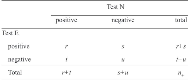
TABLE 1 - Contingency table of two tests in a paired design among those positive for the reference standard (fi ltration). Test N is a ‘new’ test, e.g., the AD12-ICT card test, and test E is an existing test, e.g., the thick blood fi lm test. Adapted from Table 2 of Hayen et al.28.
Test N
positive negative total
Test E
positive r s r+s
negative t u t+u
Total r+t s+u n+
AD12-ICT: immunochromatographic test using the monoclonal antibody AD12. Note:n
+ = r + s + t + u, where n+: number of people
with the disease; r: true positive; s: false negative; t: false positive, and u: true negative.
The performances of the AD12-ICT card and thick fi lm tests were compared according to sections 2.2 and 3.2.1 of Hayen
et al.28, with the fi ltration test used as the reference standard. There are eight possible combinations of the three tests, but
we considered only the four cells in which the fi ltration test is positive. Due to the high specifi city of fi ltration, these cells
can reliably be considered to be truly positive. The Table 1 wasadapted from Table 2 of Hayen et al.28. The true positive
fractions in the AD12-ICT card and thick blood fi lm tests can
be estimated as (r + t)/n+ and (r + s)/n+, respectively, and the McNemar test can be used to assess the difference between these values.
Ethical considerations
This study was approved by the Ethics Committee of the Aggeu Magalhães Research Center (CAEE: 0069.0.095.000-06). All of the participants with a positive result in any of the tests were treated and underwent clinical and laboratory examinations.
A total of 805 schoolchildren participated in the study and
agreed to be tested via fi nger prick. Overall, 783 children were tested by the thick blood fi lm test and 797 by the AD12-ICT card
test. Consent for taking venous blood samples was obtained from
only 554 participants, all of whom were examined by Knott's concentration method, 546 by the fi ltration technique, and 545 by the Og4C3-ELISA test. In 487 schoolchildren, fi larial infection
was assessed with all laboratory tests.
The microfi laria (MF) loads ranged from 0 to 216 MF/60μL, 0 to 1,423 MF/mL, and 0 to 1,834 MF/mL according to the
thick blood fi lm, Knott’s concentration, and fi ltration tests, respectively. No statistically signifi cant variation in MF load
was observed between age groups or genders when assessed by any of these three methods (Table 2). The mean values of
fi larial antigenemia also did not differ according to age group
or gender (Table 2).
The estimated prevalence of fi larial infection according to both fi larial antigen tests (overall and stratifi ed by age group and
gender) was similar and markedly higher than the prevalence estimated by parasitological methods. This difference in estimates was more pronounced in the younger age group (4-9 years), in whom the prevalence of filarial infection
estimated by the ICT and Og4C3-ELISA tests was approximately fi ve times higher than that obtained by parasitological methods (thick blood fi lm, Knott’s concentration, or fi ltration test)
(Table 3).
The MF prevalence estimated by either Knott's (4.6%; 95%CI: 3.1-6.8) or the fi ltration (5.2%; 95%CI: 3.5-7.5%) method was slightly higher than the prevalence estimated by the thick fi lm method. The prevalence of fi lariasis according to the parasitological methods was signifi cantly higher in the
10-15-year-old age group than in the 4-9-year-old age group, whereas no difference between age groups was found by the
immunological methods (AD12-ICT card test and Og4C3-ELISA) (Table 3). The prevalence of fi larial infection was
higher in boys than in girls according to all techniques, although
a statistically signifi cant difference in the prevalence between the groups was observed only with the AD12-ICT card test
(Table 3).
The comparative analysis of the performance of the
AD12-ICT card test and Og4C3-ELISA against the gold standard (the
combination of all parasitological test results) showed that both
immunological tests exhibited 100% sensitivity. The PPVs were approximately 30%, and the NPVs were close to 100%. The LR+ was 6.89 and 7.89 for the AD12-ICT card test and Og4C3-ELISA, respectively. Both LR- values were zero (Table 4).
Figure 1 shows the curves comparing the specifi cities of the
AD12-ICT and Og4C3-ELISA tests over the feasible range of values of infection prevalence (5% to 20%) based on the method
of Staquet et al. Although the exact values are unknown, we can conclude from these ranges that a) both tests have relatively
high specifi cities (84.4% [95% CI: 80.8-87.4] for the AD12-ICT and 86.5% [CI 95%: 83.0-89.3] for the Og4C3-ELISA), and b) the Og4C3-ELISA has a slightly higher specifi city than the AD12-ICT test.
The true positive fractions (TPFs) of the AD12-ICT card and thick blood fi lm tests, calculated with the Hayen formula, were 100% (27+0/27) and 85.2% (23+4/27), respectively. This difference of 14.8% between the TPF values was statistically signifi cant, suggesting that for each 1,000 schoolchildren with the infection, the AD12-ICT card test was able to detect 148 more cases than the thick blood fi lm test. The TPFs for the AD12-ICT card test and Og4C3-ELISA were both 100%.
Accordingly, no difference could be found in the ability of the
TA BLE 2 M ean s of mi cr ofi lar ia de ns ity an d an tige ne mi a me as ur ed b y th e th ic k fi l m, K nott con ce ntr ati on , me mb ran e fi ltr ati on , an d O g4C 3-ELI SA te sts ac cor di ng to age gr ou p an d ge nd er . Thi ck fi lm K not t Fi ltra tion O g4C3-E LIS A (U A ) exa m ine d m ea n* m in-m ax exa m ine d m ea n m in-m ax exa m ine d m ea n m in-m ax exa m ine d m ea n (n) (95%CI) (m f/60µ ) (n) (95%CI)* (m f/m l) (n) (95%CI)* (m f/m l) (n) (95% CI) T ot al 776 1.39 (0.55-3.53) 0-216 541 12.01 (4.15-34.7) 0-1423 519 15.34 (5.48-42.95) 0-1834 543 1,836 (1,216-2,454) A ge group 4-9 407 2.82 (0.45-17.7) 0-150 306 5.05 (1.27-20.0) 0-446 295 1 1.26(2.90-43.72) 0-1834 308 1,245 (592.5-1,899) 10-15 360 0.76 (0.22-2.67) 0-216 232 4.22 (0.52-34.51 0-1423 221 1.86(0.23-14.8) 0-989 231 2,655 (1,489-3,823) G ende r m al e 393 0.99 (0.15-6.36) 0-152 288 1.77 (0.21-14.72) 0-446 281 0.94 (0.12-7.41) 0-1834 290 2,364 (1,414-3,3495) fe m al e 383 1.40 (0.38-5.18) 0-216 253 8.84 (2.07-37.71) 0-1423 238 15.79 (3.90-63.95) 0-989 253 1,231 (487.8-1,994) O g4C 3-ELI SA : e nz ym e-l inke d im m unos orbe nt a ss ay w ith the 4C3 m onoc lona l a nt ibody ta rge ting O nc hoc er ca gi bs oni ; 95%C I: 95% Confi de nc e Int erva l; LR χ 2: L ike lihood ra tio chi squa re ; n : num be
r of c
hi ldre n e xa m ine d; p os : num be
r of pos
it ive re sul ts w it h t he t ec hni que us ed; *D et erm ine
d by a
ne ga ti ve bi nom ia l re gre ss ion m ode l. LR χ 2= 1.18; LR χ
2 = 1.74;
LR
χ
2 = 0.35;
K rus ka l-W al lis p-va lue =0.28 p-va lue = 0.19 p-va lue = 0.55 χ 2= 2.6; p-va lue = 0.1 1 LR χ 2= <0.01; LR χ 2= 0.28; LR χ 2< 0.01; K rus ka l-W al lis p-va lue = 0.99 p-va lue = 0.60 p-va lue = 0.95 χ 2= 0.071; p-va lue = 0.79 DISCUSSION
This study assessed the accuracy of tests in detecting bancroftian filariasis in a large and representative sample of schoolchildren from an
endemic area in northeastern Brazil before the
initiation of MDA. The performance evaluation of these tests in children is critical to evaluate the impact of the clinical and epidemiological characteristics of
this population that may infl uence test performance2,3. This evaluation is of particular importance, as this demographic group has been recommended
as a sentinel population by the WHO in whom to
assess MDA effectiveness in areas undergoing this intervention.
The prevalence of fi lariasis estimated by the
filarial antigen tests (AD12-ICT card test and
Og4C3-ELISA) was far higher than that estimated
by parasitological methods. The difference between
the results of these methods was as much as fi vefold
in the group aged below 10 years. The comparative
evaluation of the performance of the thick fi lm test in relation to the AD12-ICT card test using the Hayen
et al.28 formula also showed that in each group of 1,000 schoolchildren with filarial infection, the
AD12-ICT card test was able to detect approximately 150 more cases than the thick fi lm test. These results are in agreement with previous studies conducted in the school-age population29-39 and reinforce the
usefulness of the AD12-ICT card test in screening for fi larial infection in children for the assessment of MDA effi cacy6,40. The greater sensitivity of the antigen tests can be explained by their ability to
detect low-level microfi laremia and amicrofi laremic
cases that are typically more common in children than in adults7.
However, it is worth noting that both the AD12-ICT and Og4C3-ELISA tests may yield positive
results after treatment even when there is no viable infection16, which could overestimate prevalence estimates. Conversely, because this study population was tested before MDA, it is probable that any prevalence excess due to false-positive tests would be of low magnitude. Additionally, the data draw attention to the low sensitivity of the parasitological
methods, particularly the thick blood fi lm test, in the screening of fi larial infection at younger ages.
Several studies have shown that the sensitivity of both parasitological and antigen tests decreases as the MF loads decrease21,31,33,41. This decrease may result in differential performance across population
subgroups. In this study, the MF loads were slightly
TA
BLE 3 - P
re val en ce of mi cr ofi lar emi a amon g c hi ld re n b y p ar as itol ogi cal an d i mmu nol ogi cal te ch ni qu es ac cor di
ng to age
gr ou p an d ge nd er . O lin da, S tate of P er namb uc o, 2007-2009. Thi ck fi lm K not t Fi ltra tion (~60µ L) (1m L) (1m L) A D 12-ICT c ard O g4C3-E LIS A Cha ra ct eri st ic s N (pos ) % (95%CI) N (pos ) % (95%CI) N (pos ) % (95%CI) N (pos ) % (95%CI) N (pos ) % (9 5%CI) T ot al 776 (24) 3.1 (2.0-4.6) 541 (25) 4.6 (3.1-6.8) 519 (27) 5.2 (3.5-7.5) 796 (1 16) 14.6 (12.4-17.5) 543 (94) 17.4 (14.4-21.0) A ge group (ye ars ) 4-9 407 (8) 2.0 (0.9-4.0) 306 (7) 2.3 (1.0-4.9) 295 (9) 3.1 (1.5-5.9) 419 (55) 13.1 (10.1-16.8) 308 (47) 15.3 (1 1.5-19.9) 10-15 360 (16) 4.4 (2.6-7.3) 232 (18) 7.8 (4.8-12.2) 221 (18) 8.1 (5.0-12.8) 368 (61) 16.6 (13.0-20.9) 231 (47) 15.5 (15.5-26.2) χ2=3.87; χ2=8.91; χ2=6.61; χ2=1.85; χ2=2.37; p-va lue =0.049 p-va lue =0.003 p-va lue =0.010 p-va lue = 0.173 p-va lue = 0.124 G ende r m al e 393 (15) 3.8 (2.2-6.3) 288 (16) 5.6 (3.3-9.0) 281 (17) 6.1 (3.7-9.7) 401 (69) 17.2 (13.7-21.3) 290 (53) 18.3 (14.1-23.3) fe m al e 383 (9) 2.3 (1.1-4.6) 253 (9) 3.6 (1.7-6.9) 238 (10) 4.2 (2.2-7.8) 395 (47) 1 1.9 (8.9-15.6) 253 (41) 16.2 (12.0-21.5) χ2=1.39; χ2=1.22; χ2=0.89; χ2=4.50; χ2=0.40; p-va lue =0.238 p-va lue =0.269 p-va lue =0.345 p-va lue = 0.034 p-va lue = 0.525 A D 12-I C T: i m m unoc hrom at ogra phi c te st w it h t he m onoc lona l ant ibody A D 12; O g4C 3-ELI SA : enz ym e-l inke d im m unos orbe nt a ss ay us ing the m onoc lona l ant ibody 4C3, ta rge ting O nc hoc er ca gi bs oni ; 95%C I: 95% c onfi de nc e i nt erva l; χ 2: c hi s qua re ; n : num be
r of c
hi ldre n e xa m ine d; p os : num be
r of pos
it ive re sul ts w it h t he t ec hni que us ed.
signifi cant. Similarly, we found no signifi cant differences in
antigenemia according to age or gender. Thus, any variation in test accuracy across these subgroups of this particular population may be limited.
The prevalence estimated by Knott’s test and the fi ltration test was, in general, slightly higher than that
estimated by the thick film test. This finding implies
that the former tools are most appropriate for fi lariasis screening of school-age populations before antifi larial treatment. Knott’s technique is more laborious than the fi ltration technique but is usually less expensive42,43,and it
showed a similar performance in detecting microfi laremia
(15.3 vs. 12.0 MF/mL). Thus, it may be preferred in areas
with limited fi nancial resources.A further advantage of this
test is its ability to differentiate species of fi laria, which
makes it useful in other endemic areas where co-infection
with other fi larial parasites occurs.
The analysis according to age showed that the prevalence
was signifi cantly higher in the 10-15-year-old age group
than in the 4-9-year-old age group, as evaluated by all
parasitological methods. However, no difference in the
prevalence of antigenemia between age groups according
to the ICT and Og4C3 tests was observed. Moreover, the prevalence of infection estimated by the antigen tests (ICT
and Og4C3) was approximately seven times higher than that estimated by the parasitological methods in the younger
age group (4-9 years). In the older group (10-15 years),
although the prevalence estimated by the antigen tests was again higher than that estimated by the parasitological methods, this difference was less pronounced. These results demonstrate the lower sensitivity of the parasitological methods in the younger age groups compared with the older population, as a larger proportion of individuals in the prepatent stages or who have amicrofilaremic infections (with adult worms but with no or few reproducing females) are usually found in this population, as previously described7,33.
The performance evaluation of the AD12-ICT card test and Og4C3-ELISA against the gold standard of the
combination of the parasitological test results demonstrated
100% sensitivity for both tests. Additionally, the values of LR+ and LR- were compatible with the high accuracy
of these tests in the diagnosis of infection in this study population, even when considering that these measures may be distorted due to the imperfection of the gold standard
due to the combination of parasitological results. The LR+
for both tests may represent an underestimate because
subjects with an amicrofi laremic infection could have been misclassifi ed as free of disease according to the gold standard. Similarly, the low PPVs of the AD12-ICT card and Og4C3-ELISA tests can also be explained by the gold
standard, which is assumed to have a lower sensitivity27 than the antigen-based tests (AD12 and OG4C3 tests).
In regions with ongoing elimination programs, it is important to use high-specifi city tests to ascertain the actual
TABLE 4 - Sensitivity, positive predictive value, negative predictive value, accuracy, and likelihood ratio of the ICT card and Og4C3-ELISA tests compared with those of the combination of all parasitological tests (gold standard).
Diagnostic performance AD12-ICT card test (n=546) Og4C3-ELISA (n=542)
Sensitivity % (95% CI) 100.0 (85.4-100.0) 100.0 (85.4-100.0)
PPV % (95% CI) 27.8 (19.8-37.7) 30.8 (22-41.3)
NPV % (95% CI) 100.0 (98.9-100.0) 94.6 (98.9-100.0)
Likelihood positive (95% CI) 6.9 (5.6-8.5) 7.9 (6.3-9.9)
Likelihood negative 0.0 0.0
Accuracy % (95% CI) 86.2 (83.0-88.9) 88.0 (84.9-90.5)
AD12-ICT: immunochromatographic test with the monoclonal antibody AD12; Og4C3-ELISA: enzyme-linked immunosorbent assay with
the 4C3 monoclonal antibody, targeting Onchocerca gibsoni; 95%CI: 95% confi dence interval; PPV: positive predictive value; NPV: negative predictive value.
0,82 0,84 0,86 0,88 0,90 0,92 0,94 0,96 0,98 1,00
0,00 0,05 0,10 0,15 0,20 0,25
S
p
e
c
if
ic
it
y
Prevalence
ICT Og4c3
FIGURE 1 - Estimated specifi city of the AD12-ICT card test and Og4C3-ELISA according to the prevalence of microfi laria determined by the combination of all parasitological tests. AD12-ICT: immunochromatographic test with the monoclonal antibody AD12; Og4C3-ELISA: enzyme-linked immunosorbent assay using the monoclonal antibody 4C3, targeting Onchocerca gibsoni.
certainty. Although the parasitological methods are 100% specifi c,
they lose their usefulness after the initiation of MDA due to the
potent microfi laricide effect of the antifi larial drugs, which yield a dramatic decrease in microfi laremia16,44. However, studies have shown that patients can remain infected with detectable levels of antigenemia even after several courses of treatment16,24,45,46. These issues limit the use of the antigen-based tests, such as the AD-12 and Og4C3 tests, in the assessment of treatment effectiveness, both at the individual and population levels.
The comparative evaluation demonstrated the high
specifi city of the AD12-ICT and Og4C3-ELISA tests in the study population, although a slightly higher specifi city of the Og4C3
test was observed when the different levels of prevalence were taken into account. Similar results were also found by Gass et al.40, who demonstrated that the Og4C3-ELISA test showed
a greater specifi city. These results suggest that the
Og4C3-ELISA test is more appropriate for assessing the interruption of fi larial transmission in areas with ongoing elimination programs
because it yields more accurate negative results, in addition to providing quantitative results that enable the monitoring of MDA effectiveness18. Furthermore, the Og4C3-ELISA
test can be performed using fi lter paper, which facilitates the attainment of samples in fi eld studies as well as in venous blood samples. However, there is a need for better standardization
of the technique, as well as better quality control of the kits by the manufacturer, particularly with regard to the plates and reagents.
Based on these results, we conclude that for an initial assessment of possible transmission of W. bancrofti, the ICT
card test is the ideal tool for surveys and for mapping infection in the school-age population. The high accuracy of the
AD12-ICT card test, in addition to other features such as rapidity and
the simplicity of execution, makes this test the method of choice
for the screening of fi larial infection in this population prior to the initiation of antifi larial treatment.
Due to their low sensitivity, the parasitological methods,
particularly the thick fi lm technique, are very limited in their
usefulness for the estimation of the burden of infection in
this population even before treatment. The ICT card test is
considered the most suitable test due to its high sensitivity and,
as previously mentioned, its lower specifi city in populations that
have already been treated16,46-48. Finally, in areas with ongoing or completed MDA, the interruption of transmission, as assessed
by the tracking of incident cases of fi lariasis by the ICT card test in the school-age population without prior antifi larial treatment,
is the preferred approach.
ACKNOWLEDGMENTS
The authors thank the National Reference Service for
The authors declare that there is no confl ict of interest.
CONFLICT OF INTEREST
FINANCIAL SUPPORT
This research was funded by the Secretaria de Vigilância em Saúde/Fundação Oswaldo Cruz/Fundação para o
Desenvolvimento Tecnológico em Saúde - Vice-Presidência de Pesquisa e Laboratórios de Referência
(FIOCRUZ/FIOTEC-VPPLR)-002-LIV11-2-1 Project.
REFERENCES
1. World Health Organization. Global Programme Eliminate Lymphatic Filariasis; progress report on mass drug administration in 2012. Wkly Epidemiol Rec 2013; 88:389-400.
2. Jaeschke R, Guyatt G, Sackett DL. Users’ Guides to the Medical Literature. III How to Use an Article About a Diagnostic Test. A. Are the Results of the Study Validate. JAMA 1994; 271:389-391.
3. Reid C, Lachs MS, Feinstein AR. Use of methodological standards in diagnostic test research. JAMA 1995; 274:645-651.
4. Ramzy RMR, Hafez ON, Gad AM, Faris R, Harb M, Buck AA, et al. Effi cient assessment of fi lariasis endemicity by screening for fi larial antigenemia in a sentinel population. Trans R Soc Trop Med Hyg 1994; 88:41-44.
5. World Health Organization. Monitoring and epidemiological assessment of the programme to eliminate lymphatic fi lariasis at implementation unit level. WHO/CDS/CPE/CEE/2005.50; 2005.
6. World Health Organization. Transmission assessment surveys in the Global Programme to Eliminate Lymphatic Filariasis. WHO; 2012. 7. Witt C, Ottesen EA. Lymphatic fi lariasis: an infection of childhood.
Trop Med Int Health 2001; 6:582-606.
8. Shenoy RK. Lymphatic fi lariasis in children. J Commun Dis 2006; 38:118-123.
9. Shenoy RK, Suma TK, Kumaraswami V, Rahmah N, Dhananjayan G, Padma S, et al. Preliminary fi ndings from a cross-sectional study on lymphatic fi lariasis in children, in an area of India endemic for Brugia malayi infection. Trop Med Parasitol 2007; 101:205-213.
10. Instituto Brasileiro de Geografi a e Estatística. Censo demográfi co 2010: Características populacionais e domiciliares, Rio de Janeiro, Brasil: IBGE; 2010. [Cited 2010 June 30]. Available at: http://www.ibge.gov.br/. 11. Medeiros Z, Gomes J, Beliz F, Coutinho A, Dreyer P, Dreyer G. Screening
of army soldiers for Wuchereria bancrofti infection on metropolitan Recife region, Brazil. Trop Med Int Health 1999; 4:499-505.
12. Braga C, Dourado MI, Ximenes RA, Alves L, Brayner F, Rocha A, et al. Field evaluation of the whole blood immunocromatographic test for rapid bancroftian fi lariasis diagnosis in the northeast of Brazil. Rev Inst Med Trop Sao Paulo 2003; 45:125-129.
13. Eberhard ML, Lammie PJ. Laboratory diagnosis of fi lariasis. Clin Lab Med 1991; 11:977-1010.
14. Knott JA. Method for making microfi larial surveys on day blood. Trans R Soc Trop Med Hyg 1939; 32:191-196.
15. Dennis DT, Kaen BH. Isolation of microfi lariae: report of new method. J Parasit 1971; 57:1146-1147.
16. Weil GJ, Ramzy RMR. Diagnostic tools for fi lariasis elimination programmes. Trends Parasitol 2007; 23:78-82.
17. More SJ, Copeman DB. A highly specifi c and sensitive monoclonal antibody-based ELISA for the detection of circulating antigen in bancroftian fi lariasis. Trop Med Parasitol 1990;41:403-406.
18. Lalitha P, Ravichandra M, Suba S, Kaliraj P, Narayanan RB, Jayaraman K. Quantitative assessement of circulating antigens in human lymphatic fi lariasis: a fi eld evaluation of monoclonal antibody-based ELISA using blood collected on fi lter strips. Trop Med Int Health 1998; 3:41-45. 19. Weil GJ, Jain DC, Santhanam S, Malhotra A, Kumar H, Sethumadhavan KVP,
et al. A monoclonal antibody-based enzyme immunoassay for detecting parasite antigenemia in bancroftian fi lariasis. J Infec Dis 1987; 156:350-355.
20. Eberhard ML, Hightower AW, Addiss DG, Lammie PJ. Clearance of Wuchereria bancrofti antigen after treatment with diethylcarbamazine or ivermectin. Am J Trop Med Hyg 1997; 57:483-486.
21. Nicolas L, Plichart C, Nguyen LN, Moulia-Pelat JP. Reduction of Wuchereria bancrofti adult worm circulating antigen after annual treatments of diethylcarbamazine combined with ivermectin in French Polynesia. J Infec Dis 1997; 175:489-492.
22. Ismail MM, Weil GJ, Jayasinghe KSA, Premaratne UN, Abeyewickreme W, Rajaratnam HN, et al. Prolonged clearance of microfi laraemia in patients with bancroftian fi lariasis after multiple high doses of ivermectin or diethylcarbamazine. Trans R Soc Trop Med Hyg 1996; 90:684-688. 23. Weil GJ, Lammie PJ, Weiss N. The ICT fi lariasis test: A rapid-format
antigen test for diagnosis of bancroftian fi lariasis. Parasitol Today 1997; 13:401-404.
24. Freedman DO, Plier DA, Almeida AB, Oliveira AL, Miranda J, Braga C. Effect of aggressive prolonged diethylcarbamazine therapy on circulating antigen levels in bancroftian fi lariasis. Trop Med Int Health 2001; 6: 37-41.
25. Wilson K, Grenfell BT. Generalized linear modelling for parasitologists. Parasitol Today 1997; 13:33-38.
26. Deeks JJ, Altman DG. Statistics Notes: Diagnostic tests 4: likelihood ratios. BMJ 1994; 329:168.
27. Staquet M, Rozencweig M, Lee YJ, Muggia FM. Methodology for the assessment of new dichotomous diagnostic tests. J Chronic Dis 1981; 34:599-610.
28. Hayen A, Macaskill P, Irwig L, Bossuyt P. Appropriate statistical methods are required to assess diagnostic tests for replacement, add-on, and triage. J Clin Epidemiol 2010; 63:883-991.
29. Chanteau S, Moulia-Pelat JP, Glaziou P, Nguyen NL, Luquiaud P, Plichart C, et al. Og4C3 circulating antigen: a marker of infection and adult worm burden in Wuchereria bancrofti fi lariasis. J Infect Dis 1994; 170:247-250. 30. Lammie PJ, Hightower AW, Eberhard ML. The age-specifi c prevalence
of antigenemia in a Wuchereria bancrofti-exposed population. Am J Trop Med Hyg 1994; 51:348-355.
31. Rocha A, Addiss D, Ribeiro ME, Noroes J, Baliza M, Medeiros Z, et al. Evaluation of the Og4C3 ELISA in Wuchereria bancrofti infection: infected persons with undetectable or ultra-low microfi larial densities. Trop Med Int Health 1996; 1:859-864.
32. Freedman DO, Almeida A, Miranda J, Plier DA, Braga C. Field trial of a rapid card test for Wuchereria bancrofti. Lancet 1997; 350:1681. 33. Itoh M, Weerasooriya MV, Gunawardena NK, Mudalige MPS,
Samarawickrema WA, Kimura E. Wuchereria bancrofti antigenemia in Sri Lanka. Trop Med Int Health 1999; 4:207-210.
34. Ramzy RM, Helmy H, el-Lethy AS, Kandil AM, Ahmed ES, Weil GJ, et al. Field evaluation of a rapid-format kit for the diagnosis of bancroftian fi lariasis in Egypt. East Mediterr Health J 1999; 5:880-887.
35. Omar MS, Sheikha AK, Al-Amari OM, Abdalla SE, Musa RA. Field evaluation of two diagnostic antigen tests for Wuchereria bancrofti infection among Indian expatriates in Saudi Arabia. Southeast Asian J Trop Med Public Health 2000; 31:415-418.
36. Njenga SM, Wamae CN. Evaluation of ICT fi lariasis card test using whole capillary blood: comparison with Knott’s concentration and counting chamber methods. J Parasitol 2001; 87:1140-1143.
38. Bal MS, Beuria MK, Mandal NN, Das MK. Antigenemia in young children living in Wuchereria bancrofti endemic areas of Orissa, India. Trans R Soc Trop Med Hyg 2009; 103:262-265.
39. Rocha A, Braga C, Belém M, Carrera A, Aguiar-Santos AM, Oliveira P, et al. Comparison of tests for the detection of circulating fi larial antigen (Og4C3-ELISA and AD12-ICT) and ultrasound in diagnosis of lymphatic fi lariasis in individuals with microfi lariae. Mem Inst Oswaldo Cruz 2009; 104:621-625. 40. Gass K, Rochars MVB, Boakye D, Bradley M, Fischer PU, Gyapong J, et al.
A multicenter evaluation of diagnostic tools to defi ne endpoints for programs to eliminate bancroftian fi lariasis. PLoS Negl Trop Dis 2012, 6:e1479. 41. Rocha A, Lima G, Medeiros Z, Aguiar-Santos A, Alves S, Montarroyos U,
et al. Circulating fi larial antigen in the hydrocele fl uid from individuals living in a bancroftian fi lariasis area-Recife, Brazil: detected by the monoclonal antibody Og4C3-assay. Mem Inst Oswaldo Cruz 2004; 99:101-105.
42. Rocha A. Métodos laboratoriais disponíveis para o diagnóstico da fi lariose linfática. RBAC 2000; 32:265-270.
43. Melrose WD, Turner PF, Pisters P, Turner B. An improved Knott's concentration test for the detection of microfi lariae. Trans R Soc Trop Med Hyg 2000; 94:176.
44. Ottesen EA, Hooper PJ, Bradley M, Biswas G. The Global Programme to Eliminate Lymphatic Filariasis: Health Impact after 8 Years. PLOS Negl Trop Dis 2008; 2:1-12.
45. Schuetz A, Addiss DG, Eberhard ML, Lammie PJ. Evaluation of the whole blood fi lariasis ICT test for short-term monitoring after antifi larial treatment. Am J Trop Med Hyg 2000; 62:502-503.
46. Ramzy RM, El Setouhy M, Helmy H, Ahmed ES, Abd Elaziz KM, Farid HA, et al. Effect of yearly mass drug administration with diethylcarbamazine and albendazole on bancroftian fi lariasis in Egypt: a comprehensive assessment. Lancet 2006; 367:992-999.
47. Njenga SM, Wamae CN, Njomo DW, Mwandawiro CS, Molyneux DH. Impact of two rounds of mass treatment with diethylcarbamazine plus albendazole on Wuchereria bancrofti infection and the sensitivity of immunochromatographic test in Malindi, Kenya. Trans R Soc Trop Med Hyg 2008; 102:1017-1024.