Scientific use of the finite element method in Orthodontics
Luegya Knop1, Luiz Gonzaga Gandini Jr.2, Ricardo Lima Shintcovsk1, Marcia Regina Elisa Aparecida Schiavon Gandini3
How to cite this article: Knop L, Gandini Jr. LG, Shintcovsk RL, Gandini MREAS. Scientific use of the finite element method in Orthodontics. Dental Press J Orthod. 2015 Mar-Apr;20(2):119-25. DOI: http://dx.doi. org/10.1590/2176-9451.20.2.119-125.sar
Submitted: November 20, 2014
Revised and accepted: December 12, 2014 » The authors report no commercial, proprietary or financial interest in the
prod-ucts or companies described in this article.
1 PhD resident in Orthodontics, Universidade Estadual Paulista (UNESP), School of Dentistry, Department of Orthodontics and Pediatric Dentistry, Araraquara, São Paulo, Brazil.
2 Assistant professor, Universidade Estadual Paulista (UNESP), School of Dentistry, Department of Orthodontics and Pediatric Dentistry, Araraquara, São Paulo, Brazil.
3 Volunteer professor, Universidade Estadual Paulista (UNESP), School of Dentistry, Department of Orthodontics and Pediatric Dentistry, Araraquara, São Paulo, Brazil.
Introduction: The finite element method (FEM) is an engineering resource applied to calculate the stress and deformation of complex structures, and has been widely used in orthodontic research. With the advantage of be-ing a non-invasive and accurate method that provides quantitative and detailed data on the physiological reactions possible to occur in tissues, applying the FEM can anticipate the visualization of these tissue responses through the observation of areas of stress created from applied orthodontic mechanics. Objective: This article aims at reviewing and discussing the stages of the finite element method application and its applicability in Orthodontics. Results:
FEM is able to evaluate the stress distribution at the interface between periodontal ligament and alveolar bone, and the shifting trend in various types of tooth movement when using different types of orthodontic devices. Therefore, it is necessary to know specific software for this purpose. Conclusions: FEM is an important experimental method to answer questions about tooth movement, overcoming the disadvantages of other experimental methods.
Keywords:Bioengineering. Finite element method. Orthodontics.
DOI: http://dx.doi.org/10.1590/2176-9451.20.2.119-125.sar
Contact address: Luegya Amorim Henriques Knop Rua Magno Valente 110, apto 1401 A, Pituba CEP: 41810-620 - Salvador, BA, Brazil E-mail: [email protected]
Introdução: o Método de Elementos Finitos (MEF) é um recurso da Engenharia empregado para calcular o estresse e a deformação de estruturas complexas, e tem sido amplamente utilizado nas pesquisas em Ortodontia. Apresenta a vantagem de ser um método não-invasivo e preciso, que fornece dados quantitativos e detalhados acerca das reações fisiológicas que podem ocorrer nos tecidos. Objetivo: esse artigo pretende realizar uma revisão da literatura sobre as etapas para realização do Método de Elementos Finitos, bem como de sua aplicabilidade na Ortodontia. Resulta-dos: o MEF é capaz de avaliar a distribuição do estresse na interface entre o ligamento periodontal e o osso alveolar, bem como a tendência de deslocamento em diversos tipos de movimentos dentários, quando utilizados diferentes tipos de aparelhos. Para tanto, é necessário conhecimento de softwares específicos para esse fim.Conclusões: o MEF é um importante método experimental que pode esclarecer questionamentos acerca da movimentação dentária, superando as desvantagens de outros métodos experimentais.
INTRODUCTION
Doubts and questions on the use of the inite ele-ment method (FEM) for health researches, especially re-garding Dentistry and Orthodontics, are very frequent. In this context, we present this special topic to elucidate concepts, principles, objectives and application of this method as part of a line of research included in the post-graduate studies of the School of Dentistry of Universi-dade Estadual Paulista (UNESP) — Araraquara.
The FEM is an engineering resource used to calcu-late stress and deformations in complex structures, and it has been widely applied in biomedical research.1,2
On the scope of Structural Engineering, the use of the FEM aims at establishing the state of tension and deformation of an arbitrary-geometry solid submitted to exterior actions. This type of calculation has the ge-neric designation of analyzing structures, and it is com-mon in studies on buildings, bridges, dams, etc. When a structure is required to be projected, it is common to proceed with a succession of analyses and alterations of its characteristics in order to reach a satisfactory solu-tion regarding either the economy or the veriicasolu-tion of functional and regulatory requirements.3
According to Azevedo,3 Ray Clough is the author of
the oldest written record using the designation “inite element”, in 1960; a few other techniques previously known had been incorporated into the FEM. The 60s and the early 70s were the landmark of the major steps towards the FEM development that directed the meth-od to reach its current widely accepted format.
By applying the FEM, Orthodontics is able to shape and analyze any material or dentomaxillofacial structures.4
The FEM principle is based on the division of a com-plex structure into smaller sections called elements5 in
which physical properties, such as the modulus of elas-ticity, are applied to indicate the object response against an external stimulus such as an orthodontic force. It rep-resents a great advantage of the method, since the degree of simpliication can be controlled.6
LITERATURE REVIEW AND DISCUSSION
Several studies on orthodontic-force-induced tooth movement were conducted using experimental animal models.7-11 These studies provide indications on the
consequences of applying orthodontic forces to human tissues.6 Since this type of experiment requires the use
of living animals in laboratory, it is frequent that ethics
committees on animal research have objections. With FEM, it is possible to anticipate the tissue responses to orthodontic mechanics applied. Alternative experimen-tal models used to analyze the biomechanics of tooth movement include photoelastic models;12 however, they
have the disadvantage of exploring only the surface of the model, leaving internal structures, such as the peri-odontal ligament, behind.
For overcoming the aforementioned disadvantages, the FEM has reformed biomechanical research in Orthodontics. It represents a non-invasive, accurate method that provides quantitative and detailed data re-garding the physiological responses occurring in tissues, such as the periodontal ligament and the alveolar bone.13
According to Middleton et al,14 this accurate analysis of
potential stress and tension occurring in tooth tissues is diicult to be obtained through any other experimental technique due to the interaction between surrounding tissues and the individual response. Another advantage of the FEM is the possibility to study a homogenous sample while controlling all study variables.2
Several studies have investigated the action of orthodontic forces on the craniofacial complex using the FEM.2,13,15-20 For instance, the method enables the
calculation of stress and deformation produced dur-ing the translation or distal tippdur-ing of an upper right canine in the exodontia area of a premolar.2
Mc Guinness et al15 applied the FEM to assess
the distribution of orthodontic forces released by the Edgewise appliance. The authors used an upper canine bracket with slot 0.022-in, and a wire filling the slot. A force of 98.1 gF was applied exclusively on mesiodistal direction, parallel to the orthodontic wire. The authors observed that stress concentration was higher at the cervical margin of the periodontal ligament and on the tooth apex.
Kojima and Fukui17 sought to investigate possible
orthodontic movements for anchorage teeth with the application of a passive BTP. The FEM results indicated that passive BTP presented almost no efect on anchorage maintenance due to the occurrence of mesial movement of molars when a mesial force was applied.
Tominaga et al18 proposed to analyze the en masse
to unsatisfactory results of the resources of automatic segmentation structures in the reconstruction sotware, which makes the limits of structures, such as the peri-odontal ligament, enamel or even the medullary and cortical bone, impossible to be established.
Micro CTs would enable the capture of details on gauge scale; however, the radiation dose emitted by mi-cro CTs is above the limits recommended for humans, in addition to involving high costs and diicult access, which justiies the use of computed tomographies.21
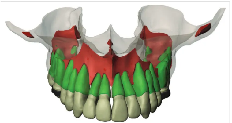
Figure 1 illustrates a virtual model of the maxilla built through computed axial tomography (iCAT, Xo-ran Technologies, Ann Arbor, USA) totaling 218 sec-tions of 640 x 640 voxels each.
The model presented a maxilla with 956,196 faces and each tooth with dozens of hundreds of polyhedral faces. Considering that, at this stage, the model had not been inished, we reduced the number of faces to a maximum of a few hundreds so as to carry out its edi-tion. The simple reduction in the number of nonpara-metric faces leads to great distortion of the model, since the former are exclusively triangular and lat. In order to Kanjanaouthaia et al19 employed the FEM to
demon-strate that ater having received force of 1 N in lingual di-rection, upper incisors that were more inclined presented higher concentration of stress on the apex when compared with incisors well positioned in buccolingual direction.
To conduct this experimental method it is interesting to use a resource with anatomical records and modiica-tions in CAD sotware so as to build geometrically supe-rior and accurate models. To that purpose, it is necessary to build a virtual model using an image-processing and digital reconstruction sotware, such as Mimics (Mate-rialize, Leuven Belgium) or Simpleware 4 (Simpleware Ltd., Exeter, United Kingdom). In general, regarding the maxillomandibular complex, these reconstructions are carried out through computed tomography.
Computed tomography should be obtained with cross-sections of at least 0.25 mm distance so as to achieve improved resolution. The sections will be re-corded on DICOM format (Digital Imaging and Com-munications in Medicine) and imported into an image-processing and digital reconstruction sotware. The lev-el of contrast and deinition of clinical tomography lead
enable further edition without signiicant distortion, the models should be parameterized using Solidworks Pre-mium sotware “scan to 3D” (Dassault Systemes, Solid-works Corps, USA), thereby making the transformation of nonparametric models into parametric models with NURBS faces (Non Uniform Rational Bases Splines), with minimum distortion, possible. Orthodontic com-ponents should also be virtually reconstructed with the aid of a digital calliper (Litz professional, Germany) and a digital microscope.
The more structures are modelled, the more accurate are the results; however, it makes the model more diicult to be obtained and the analysis of the results more com-plex. Therefore, simpler models should be applied in order to obtain the same quantitative results. Modelling should be carefully assessed so as to simplify the model according to its actual needs and without compromising the results.4
In order to enable analysis by means of FEM, we also used the aforementioned sotware to conduct the
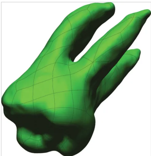
transformation of the solid model into a mesh of bonds and elements, which is the discretization of the model.
The elements represent coordinates in space and may present with several formats, in which case tetrahe-drons and hexahetetrahe-drons are the most common. The ex-tremities of each element present points, or bonds, that connect the elements to each other forming an arranged mesh.4 It is the bonds that transmit information
be-tween elements (Fig 2).
To reach the ideal mesh of inite elements, we use a process of mesh reinement to verify the convergence of results with gradual increase in the number of bonds and elements until the diference of voltage peaks be-tween mesh reinements is 5% or lower. These mea-sures minimize the geometric error peculiar to a process of mesh discretization.
After the entire virtual model is reconstruct-ed and transformreconstruct-ed into this mesh of finite ele-ments, it is exported from Solidworks software to
Ansys Workbench V11 software for finite elements simulation. (Ansys Inc., Canonsburg, PA, USA). This software requires the correct representation of the mechanical behavior of each component; thus, the model is set with a modulus of elasticity (Young) and Poisson’s coefficient. Poisson’s coefficient refers to the absolute value of the relationship between transverse and longitudinal deformations in an axial traction axis; whereas Young’s model represents the inclination of the linear portion of (material) the stress-deformation diagram.4 Subsequently, we
con-duct the activation of the system with the applica-tion of charges using the aforemenapplica-tioned software.
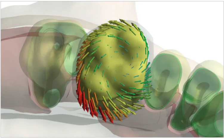
The results obtained by means of the FEM enable analysis of stress distribution produced by forces be-tween the periodontal ligament and the bone, thereby demonstrating the areas of stress and, thus, the location where tooth movement occurs. It also enable us to in-fer about areas that are more prone to root resorption.
These results are revealed by means of colors and ar-rows that are able to indicate the direction of tooth dis-placement ater force application.
By verifying the colored illustration (Fig 3) of this experimental model, which is part of a research con-ducted at School of Dentistry/Unesp-Araraquara, we found that the region near the tooth fulcrum (red) holds a higher concentration of forces, and that the stress gradually decreases towards the apex (blue). In addition, using a resource of Ansys 14 Software, we assessed the tendency of movement to which the tooth is submitted against the application of the orthodontic force by means of colored arrows that demonstrate (red) the location with higher tooth displacement (Fig 4).
In addition to viewing the stress distribution on the periodontal ligament, it is possible to observe the deformation of the orthodontic wire, and its region with higher stress concentration (Fig 5).
Figure 4. Arrows indicate the direction of tooth displacement and its intensity (red for higher displacement; green for lower displacement).
1. Sameshima GT, Melnick M. Finite element-based cephalometric analysis. Angle Orthod. 1994;64(5):343-50.
2. Viecilli RF. Self-corrective T-loop design for differential space closure. Am J Orthod Dentofacial Orthop. 2006;129(1):48-53.
3. Azevedo AFM. Método dos Elementos Finitos. [Acesso em: 2014 Oct
29]. Disponível em: http://civil.fe.up.pt/pub/apoio/ano5/aae/pdf/ Apontamentos/Livro_MEF_AA.pdf.
4. Lotti RS, Machado AW, Mazzieiro ET, Landre Jr J. Raquel S.
Aplicabilidade cientifica do métodos dos elementos finitos. Rev Dent Press Ortod Ortop Facial. 2006;11(2):35-43.
5. Shaw AM, Sameshima GT, Vu HV. Mechanical stress generated by
orthodontic forces on apical root cementum: a finite element model. Orthod Craniofacial Res. 2004;7(2):98-107.
6. Jones ML, Hickman J, Middleton J, Knox J, Volp C. A validated finite
element method study of orthodontic tooth movement in the human subject. Am J Orthod. 2001;28(1):29-38.
7. Reitan K. Tissue behavior during orthodontic tooth movement. Am J
Orthod. 1960;46(12):881-900.
8. Sari E, Olmez H, Gurton U. Comparison of some effects of
acetylsalicylic acid and rofecoxib during orthodontic tooth movement. Am J Orthod Dentofacial Orthop. 2004;125(3):310-5.
9. Kalia S, Melsen B, Verna C. Tissue reaction to orthodontic tooth movement in acute and chronic corticosteroid treatment. Orthod Craniofac Res. 2004;7(1):26-34.
10. Jäger A, Zhang D, Kawarizadeh A, Tolba R, Braumann B, Lossdörfer S, Götz W. Soluble cytokine receptor treatment in experimental orthodontic tooth movement in the rat. Eur J Orthod. 2005;27(1):1-11. 11. Ong CK, Walsh LJ, Harbrow D, Taverne AA, Symons AL. Orthodontic
tooth movement in the prednisolone-treated rat. Angle Orthod. 2000;70(2):118-25.
12. Caputo A, Chaconis SJ, Hayashi RK. Photoelastic visualization of orthodontic forces during canine retraction. Am J Orthod. 1974;65(3):250-9.
REFERENCES
13. Kamble RH, Lohkare S, Hararey PV, Mundada RD. Stress distribution pattern in a root of maxillary central incisor having various root morphologies: a finite element study. Angle Orthod. 2012;82(5):799-805.
14. Middleton J, Jones M, Wilson A. The role of the periodontal ligament in bone modeling: the initial development of a time- dependent finite element model. Am J Orthod Dentofacial Orthop. 1996;109(2):155-62. 15. McGuinness N, Wilson AN, Jones M, Middleton J, Robertson NR.
Stresses induced by edgewise appliances in the periodontal ligament-a finite element study. Angle Orthod. 1992;62(1):15-22.
16. Shaw AM, Sameshima GT, Vu HV. Mechanical stress generated by orthodontic forces on apical root cementum: a finite element model. Orthod Craniofac Res. 2004;7(2):98-107.
17. Kojima Y, Fukui H. Effects of transpalatal arch on molar movement produced by mesial force: a finite element simulation. Am J Orthod Dentofacial Orthop. 2008;134(3):335.e1-7.
18. Tominaga JY, Tanaka M, Koga Y, Gonzales C, Kobayashi M, Yoshida N. Optimal loading conditions for controlled movement of anterior teeth in sliding mechanics. Angle Orthod. 2009;79(6):1102-10.
19. Kanjanaouthai A, Mahatumarat K, Techalertpaisarn P, Versluis A. Effect of the inclination of a maxillary central incisor on periodontal stress Finite element analysis Angle Orthod. 2012;82(5):812-9.
20. Choi DS, Cha BK, Jang I, Kang KH, Kim SC. Three-dimensional finite element analysis of occlusal stress distribution in the human skull with premolar extraction Angle Orthod. 2013;83(2):204-11.
21. Andreaus U, Colloca M, Iacoviello D. Coupling image processing and stress analysis for damage identification in a human premolar tooth. Comput Methods Programs Biomed. 2011;103(2):61-73.
FINAL CONSIDERATIONS
The Finite Element Method (FEM) proves to be an important instrument in orthodontic research, highlighting several points, such as: stress distribu-tion areas in the periodontal ligament and alveo-lar bone during tooth movements; direction of the tooth displacement; the ideal position of orthodon-tic appliances during a specific mechanics; areas most likely to present root resorption; In addition