Acute Myeloid Leukemia (AML) with Normal
Cytogenetics
Marta Fernandez-Mercado1, Bon Ham Yip1, Andrea Pellagatti1, Carwyn Davies1, Marı´a Jose´ Larrayoz2, Toshinori Kondo3, Cristina Pe´rez4, Sally Killick5, Emma-Jane McDonald5, Marı´a Dolores Odero2,6, Xabier Agirre7, Felipe Pro´sper7, Marı´a Jose´ Calasanz2, James S. Wainscoat1, Jacqueline Boultwood1* 1LLR Molecular Haematology Unit, NDCLS, John Radcliffe Hospital, Oxford, United Kingdom,2Department of Genetics, University of Navarra, Pamplona, Spain,3Division of Hematology, Kawasaki Medical School, Okayama, Japan,4Laboratory of Myeloproliferative Syndromes, Oncology Area, Foundation for Applied Medical Research, Clı´nica Universitaria, Universidad de Navarra, Pamplona, Spain,5Department of Haematology, Royal Bournemouth Hospital, Bournemouth, United Kingdom,6Division of Oncology, Center for Applied Medical Research, Universidad de Navarra, Pamplona, Spain,7Division of Cancer and Area of Cell Therapy and Hematology Service, Foundation for Applied Medical Research, Clı´nica Universitaria, Universidad de Navarra, Pamplona, Spain
Abstract
Acute myeloid leukemia patients with normal cytogenetics (CN-AML) account for almost half of AML cases. We aimed to study the frequency and relationship of a wide range of genes previously reported as mutated in AML (ASXL1,NPM1,FLT3, TET2,IDH1/2,RUNX1,DNMT3A,NRAS,JAK2,WT1,CBL,SF3B1,TP53,KRASandMPL) in a series of 84 CN-AML cases. The most frequently mutated genes in primary cases wereNPM1(60.8%) andFLT3(50.0%), and in secondary casesASXL1(48.5%) and TET2(30.3%). We showed that 85% of CN-AML patients have mutations in at least one ofASXL1,NPM1,FLT3,TET2,IDH1/2 and/orRUNX1. Serial samples from 19 MDS/CMML cases that progressed to AML were analyzed forASXL1/TET2/IDH1/2 mutations; seventeen cases presented mutations of at least one of these genes. However, there was no consistent pattern in mutation acquisition during disease progression. This report concerns the analysis of the largest number of gene mutations in CN-AML studied to date, and provides insight into the mutational profile of CN-AML.
Citation:Fernandez-Mercado M, Yip BH, Pellagatti A, Davies C, Larrayoz MJ, et al. (2012) Mutation Patterns of 16 Genes in Primary and Secondary Acute Myeloid Leukemia (AML) with Normal Cytogenetics. PLoS ONE 7(8): e42334. doi:10.1371/journal.pone.0042334
Editor:Ralf Krahe, University of Texas MD Anderson Cancer Center, United States of America ReceivedApril 13, 2012;AcceptedJuly 3, 2012;PublishedAugust 9, 2012
Copyright:ß2012 Fernandez-Mercado et al. This is an open-access article distributed under the terms of the Creative Commons Attribution License, which permits unrestricted use, distribution, and reproduction in any medium, provided the original author and source are credited.
Funding:This work was supported by Leukaemia and Lymphoma Research and the Kay Kendall Leukaemia Fund of the United Kingdom. The funders had no role in study design, data collection and analysis, decision to publish, or preparation of the manuscript.
Competing Interests:The authors have declared that no competing interests exist.
* E-mail: [email protected]
Introduction
Acute myeloid leukemia (AML) is a heterogeneous disease in terms of karyotype and molecular abnormalities. The discovery of the classic karyotype abnormalities in AML such as the t(15;17) has been invaluable in enabling more accurate prognostic estimates, the development of specific therapies and the molecular monitoring of disease. However, approximately half of AML patients have no karyotype abnormality (CN-AML). This group of AML cases is presumably heterogeneous in all respects, and molecular monitoring is not possible unless there is an associated mutation. Recently it has been demonstrated that mutations of
FLT3, NPM1and CEBPA genes are preferentially found in CN-AML. [1] Nevertheless many cases do not possess such mutations and this imposes a severe limitation in understanding their specific pathophysiology and monitoring disease progression. We have chosen to study CN-AML with the aim of finding a restricted panel of genes which are mutated in the majority of cases. In a series of 84 CN-AML patients, we examined 16 genes with mutations that had previously been described in cases of CN-AML (Table S1). [2,3,4,5,6,7,8,9,10,11,12,13,14,15,16,17] The charac-terisation of cases by the presence or absence of mutations in these selected genes should allow a molecular dissection of cases of
CN-AML into different biological and prognostic groups, as well as achieving the long sought after goal of molecular monitoring of CN-AML.
Design and Methods
Patients
karyotype were subjected to confirmation by molecular tech-niques. This study was approved by the ethics committees of the institutes involved: the John Radcliffe Hospital (Oxford 06/ Q1606/110), the Royal Bournemouth Hospital (Bournemouth
9991/03/E) and the University of Navarre (Pamplona IRB00006933); written informed consent was received from all patients.
Figure 1. Concurrence of mutations in 16 genes analyzed in CN-AML samples.Columns show results for each of the 84 analysed cases. Solid boxes indicate mutated cases. Grey boxes mark unavailable data.FLT3-ITDmutations are indicated with top-half solid boxes andFLT3-TKDwith
bottom-half solid boxes. Similarly,IDH2-R140Qmutations are shown with top-half solid boxes andIDH2-R172Kwith bottom-half solid boxes. doi:10.1371/journal.pone.0042334.g001
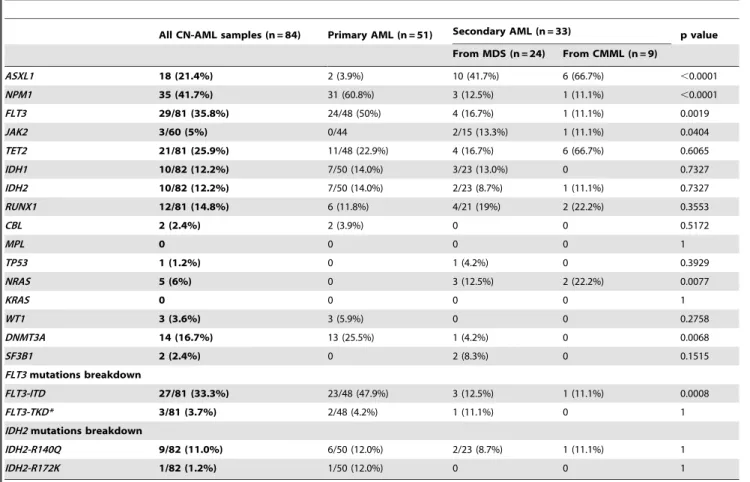
Table 1.Frequency of mutations in normal karyotype AML samples.
All CN-AML samples (n = 84) Primary AML (n = 51) Secondary AML (n = 33) p value
From MDS (n = 24) From CMML (n = 9)
ASXL1 18 (21.4%) 2 (3.9%) 10 (41.7%) 6 (66.7%) ,0.0001
NPM1 35 (41.7%) 31 (60.8%) 3 (12.5%) 1 (11.1%) ,0.0001
FLT3 29/81 (35.8%) 24/48 (50%) 4 (16.7%) 1 (11.1%) 0.0019
JAK2 3/60 (5%) 0/44 2/15 (13.3%) 1 (11.1%) 0.0404
TET2 21/81 (25.9%) 11/48 (22.9%) 4 (16.7%) 6 (66.7%) 0.6065
IDH1 10/82 (12.2%) 7/50 (14.0%) 3/23 (13.0%) 0 0.7327
IDH2 10/82 (12.2%) 7/50 (14.0%) 2/23 (8.7%) 1 (11.1%) 0.7327
RUNX1 12/81 (14.8%) 6 (11.8%) 4/21 (19%) 2 (22.2%) 0.3553
CBL 2 (2.4%) 2 (3.9%) 0 0 0.5172
MPL 0 0 0 0 1
TP53 1 (1.2%) 0 1 (4.2%) 0 0.3929
NRAS 5 (6%) 0 3 (12.5%) 2 (22.2%) 0.0077
KRAS 0 0 0 0 1
WT1 3 (3.6%) 3 (5.9%) 0 0 0.2758
DNMT3A 14 (16.7%) 13 (25.5%) 1 (4.2%) 0 0.0068
SF3B1 2 (2.4%) 0 2 (8.3%) 0 0.1515
FLT3mutations breakdown
FLT3-ITD 27/81 (33.3%) 23/48 (47.9%) 3 (12.5%) 1 (11.1%) 0.0008
FLT3-TKD* 3/81 (3.7%) 2/48 (4.2%) 1 (11.1%) 0 1
IDH2mutations breakdown
IDH2-R140Q 9/82 (11.0%) 6/50 (12.0%) 2/23 (8.7%) 1 (11.1%) 1
IDH2-R172K 1/82 (1.2%) 1/50 (12.0%) 0 0 1
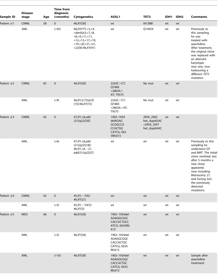
Table 2.Mutational analysis of serial samples from patients in transformation.
Sample ID
Disease
stage Age
Time from diagnosis
(+months) Cytogenetics ASXL1 TET2 IDH1 IDH2 Comments
Patient#1 CMML 58 0 46,XY[30] wt M1388I wt wt
AML (+45) 68,XXYYY,+3,+4, +der(6)x3,+7,+8, +8,+9,+11,+11, +12,+13,+17,+19, +19,+20,+21,+21, +22[9]/46,XY[41]
wt Q1445X wt wt Previously to
this sampling, he was treated with azacitidine. After treatment, the original clone was replaced with an aberrant karyotype new one, now harbouring a differentTET2
mutation.
Patient#2 CMML 65 0 46,XY[30] 2242C.CT;
Q748X +2863A.
AT; T957S
No mut wt wt
AML (+9) 46,XY,i(17)(q10)
[15]/46,XY[15]
2242C.CT; Q748X +2863A.AT; T957S
No mut wt wt
Patient#3 CMML 65 0 47,XY,+8,add
(21)(q22)[30]
1902–1924 delAGAG GCGGCCA CCACTGC CATCG; E63 5RfsX15
2959_2962 het_dupAGAC +2954_2957 het_dupAAAC
wt wt
AML (+6) 47,XY,+8,add
(21)(q22)[18]/ 46,XY,+8,221, add(21)(q22)[7]
wt wt wt wt Previously to this
sampling he underwent QT and BMT. The initial clone remitted, but after 5 months a new clone appeared, now including Monosomy 21, and having lost the previously detected mutations.
Patient#4 CMML 42 0 45,XY,27[4]/
46,XY[21]
wt wt wt wt
AML (+5) 45,XY,27[47]/
46,XY[3]
wt wt wt wt
Patient#5 MDS 66 0 46,XY[30] 1902–1924del
AGAGGCGGC CACCACTGCC ATCG; E635Rfs X15
wt wt wt
AML (+5) 46,XY[30] 1902–1924del
AGAGGCGGC CACCACTGC CATCG; E635 RfsX15
wt wt wt
AML (+10) 46,XY[30] 1902–1924del
AGAGGCGGC CACCACTGC CATCG; E635 RfsX15
wt wt wt Sample after
Table 2.Cont.
Sample ID
Disease
stage Age
Time from diagnosis
(+months) Cytogenetics ASXL1 TET2 IDH1 IDH2 Comments
AML (+12) 46,XY[30] 1902–1924del
AGAGGCGGC CACCACTGC CATCG; E635 RfsX15
wt wt wt Sample after
azacitidine treatment.
Patient#6 MDS 72 0 46,XY[30] 1925het_insA;
G643RfsX13
L34F wt wt
AML (+11) 46,XY[30] 1925het_insA;
G643RfsX13
L34F wt wt AdditionalJAK2,
RUNX1andNRAS
mutations.
Patient#7 MDS 70 0 46,XY[30] 1934dupG;
G646WfsX12
wt wt wt
MDS (RAEB) (+8) 46,XY[30] 1934dupG;
G646WfsX12
wt wt wt
AML (+15) 46,XY[30] 1934dupG;
G646WfsX12
wt wt wt Additional
NRASmutation.
Patient#8 MDS 76 0 46,XX[30] 1902–1924del
AGAGGCGGC CACCACTGC CATCG E635 RfsX15
wt wt wt
AML (+12) 46,XX[30] 1902–1924del
AGAGGCGGC CACCACTGCC ATCG E635 RfsX15
wt R132C wt
Patient#9 MDS (RA) 77 0 46,XX[30] wt Q403X wt wt
CMML (+8) 46,XX[30] 1934dupG
G646WfsX12
Q403X wt wt AML-transformed
at (+40).
Patient#10 MDS (RA) ND 0 46,XX[30] 1934dupG
G646WfsX12
wt wt wt
CMML (+12) 46,XX[30] 1934dupG
G646WfsX12
ND wt wt
Patient#11 MDS 84 0 46,XX[30] 1934dupG
G646WfsX12
L1151P wt wt
MDS (RAEB) (+12) 46,XX[30] 1934dupG
G646WfsX12
L1151P wt wt
Patient#12 MDS 76 0 46,XX[30] 1934dupG
G646WfsX12
Y867H wt R140Q
MDS (RAEB) (+3) 46,XX[30] 1934dupG
G646WfsX12
Y867H wt R140Q Previously to this sample, she underwent one course of AraC.
Patient#13 MDS 64 0 46,XX[30] 1934dupG wt wt R140Q
MDS (RAEB) (+12) 46,XX[30] 1934dupG wt wt R140Q
Patient#14 MDS 79 (+20) 46,XX,del(5) (q15:q33)[30]
1934dupG G646WfsX12
S794X wt wt
MDS (RAEB) (+53) 46,XX,del(5) (q15:q33)[30]
1934dupG G646WfsX12
S794X wt wt
Patient#15 MDS 80 0 46,XX[30] wt wt wt wt
MDS (+1) 46,XX[30] wt wt wt wt
AML (+2) 46,XX[30] 2893C.
C/T; R965X
wt wt wt Non mutated
DNA sequencing and analysis
Genomic DNA was isolated from patient bone marrow or peripheral blood samples. Primers and PCR conditions for the 16 genes analyzed are detailed in Table S2. Relevant regions were selected for analysis (Table S2): exons 12 of ASXL1
(NM_015338.5) and NPM1 (NM_002520), exons 11 and 17 of
FLT3 (NM_004119), exon 14 of JAK2 (NM_004972), entire coding region of TET2 (NM_001127208.2), Exons 4 of IDH1
(NM_005896) andIDH2(NM_002168), exons 3 to 8 ofRUNX1
(NM_001001890), exons 7–9 ofCBL(NM_005188), exons 9 and 10 of MPL (NM_005373), exons 3 to 9 ofTP53(NM_000546), exons 2 and 3 ofNRAS(NM_002524.4) andKRAS(NM_033360), Exons 4 to 9 ofWT1(NM_024426), exons 7 to 23 ofDNMT3A
(NM_022552) and exons 12 to 16 ofSF3B1(NM_012433.2). PCR was performed using ThermoStart PCR Master Mix (Thermo Fisher Scientific), following the manufacturer’s protocol. PCR products were purified and bidirectionally sequenced using the BigDye Terminator v1.1 cycle sequencing kit (Applied Biosystems, Foster City, CA, USA) and an ABI 3100 Genetic Analyzer. Sequence data were analyzed using Mutation Surveyor V3.25 (Softgenetics, State College, PA, USA). Two sided Fisher’s exact test was performed to compare mutation frequencies in primary versus secondary cases, and in the analysis of cooperating mutations.
Results and Discussion
A total of 84 CN-AML patients were recruited for mutational analysis, including 51 primary cases and 33 cases secondary to either MDS (n = 24) or CMML (n = 9). The 16 genes analyzed were: ASXL1, NPM1, FLT3, TET2, IDH1, IDH2, RUNX1,
DNMT3A, NRAS, JAK2, WT1, CBL, SF3B1, TP53, KRAS and
MPL. The regions analysed for each gene are detailed in Table S2. The frequencies of mutation are shown in Table 1. The most frequently mutated genes in primary cases wereNPM1 (60.8%) and FLT3 (50.0%), and in secondary cases ASXL1(48.5%) and
TET2(30.3%).
An analysis of the mutations occurring in more than 10% of cases revealed statistically significant associations (Figure 1, Table S3). In agreement with previous reports, FLT3 and DNMT3A
mutations were significantly associated withNPM1mutations, [12] whereas patients with ASXL1 mutations had significantly lower incidence of NPM1 and DNMT3A mutations. [8,9] IDH1 and
IDH2mutations were mutually exclusive. With the exception of one patient, no cases with IDH1/2 mutation also had a TET2
mutation.IDH1andIDH2mutations were less frequent inTET2 -mutated than in TET2-wt patients, and this has been reported before. [19,20] Concurrence ofIDH1/2andASXL1mutations was also a relatively infrequent event in our patient cohort (Figure 1). This observation is in agreement with a report on a series of 63 AML secondary to MPN cases. [3]
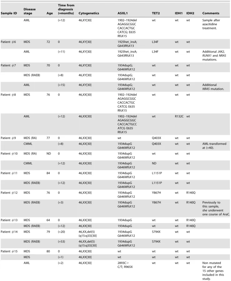
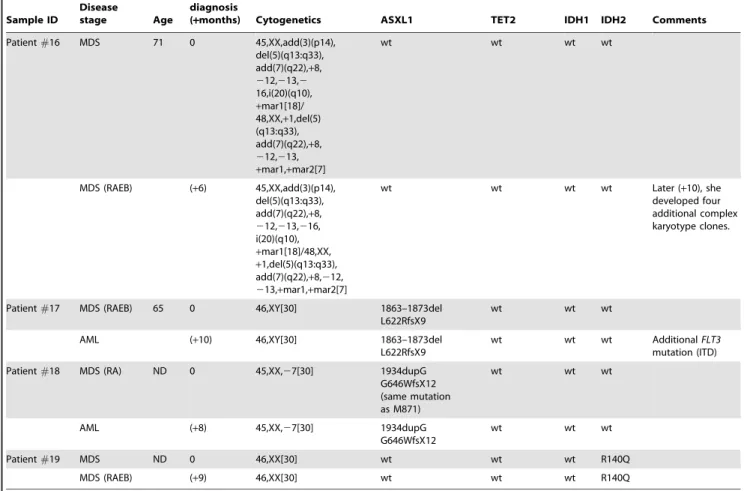
Table 2.Cont.
Sample ID
Disease
stage Age
Time from diagnosis
(+months) Cytogenetics ASXL1 TET2 IDH1 IDH2 Comments
Patient#16 MDS 71 0 45,XX,add(3)(p14), del(5)(q13:q33), add(7)(q22),+8,
212,213,2
16,i(20)(q10), +mar1[18]/ 48,XX,+1,del(5) (q13:q33), add(7)(q22),+8,
212,213, +mar1,+mar2[7]
wt wt wt wt
MDS (RAEB) (+6) 45,XX,add(3)(p14), del(5)(q13:q33), add(7)(q22),+8,
212,213,216, i(20)(q10), +mar1[18]/48,XX, +1,del(5)(q13:q33), add(7)(q22),+8,212,
213,+mar1,+mar2[7]
wt wt wt wt Later (+10), she
developed four additional complex karyotype clones.
Patient#17 MDS (RAEB) 65 0 46,XY[30] 1863–1873del
L622RfsX9
wt wt wt
AML (+10) 46,XY[30] 1863–1873del
L622RfsX9
wt wt wt AdditionalFLT3
mutation (ITD)
Patient#18 MDS (RA) ND 0 45,XX,27[30] 1934dupG G646WfsX12 (same mutation as M871)
wt wt wt
AML (+8) 45,XX,27[30] 1934dupG
G646WfsX12
wt wt wt
Patient#19 MDS ND 0 46,XX[30] wt wt wt R140Q
MDS (RAEB) (+9) 46,XX[30] wt wt wt R140Q
ASXL1mutations were significantly more frequent in secondary AML compared tode novoAML cases (primary cases: 2/51, 3.9%; secondary to MDS/CMML: 16/33, 48.5%, p,0.0001). We have previously reported a high prevalence of ASXL1 mutations in advanced MDS. [18]NPM1,FLT3, andDNMT3Amutations were significantly more common in primary CN-AML than in secondary AML cases (Table 1).NRAS, JAK2, SF3B1and TP53
mutations were exclusively present in secondary AML samples (Table 1). Only 9.5% of the samples analyzed (8/84, 6de novoand 2 post-MDS cases) showed no mutation in any of the genes tested. When considering only theASXL1,NPM1,FLT3,TET2,IDH1/2
and RUNX1gene analysis, 88% ofde novoCN-AML included in this series presented at least one molecular marker. For secondary cases, 85% of patients carried mutations in at least one of these 7 genes.
Recent reports showed thatDNMT3Amutations are associated with a poor outcome in AML, [21,22] and that the location of the mutations could have an impact in age-related risk classification. [23] It is worth noting that in our series,DNMT3Awas not found as a sole mutation suggesting that additional aberrations are needed to sustain leukemogenic development.
Approximately 70% of CN-AML cases secondary to either MDS or CMML presented mutations in at least one of ASXL1,
TET2, IDH1orIDH2genes. Therefore, we chose to assess the presence and chronology of ASXL1, TET2 and IDH1/2
mutational events, in order to investigate whether they could have a role in disease development or evolution. We studied 15 MDS and 4 CMML cases that progressed to AML, for which at least two samples at different time-points were available. Remarkably, with the exception of two patients all of them possessed at least one gene mutation at some stage of the disease. The majority showed the same mutations at early and later stages of the disease, except one patient who developed an IDH1
mutation at transformation, a second patient with a TET2
mutation who acquired an additional ASXL1 mutation at transformation, and another patient who developed a nonsense mutation of ASXL1 at AML stage, and showed rapid disease evolution (Table 2). On the basis of this study we therefore did not find any consistent patterns in mutation acquisition. A sensitive mutation analysis (such as allele-specific PCR) at early stages of AML in future studies could help clarify whether the mutations found in cases from later stage AML were already present as a minor pre-existing clone at the earlier stage, and if so, how it evolved as the disease progressed to AML.
In order to investigate whether the observed low incidence of
ASXL1 mutations is a specific characteristic of karyotypically normalde novocases, or is a common feature of other subtypes of primary AML, we screened an additional cohort of 100 primary AML, including the most common karyotypic subgroups. Overall,
only 8 out of 100 cases showed nonsense or frameshift mutations (8%) (Table S4), confirming that ASXL1 mutations are less common in primary AML than in secondary AML.
This report concerns the analysis of the largest number of gene mutations in CN-AML studied to date. Our results show that 85% of CN-AML patients have mutations in one or more of 7 selected genes (ASXL1, NPM1, FLT3,TET2,IDH1/2 andRUNX1). This finding will facilitate further analysis of this important group of patients by enabling CN-AML patients to be subdivided into groups with common mutation patterns. Detailed studies of the CN-AML subgroups in regard to their hematological features, prognosis, disease progression and treatment response will now be facilitated.
Supporting Information
Table S1 Relevant literature on mutations of AML patients. ND = not done. CN = cytogenetically normal. MPN = myelopro-liferative neoplasm. Yo = years old. CBF = core binding factor. APL = acute promyelocytic leukemia.
(PDF)
Table S2 Primers and PCR conditions. PCR was performed using ThermoStart PCR Master Mix (Thermo Fisher Scientific), following the manufacturer’s protocol, 35 cycles, unless otherwise stated, using indicated annealing temperature. The same primers were used for Sanger sequencing unless otherwise stated. (PDF)
Table S3 Double-sided Fisher’s exact test analysis of coopera-tion between most frequent mutacoopera-tions in normal karyotype AML samples.
(PDF)
Table S4 ASXL1 mutations in 100 de novo AML cases with aberrant cytogenetics.
(PDF)
Acknowledgments
The authors would like to thank the patients who accepted to participate in this study. The authors would also like to thank all co-workers in their laboratories for their technical assistance as well as all physicians for referring patient material to their centers.
Author Contributions
Conceived and designed the experiments: JB JSW. Performed the experiments: MFM BHY AP CD MJL TK CP. Analyzed the data: MFM BHY AP CD MJL TK CP. Contributed reagents/materials/analysis tools: SK EJM MJC MDO XA FP. Wrote the paper: MFM AP CD JSW JB.
References
1. Mrozek K, Marcucci G, Paschka P, Whitman SP, Bloomfield CD (2007) Clinical relevance of mutations and gene-expression changes in adult acute myeloid leukemia with normal cytogenetics: are we ready for a prognostically prioritized molecular classification? Blood 109: 431–448.
2. Rocquain J, Carbuccia N, Trouplin V, Raynaud S, Murati A, et al. (2010) Combined mutations of ASXL1, CBL, FLT3, IDH1, IDH2, JAK2, KRAS, NPM1, NRAS, RUNX1, TET2 and WT1 genes in myelodysplastic syndromes and acute myeloid leukemias. BMC Cancer 10: 401.
3. Abdel-Wahab O, Manshouri T, Patel J, Harris K, Yao J, et al. (2010) Genetic analysis of transforming events that convert chronic myeloproliferative neoplasms to leukemias. Cancer Res 70: 447–452.
4. Ishikawa Y, Kiyoi H, Tsujimura A, Miyawaki S, Miyazaki Y, et al. (2009) Comprehensive analysis of cooperative gene mutations between class I and class II in de novo acute myeloid leukemia. Eur J Haematol 83: 90–98.
5. Abbas S, Rotmans G, Lowenberg B, Valk PJ (2008) Exon 8 splice site mutations in the gene encoding the E3-ligase CBL are associated with core binding factor acute myeloid leukemias. Haematologica 93: 1595–1597.
6. Couronne L, Lippert E, Andrieux J, Kosmider O, Radford-Weiss I, et al. (2010) Analyses of TET2 mutations in post-myeloproliferative neoplasm acute myeloid leukemias. Leukemia 24: 201–203.
7. Schlenk RF, Dohner K, Krauter J, Frohling S, Corbacioglu A, et al. (2008) Mutations and treatment outcome in cytogenetically normal acute myeloid leukemia. N Engl J Med 358: 1909–1918.
8. Carbuccia N, Trouplin V, Gelsi-Boyer V, Murati A, Rocquain J, et al. (2010) Mutual exclusion of ASXL1 and NPM1 mutations in a series of acute myeloid leukemias. Leukemia 24: 469–473.
10. Flach J, Dicker F, Schnittger S, Schindela S, Kohlmann A, et al. (2011) An accumulation of cytogenetic and molecular genetic events characterizes the progression from MDS to secondary AML: an analysis of 38 paired samples analyzed by cytogenetics, molecular mutation analysis and SNP microarray profiling. Leukemia 25: 713–718.
11. Dicker F, Haferlach C, Sundermann J, Wendland N, Weiss T, et al. (2010) Mutation analysis for RUNX1, MLL-PTD, FLT3-ITD, NPM1 and NRAS in 269 patients with MDS or secondary AML. Leukemia 24: 1528–1532. 12. Shen Y, Zhu YM, Fan X, Shi JY, Wang QR, et al. (2011) Gene mutation
patterns and their prognostic impact in a cohort of 1185 patients with acute myeloid leukemia. Blood 118: 5593–5603.
13. Thol F, Damm F, Ludeking A, Winschel C, Wagner K, et al. (2011) Incidence and prognostic influence of DNMT3A mutations in acute myeloid leukemia. J Clin Oncol 29: 2889–2896.
14. Beer PA, Delhommeau F, LeCouedic JP, Dawson MA, Chen E, et al. (2010) Two routes to leukemic transformation after a JAK2 mutation-positive myeloproliferative neoplasm. Blood 115: 2891–2900.
15. Pratcorona M, Abbas S, Sanders M, Koenders J, Kavelaars F, et al. (2011) Acquired mutations in ASXL1 in acute myeloid leukemia: prevalence and prognostic value. Haematologica.
16. Metzeler KH, Becker H, Maharry K, Radmacher MD, Kohlschmidt J, et al. (2011) ASXL1 mutations identify a high-risk subgroup of older patients with primary cytogenetically normal AML within the ELN Favorable genetic category. Blood 118: 6920–6929.
17. Jankowska AM, Makishima H, Tiu RV, Szpurka H, Huang Y, et al. (2011) Mutational spectrum analysis of chronic myelomonocytic leukemia includes
genes associated with epigenetic regulation: UTX, EZH2, and DNMT3A. Blood 118: 3932–3941.
18. Boultwood J, Perry J, Pellagatti A, Mercado M, Fernandez-Santamaria C, et al. (2010) Frequent mutation of the polycomb-associated gene ASXL1 in the myelodysplastic syndromes and in acute myeloid leukemia. Leukemia 24: 1062–1065.
19. Figueroa ME, Abdel-Wahab O, Lu C, Ward PS, Patel J, et al. (2010) Leukemic IDH1 and IDH2 mutations result in a hypermethylation phenotype, disrupt TET2 function, and impair hematopoietic differentiation. Cancer Cell 18: 553– 567.
20. Metzeler KH, Maharry K, Radmacher MD, Mrozek K, Margeson D, et al. (2011) TET2 mutations improve the new European LeukemiaNet risk classification of acute myeloid leukemia: a Cancer and Leukemia Group B study. J Clin Oncol 29: 1373–1381.
21. Ley TJ, Ding L, Walter MJ, McLellan MD, Lamprecht T, et al. (2010) DNMT3A mutations in acute myeloid leukemia. N Engl J Med 363: 2424–2433. 22. Renneville A, Boissel N, Nibourel O, Berthon C, Helevaut N, et al. (2012) Prognostic significance of DNA methyltransferase 3A mutations in cytogenet-ically normal acute myeloid leukemia: a study by the Acute Leukemia French Association. Leukemia.