Effects of centrally acting
antihypertensive drugs on the
microcirculation of spontaneously
hypertensive rats
1Departamento de Fisiologia e Farmacodinâmica, Instituto Oswaldo Cruz,
FIOCRUZ, Rio de Janeiro, RJ, Brasil
2Laboratoire de Neurobiologie et Pharmacologie Cardiovasculaire,
Faculté de Médecine, Université Louis Pasteur, Strasbourg, France V. Estato1,
C.V. Araújo1,
P. Bousquet2 and
E. Tibiriçá1
Abstract
We investigated the acute effects of centrally acting antihypertensive drugs on the microcirculation of pentobarbital-anesthetized spontane-ously hypertensive rats (SHR). The effects of the sympatho-inhibitory agents clonidine and rilmenidine, known to activate both α2 -adreno-ceptors and nonadrenergic I1-imidazoline binding sites (I1BS) in the
central nervous system, were compared to those of dicyclopropyl-methyl-(4,5-dimethyl-4,5-dihydro-3H-pyrrol-2-yl)-amine hydrochlo-ride (LNP 509), which selectively binds to the I1BS. Terminal
mesen-teric arterioles were observed by intravital microscopy. Activation of the central sympathetic system with L-glutamate (125 µg, ic) induced marked vasoconstriction of the mesenteric microcirculation (27 ± 3%; N = 6, P < 0.05). In contrast, the marked hypotensive and bradycardic effects elicited by intracisternal injection of clonidine (1 µg), rilmeni-dine (7 µg)and LNP 509 (60 µg) were accompanied by significant increases in arteriolar diameter (12 ± 1, 25 ± 10 and 21 ± 4%, respectively; N = 6, P < 0.05). The vasodilating effects of rilmenidine and LNP 509 were two-fold higher than those of clonidine, although they induced an identical hypotensive effect. Central sympathetic inhibition elicited by baclofen (1 µg, ic), a GABAB receptor agonist,
also resulted in vasodilation of the SHR microvessels. The acute administration of clonidine, rilmenidine and LNP 509 also induced a significant decrease of cardiac output, whereas a decrease in systemic vascular resistance was observed only after rilmenidine and LNP 509. We conclude that the normalization of blood pressure in SHR induced by centrally acting antihypertensive agents is paralleled by important vasodilation of the mesenteric microcirculation. This effect is more pronounced with substances acting preferentially (rilmenidine) or exclusively (LNP 509) upon I1BS than with those presenting
impor-tant α2-adrenergic activity (clonidine).
Correspondence
E. Tibiriçá
Departamento de Fisiologia e Farmacodinâmica
Instituto Oswaldo Cruz, FIOCRUZ Av. Brasil, 4365
21045-900 Rio de Janeiro, RJ Brasil
Fax: +55-21-2598-4451 E-mail: etibi@ioc.fiocruz.br
Research supported by CNPq and FAPERJ, as well as FIOCRUZ (Fundação Oswaldo Cruz).
Received January 21, 2004 Accepted June 24, 2004
Key words
•Mesenteric microcirculation •Arterial hypertension •Clonidine
Introduction
Chronic elevation of peripheral vascular resistance is considered to be the major he-modynamic alteration in the established phase of human essential hypertension (1). It is also well known that most of this increased vascular resistance is determined at the mi-crovascular level, resulting mainly from func-tional (changes in vascular reactivity) and/or structural (increased arteriolar wall-to-lumen ratio) abnormalities (2,3). Moreover, several lines of evidence suggest that a reduction in the density per volume of tissue (rarefaction) of small arterioles and capillaries contri-butes significantly to the elevation of resis-tance and consequently of blood pressure in essential hypertension (4,5).
Although the microcirculation plays a causative role in certain forms of hyperten-sion, it may also represent a preferential target of this disease (6). Thus, in addition to the apparent blood pressure lowering ef-fects, which are similar between the differ-ent classes of drugs used in the treatmdiffer-ent of high blood pressure, antihypertensive thera-py should also be able to prevent and/or reverse functional and structural changes of the microcirculation (7,8).
The central sympathetic nervous system plays a major role in the control of vascular resistance (9). Moreover, several experimen-tal and clinical evidence support the hypo-thesis that an elevated sympathetic control of vascular tone is one of the major causal factors in the development of hypertension, as well as in the induction of trophic effects such as cardiac and vascular hypertrophy/remodeling (for a review, see Ref. 10). Sympathetic hyper-activity can be effectively modulated by drugs acting directly on its site of origin, i.e., the central nervous system (CNS). In this context, first generation centrally acting antihyperten-sive drugs such as clonidine have long been used in the treatment of essential arterial hy-pertension (11). Nevertheless, the antihyper-tensive effect of this class of drugs was
fre-quently accompanied by important central side effects such as sedation and dry mouth result-ing in a loss of interest in its clinical use (11). Since then, the existence of specific bind-ing sites for these drugs characterized by their lack of sensitivity to catecholamines has been demonstrated in the CNS, i.e., the nonadrener-gic I1-imidazoline binding sites (I1BS) (12,13).
The dissociation of the pharmacological mechanisms involved in the hypotensive ef-fect of clonidine-like drugs (imidazoline bind-ing sites in the ventrolateral medulla) (14-16) and the one responsible for their sedative ac-tion (α2-adrenoceptors in the locus coeruleus)
(14,17) was also established. As a result, a second generation of centrally acting antihy-pertensive drugs has been developed. In fact, new drugs such as rilmenidine and moxoni-dine have proved to be effective in the treat-ment of mild to moderate arterial hypertension without significant sedative effects (18). In the present study we also used a new pharmacolo-gical tool, dicyclopropylmethyl-(4,5-dimethyl-4,5-dihydro-3H-pyrrol-2-yl)-amine hydrochlo-ride (LNP 509), which has been shown to bind selectively to the I1BS, having no affinity for α2- and α1-adrenoceptors or activity on α2
-adrenoceptors (19,20).
The present study was designed to inves-tigate the in vivo microcirculatory modifica-tions induced by centrally acting antihyper-tensive agents in anesthetized spontaneously hypertensive rats (SHR). We hypothesized that the antihypertensive effects of these drugs could be associated to significant va-sodilation of the SHR microvessels, result-ing from the inhibition of sympatho-excita-tory neurons in the brainstem. Intravital video microscopy was used to assess the microcir-culatory parameters of the rat mesentery dur-ing central administration of drugs.
Material and Methods
General procedures
Animal Welfare Committee of the Oswaldo Cruz Foundation and were consistent with the USA National Institutes of Health Guide for the Care and Use of Laboratory Animals (NIH Publication No. 85-23, revised 1996). Twelve- to 16-week-old male SHR derived from the Okamoto-Aoki strain (UNIFESP, São Paulo, SP, Brazil) were housed 4 per cage with a 12-h light/dark cycle and had free access to tap water and standard pellet food. Before the experiment, animals were fasted overnight but given water ad libitum
in order to minimize peristaltic movements of the intestine. The animals were anesthe-tized with sodium pentobarbital (50 mg/kg,
ip), tracheostomized, immobilized with pan-curonium bromide (1 mg/kg, iv), and artifi-cially ventilated with room air (tidal volume, 10 ml/kg, stroke rate 45/min); anesthesia was complemented hourly with ip injections of 5 mg/kg pentobarbital. The rats were then placed on a surgical table and a rectal ther-mometer was inserted. The therther-mometer was connected to a temperature monitor (Effenberger, Kirchheim, Germany), which maintained temperature constant at 37ºC with a thermal pad during all procedures. The right femoral vein was catheterized to permit
ip injections. Arterial pressure was continu-ously monitored with a catheter placed in the right carotid artery connected to a Hewlett Packard (Palo Alto, CA, USA) quartz trans-ducer (1290 A), which in turn was connected to a pressure processor and recorder (Hewlett Packard 7754 system with 8805B amplifier). Systolic (SAP) and diastolic (DAP) arterial pressures were obtained directly from the recordings. The mean arterial pressure (MAP) was calculated as diastolic pressure plus one third of the differential pressure; heart rate (HR) was counted from the blood pressure waves by rapid running of the pressure re-cording. The abdomen was carefully opened by a small midline incision, the rat was then placed on its side on a plate support and the ileojejunal part of the mesentery was exteri-orized carefully and arranged in such a way
that the mesentery came to lie over a trans-parent plate set, exactly above the window light source of the microscope. To prevent drying of the exposed mesentery, we used a plate support containing a continuous water circulating system inside at 37ºC and cov-ered it with plastic film.
Cardiac output measurements
In separate groups of animals a thora-cotomy was performed via a left intercostal incision between the second and third ribs. The heart was exposed by incising the peri-cardium and the aorta was isolated from contiguous structures and freed from adven-titia and adipose tissue at the site of the flow measurements. An electromagnetic flow probe was then placed around the ascending aorta and connected to a blood flowmeter (Skalar model MDL 1401, Litchfield, Ca-nada) and cardiac output (CO, ml/min) was recorded continuously with the above-men-tioned recorder. Systemic vascular resistance (SVR) was calculated as the quotient of the MAP and the CO multiplied by a conversion factor (80) and reported as dyn s-1 (cm5)-1.
Intravital microscopy
Cyber-netics Inc., Silver Springs, MD, USA), and suitable unbranched arterioles with diam-eters between 15 and 30 µm were selected. Arteriolar diameters were measured as the distance between the two clearly defined muscular walls and expressed in µm. The data were automatically transferred to the Microsoft® Excel for calculations.
Intracisternal injections
The head of the animal was fixed in a stereotaxic apparatus (Stoelting, Wood Dale, IL, USA). A craniotomy was performed, the cisterna magna was localized with stereo-taxic coordinates and the needle was fixed on the skull with dental cement for drug administration (10 µl). At the end of the experiment, the same volume of Evans blue dye was injected under the same conditions. The brain was removed post mortem and dissected to determine if the drugs had dif-fused properly and which structures were reached.
Experimental protocol
After completion of the surgical proce-dures, the animals were allowed to equili-brate for 30 min or until a stable tracing had been obtained (control period). Before drug injection, the means of three arterial pres-sure meapres-surements (SAP, DAP and MAP), HR and CO, recorded at 5-min intervals, were calculated and considered as the basal hemodynamic values. The microvascular parameters of the arterioles and venules were recorded for 2 min at 10-min intervals throughout the experimental period (≈120
min).
Drugs
The following drugs were used: sodium pentobarbital, pancuronium bromide, L-glu-tamate, baclofen, and clonidine hydrochlo-ride, purchased from Sigma (St. Louis, MO,
USA). Rilmenidine (2-[N-(dicyclopropyl-methyl)amino]oxazoline) was kindly provid-ed by Institut de Recherches Internationales Servier (Courbevoie, France). LNP 509 (dicy- clopropylmethyl-(4,5-dimethyl-4,5-dihydro-3H-pyrrol-2-yl)-amine hydrochloride was from the Laboratoire de Neurobiologie et Pharmacologie Cardiovasculaire (LNPCV), Université Louis Pasteur (Strasbourg, France). Drugs were dissolved in saline (0.9% (w/v) NaCl).
Statistical analysis
All data are reported as mean ± SEM for each group of 6 rats. Comparisons between the groups were made with one-way ANOVA and within-group variations with time were analyzed by repeated measures ANOVA. When an overall difference was detected by ANOVA, the Student-Newman-Keuls test was used to localize the statistically signifi-cant differences. P values of less than 0.05 were considered to be significant. All calcu-lations were made by computer-assisted analyses using a commercially available sta-tistical package (Graphpad Instat, Graphpad Software, University of London, UK).
Results
Central cardiovascular effects of clonidine, rilmenidine and LNP 509
also induced a significant decrease of CO (50 ± 10 and 39 ± 2%, respectively; N = 6, P < 0.05) whereas there was a significant de-crease in SVR of 24 ± 7% (N = 6, P < 0.05) only after rilmenidine (Figure 1 and Table 1). The central administration of 60 µg LNP 509 evoked effects similar to those of cloni-dine and rilmenicloni-dine on MAP, the maximum
reduction being of 56 ± 4% (N = 6, P < 0.05). The effects of LNP 509 on CO (-22 ± 3%, N = 6, P < 0.05) and on SVR (-28 ± 6%; N = 6, P < 0.05) were significantly different from those observed after clonidine treatment (Fig-ure 1 and Table 1). On the other hand, 60 µg
ic LNP 509 had no significant effect on HR (Figure 1 and Table 1).
Figure 1. Time course of the he-modynamic changes in re-sponse to intracisternal injection of saline (SAL, control group), clonidine (CLO, 1 µg), rilmeni-dine (RIL, 7 µg), or LNP 509 (LNP, 60 µg) in anesthetized spontaneously hypertensive rats. Each set of points represents the mean ± SEM for 6 experi-ments. *P < 0.05 compared to the control group. +P < 0.05 compared to the clonidine-treated group (ANOVA).
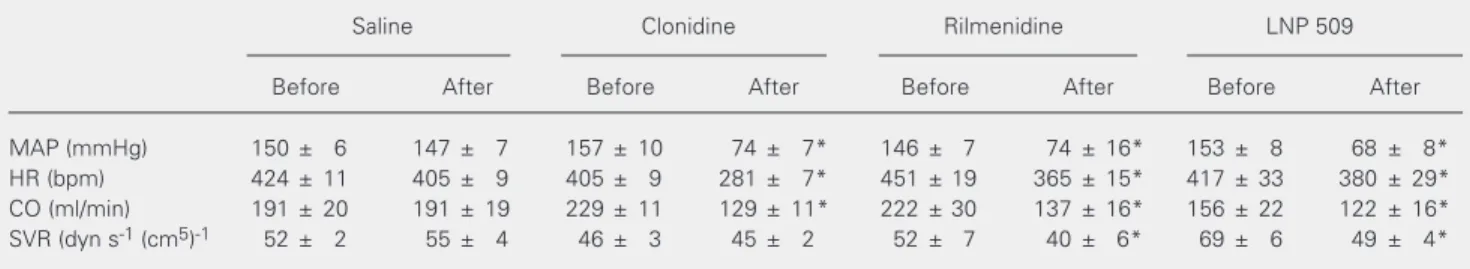
Table 1. Maximum effects of the intracisternal injection of clonidine (1 µg), or LNP 509 (60 µg) on the macrohemodynamic parameters of pentobarbital-anesthetized spontaneously hypertensive rats.
Saline Clonidine Rilmenidine LNP 509
Before After Before After Before After Before After
MAP (mmHg) 150 ± 6 147 ± 7 157 ± 10 74 ± 7* 146 ± 7 74 ± 16* 153 ± 8 68 ± 8*
HR (bpm) 424 ± 11 405 ± 9 405 ± 9 281 ± 7* 451 ± 19 365 ± 15* 417 ± 33 380 ± 29*
CO (ml/min) 191 ± 20 191 ± 19 229 ± 11 129 ± 11* 222 ± 30 137 ± 16* 156 ± 22 122 ± 16*
SVR (dyn s-1 (cm5)-1 52 ± 2 55 ± 4 46 ± 3 45 ± 2 52 ± 7 40 ± 6* 69 ± 6 49 ± 4*
CO = cardiac output; HR = heart rate; MAP = mean arterial pressure; SVR = systemic vascular resistance. Each value represents the mean ± SEM for 6 experiments.
*P < 0.05 compared to basal values (ANOVA).
Mean arterial pressure
(% variation)
125
100
75
50
25 Cardiac output (% variation)
125
100
75
50
25
Heart rate (% variation)
125
100
75
50
25 Systemic vascular resistance
(% variation)
125
100
75
50
25
-10 0 10 20 30 40 50 60 70
-20
Time (min)
-10 0 10 20 30 40 50 60 70
-20
Time (min)
*
* *
*
* *
* *
* *
* *
*
*
*
* * * *
*+
*+ *+ *+ *
*+ *+
*+ *+
*+ *+ *+
* * * *
* *
* * * * *
SAL CLO RIL LNP
*
* *
Microcirculatory effects induced by ic injection of clonidine, rilmenidine and LNP 509
The hypotensive effect induced by ic in-jection of 1 µg clonidine, 7 µg rilmenidine and 60 µg LNP 509 was accompanied by mesenteric arteriolar vasodilation, charac-terized by a maximum increase in arteriolar diameter of 12 ± 1% (N = 6, P < 0.05), 25 ± 10% (N = 6, P < 0.05) and 21 ± 4% (N = 6, P < 0.05; Figure 2) from the basal values of 19 ± 1.9, 18.8 ± 1.8 and 22 ± 1 µm, respectively.
Cardiovascular and microcirculatory effects of ic injection of baclofen and L-glutamate
The central ic injection of 1 µg baclofen elicited marked and long-lasting decreases in MAP, with a maximum of 63 ± 8% (from 155 ± 7 to 62 ± 8 mmHg; N = 6,P < 0.05; data not shown). HR decreased by 18 ± 5% (from 390 ± 11 to 316 ± 27 bpm; N = 6, P > 0.05; data not shown). The hypotensive effect of baclofen was accompanied by significant vasodilation of the mesenteric microcircula-tion (Figure 2). The maximum increase of arteriolar diameter after baclofen was 11 ± 4% (from 30 ± 5 to 33.5 ± 5 µm; N = 6, P > 0.05). On the other hand, the central ic ad-ministration of 125 µg L-glutamate evoked a marked but short-lasting increase in MAP, reaching a maximum of 24 ± 5% (from 161 ± 9 to 198 ± 9 mmHg; N = 6,P < 0.05; data not shown). HR increased only by 6 ± 2% (from 432 ± 9 to 458 ± 5 bpm; N = 6, P > 0.05; data not shown). The hypertensive effect of 125
µg L-glutamate was accompanied by mesen-teric arteriolar vasoconstriction, reaching the maximum of 27 ± 3% (N = 6, P < 0.05) from the basal value of 19.6 ± 1.4 µm (Figure 2).
Discussion
The present study is the first to demon-strate that acute administration of centrally acting antihypertensive drugs such as cloni-dine and rilmenicloni-dine significantly dilates the microcirculation of SHR. It is noteworthy that both drugs were injected directly into the CNS (intracisternally) in doses that do not present significant cardiovascular effects when administered systemically (21,22). Thus, although the activation of peripheral
α2-adrenoceptors in SHR also induces
dila-tion of terminal arterioles (23), the arteriolar vasodilation observed in the present study cannot be due to the peripheral effects of clonidine.
The small arteries and arterioles of the microcirculation are well known to be the most important site of the increased vascular resistance in hypertensive patients (7). In SHR, the progressive development of hyper-tension is associated with an increase in arteriolar tone in different vascular beds (24) that reaches even the terminal arterioles (lu-men diameter of about 20 µm) (25,26). More-over, compared to normotensive control rats (Wistar-Kyoto), the vascular tone of SHR is set at a higher steady-state level (27). In this context, it has already been shown that a decrease of only 13% in arteriolar diameter is sufficient to produce an increase in sys-temic blood pressure of about 50 mmHg (26). Moreover, since sympathetic hyperac-tivity is implicated in the development of arterial hypertension (28), modulation of the central sympathetic activity with centrally acting antihypertensive agents turns out to be a reasonable therapeutic target in the treatment of this disease.
We evaluated the microcirculatory be-havior of SHR using the microcirculation of
Arteriolar diameter (% variation)
140 130 120 110 100 90 80 70 60
SAL CLO RIL LNP BAC
GLU
*
12345 12345 12345 12345
12345 12345 12345 12345 12345 12345
12345 12345 12345 12345 12345
1234 1234 123412345
12345 12345 12345 12345 12345 12345 *
*+
*
Figure 2. Maximum changes (percent of baseline) of mesen-teric arteriolar diameter. Varia-tions observed after intracister-nal injection of saline (SAL, con-trol group), clonidine (CLO, 1 µg), rilmenidine (RIL, 7 µg), LNP 509 (LNP, 60 µg), baclofen (BAC, 1 µg), or L-glutamate (GLU, 125 µg) in anesthetized spontane-ously hypertensive rats. Each set of points represents the mean ± SEM for 6 experiments. *P < 0.05 compared to the con-trol group. +P < 0.05 compared to the clonidine-treated group
the mesentery, which is known to respond to peripheral or central sympathetic stimula-tion (29,30). In fact, the mesenteric micro-vascular network appears to have a dense sympathetic innervation (31), which is in-creased in arterioles of SHR (32). Functional studies in the rat demonstrated that the elec-trical stimulation of the posterior hypothala-mus induces an increase in MAP accompa-nied by a significant vasoconstrictor response of mesenteric arterioles (31), thus demon-strating that this particular vascular bed is under sympathetic control. Moreover, Le Noble et al. (33) showed that pentobarbital is the anesthetic of choice to be used in exper-imental studies assessing the mesenteric mi-crocirculation of the rat, since it does not interfere with microvascular reactivity to adrenergic stimulation. Our results are con-sistent with these observations, since the pharmacological activation of the central sympathetic nervous system with intracis-ternal glutamate, the main excitatory neuro-transmitter in the mammalian CNS, elicited marked arteriolar constriction in the mesen-tery.
Our results also demonstrate that the clas-sical acute hemodynamic effects of centrally acting antihypertensive drugs (i.e., hypoten-sion and bradycardia) are accompanied by significant dilatation of terminal arterioles in SHR. It is noteworthy that the vasodilating effects of rilmenidine and LNP 509 were much more pronounced than that of cloni-dine, thus supporting the view that different mechanisms might be involved in the antihy-pertensive effects of first- and second-gen-eration drugs (11). Nevertheless, we did not investigate the pharmacological mechanisms involved in the microcirculatory effects of these drugs, which could be clarified by the use of selective antagonists. In fact, the pres-ent study was designed to test our hypothesis that the inhibition of the central sympathetic system could be accompanied by a signifi-cant dilation of the microcirculation in hy-pertensive animals.
The central acute administration of anti-hypertensive drugs also induced significant decreases in CO, which were of greater mag-nitude in response to clonidine than dine or LNP 509. As a result, only rilmeni-dine and LNP 509 produced a significant fall in systemic vascular resistance. These re-sults are consistent with those reported by Azevedo et al. (34), who showed that acute intravenous administration of clonidine elic-its negative inotropic effects associated with the inhibition of cardiac-specific sympathetic outflow, evaluated by cardiac norepineph-rine spillover in patients with congestive heart failure. The acute hypotensive effect of clonidine in anesthetized rats had been at-tributed mainly to a reduction in CO rather than in total peripheral resistance (35). On the other hand, in conscious and freely mov-ing SHR, clonidine-induced hypotension seems to depend mainly on reduction of vascular resistance (36). The greater selec-tivity of rilmenidine for the I1BS, when
com-pared to the α2-adrenergic receptors (11,15),
could explain this preferential action on vas-cular resistance since LNP 509, which is devoid of α2-adrenergic-mediated
cardiovas-cular activity (19,20), elicited a hemody-namic profile similar to that of rilmenidine after intracisternal administration.
Our results also showed that the central administration of baclofen, a GABAB
signifi-cant vasoconstriction of the microcircula-tion (39,40), thus suggesting that the arteri-olar vasodilation in the mesentery observed in the present study did not result from the large drop in arterial pressure induced by the antihypertensive drugs.
Perspectives
The results of the present study show that the reduction of blood pressure in the ge-netic model of arterial hypertension induced by centrally acting antihypertensive agents is paralleled by important vasodilation of the mesenteric microcirculation. This effect is more pronounced with substances acting
preferentially (rilmenidine) or exclusively (LNP 509) upon nonadrenergic imidazoline binding sites than with those presenting im-portant α2-adrenergic selectivity (clonidine).
Since important functional and structural alterations of the microcirculation are in-volved in the pathophysiology of primary arterial hypertension, new therapeutic ap-proaches should be able to prevent or even reverse these major features of the disease.
Acknowledgments
The authors gratefully acknowledge Mr. R. Cavalheiro-Silva for skillful technical as-sistance.
References
1. Conway J (1984). Hemodynamic aspects of essential hypertension in humans. Physiological Reviews, 64: 617-660.
2. Cooper A & Heagerty A (1994). Small arteries and hypertension.
Journal of Hypertension, 12: S33-S35.
3. Vicaut E (1999). Microcirculation and arterial hypertension. Drugs, 58 (Special issue 1): 1-10.
4. Struijker-Boudier HA, Le Noble JL, Messing MW, Huijberts MS, Le Noble FA & van Essen H (1992). The microcirculation and hyperten-sion. Journal of Hypertension, 10: S147-S156.
5. Antonios TF, Singer DR, Markandu ND, Mortimer PS & MacGregor GA (1999). Structural skin capillary rarefaction in essential hyperten-sion. Hypertension, 33: 998-1001.
6. Mulvany MJ (2002). Small artery remodeling and significance in the development of hypertension. News in Physiological Sciences, 17: 105-109.
7. Levy BI, Ambrosio G, Pries AR & Struijker-Boudier HA (2001). Micro-circulation in hypertension: a new target for treatment? Circulation, 104: 735-740.
8. Ruilope LM & Schiffrin EL (2001). Blood pressure control and ben-efits of antihypertensive therapy: does it make a difference which agents we use? Hypertension, 38 (Part 2): 537-542.
9. Chalmers JP, Kapoor V, Liewellyn-Smith IJ, Minson JB & Pilowsky PM (1992). Central control of blood pressure. European Heart Jour-nal, 13: 2-9.
10. Esler M (2000). The sympathetic system and hypertension. Ameri-can Journal of Hypertension, 13 (Part 2): 99S-105S.
11. Bousquet P & Feldman J (1999). Drugs acting on imidazoline recep-tors: a review of their pharmacology, their use in blood pressure control and their potential interest in cardioprotection. Drugs, 58: 799-812.
12. Bousquet P, Feldman J & Schwartz J (1984). Central cardiovascular effects of alpha-adrenergic drugs: difference between catechol-amines and imidazolines. Journal of Pharmacology and Experimen-tal Therapeutics, 230: 232-236.
13. Reis DJ, Regunathan S, Wang H, Feinstein DL & Meeley MP (1992).
Imidazoline receptors in the nervous system. Fundamental and Clinical Pharmacology, 6: 23S-29S.
14. Tibiriçá E, Feldman J, Mermet C, Gonon F & Bousquet P (1991). An imidazoline-specific mechanism for the hypotensive effect of cloni-dine: a study with yohimbine and idazoxan. Journal of Pharmacolo-gy and Experimental Therapeutics, 256: 606-613.
15. Tibiriçá E, Feldman J, Mermet C, Monassier L, Gonon F & Bousquet P (1991). Selectivity of rilmenidine for the nucleus reticularis lateralis, a ventrolateral medullary structure containing imidazoline-preferring receptors. European Journal of Pharmacology, 209: 213-221. 16. Chan CK, Sannajust F & Head GA (1996). Role of imidazoline
recep-tors in the cardiovascular actions of moxonidine, rilmenidine and clonidine in conscious rabbits. Journal of Pharmacology and Experi-mental Therapeutics,276: 411-420.
17. De Sarro GB, Ascioti C, Froio F, Libri V & Nistico G (1987). Evidence that locus coeruleus is the site where clonidine and drugs acting at alpha1- and alpha2-adrenoceptors affect sleep and arousal mechan-isms. British Journal of Pharmacology, 90: 675-685.
18. Reid JL (2001). Update on rilmenidine: clinical benefits. American Journal of Hypertension, 14 (Part 2): 322S-324S.
19. Schann S, Bruban V, Pompermayer K, Feldman J, Pfeiffer B, Renard P, Scalbert E, Bousquet P & Ehrhardt JD (2001). Synthesis and biological evaluation of pyrrolinic isosteres of rilmenidine: discovery of cis-/trans-dicyclopropylmethyl-(4,5-dimethyl-4,5-dihydro-3H -pyrrol-2-yl)-amine (LNP 509), an I1 imidazoline receptor selective ligand with hypotensive activity. Journal of Medical Chemistry, 44: 1588-1593.
20. Bruban V, Estato V, Schann S, Ehrhardt JD, Monassier L, Renard P, Scalbert E, Feldman J & Bousquet P (2002). Evidence for synergy between α2-adrenergic and nonadrenergic mechanisms in central blood pressure regulation. Circulation, 105: 1116-1121.
21. Kobinger W (1978). Central alpha-adrenergic systems as targets for hypotensive drugs. Reviews of Physiology, Biochemistry and Phar-macology,81: 39-100.
the cardiovascular actions of clonidine in the rabbit. British Journal of Pharmacology, 47: 206-216.
23. Struijker-Boudier HA, Messing MW & van Essen H (1996). Alpha-adrenergic reactivity of the microcirculation in conscious spontane-ously hypertensive rats. Molecular and Cellular Biochemistry, 157: 239-244.
24. Evenwel RT, Kasbergen CM & Struyker-Boudier HAJ (1983). Central and regional hemodynamics and plasma-volume distribution during the development of spontaneous hypertension in rats. Clinical and Experimental Hypertension. Part A, Theory and Practice, 5: 1511-1536.
25. Suzuki H, Zweifach BW & Schimid-Schönbein GW (1996). Glucocor-ticoid modulates vasodilator response of mesenteric arterioles in spontaneously hypertensive rats. Hypertension, 27: 114-118. 26. Schimid-Schönbein GW, Zweifach BW, DeLano FA & Chen P (1987).
Microvascular tone in a skeletal muscle of spontaneously hyperten-sive rats. Hypertension, 9: 164-171.
27. Suzuki H, Zweifach BW & Schimid-Schönbein GW (1995). Vasodila-tor response of mesenteric arterioles to histamine in spontaneously hypertensive rats. Hypertension, 26: 397-400.
28. Brook RD & Julius S (2000). Autonomic imbalance, hypertension, and cardiovascular risk. American Journal of Hypertension, 13 (Part 2): 112S-122S.
29. Gootman MP, Gootman N & Buckley BJ (1983). Maturation of central autonomic control of the circulation. Federation Proceed-ings, 42: 1648-1655.
30. Nyhof RA, Laine GA, Meiniger GA & Granger HJ (1983). Splanchnic circulation in hypertension. Federation Proceedings, 42: 1690-1993. 31. Le Noble LML, Tangelder GJ, Slaaf DW, Smits JFM & Struyker-Boudier HAJ (1987). Adrenergic stimulation of the rat mesenteric vascular bed: a combined micro- and macrocirculatory study.
Pflügers Archiv,410: 250-256.
32. Lee RM, Forrest JB, Garfield RE & Daniel EE (1983). Ultrastructural
changes in mesenteric arteries from spontaneously hypertensive rats. A morphometric study. Blood Vessels,20: 72-91.
33. Le Noble LML, Smits JFM, Slaaf DW, Tangelder GJ & Struyker-Boudier HAJ (1984). Effects of anesthesia on regional and microcir-culatory hemodynamic effects of vasopressor substances in the rat. International Journal of Microcirculation, Clinical and Experi-mental, 3: 314-316.
34. Azevedo ER, Newton GE & Parker JD (1999). Cardiac and systemic sympathetic activity in response to clonidine in human heart failure.
Journal of the American College of Cardiology, 33: 186-191. 35. Dabire H & Richer C (1991). Implication of the central nervous
system in the systemic and regional hemodynamics of two cen-trally acting hypotensive drugs, flesinoxan and clonidine, in the rat.
Journal of Cardiovascular Pharmacology, 18: 605-613.
36. El-Mas MM & Abdel-Rahman AA (1999). Role of the sympathetic control of vascular resistance in ethanol-clonidine hemodynamic interaction in SHRs. Journal of Cardiovascular Pharmacology, 34: 589-596.
37. Bowery NG, Bettler B, Froestl W, Gallagher JP, Marshall F, Raiteri M, Bonner TI & Enna SJ (2002). International Union of Pharmacolo-gy. XXXIII. Mammalian gamma-aminobutyric acid (B) receptors: structure and function. Pharmacological Reviews, 54: 247-264. 38. Williford DJ, DiMicco JA & Gillis RA (1980). Evidence for the
pres-ence of a tonically active forebrain GABA system influencing central sympathetic outflow in the cat. Neuropharmacology, 19: 245-250. 39. Paes-da-Silva F, Gonzalez AP & Tibiriçá E (2003). Effects of fluid
resuscitation on mesenteric microvascular blood flow and lym-phatic activity after severe hemorrhagic shock in rats. Shock, 19: 55-60.