University of Lisbon
Faculty of Medicine of Lisbon
Venous Circulation in Glaucoma
Luís Alexandre Pereira Abegão Pinto
Thesis supervised by Prof. Dr. Carlos
Marques-Neves and Prof. Dr. Ingeborg Stalmans
Field of Medicine
Specialty Ophthalmology
The printing of this thesis has been approved by
the Scientific Council of the Faculty of Medicine
Acknowledgements
This thesis submitted to appreciation of the Faculty of Medicine of Lisbon’s University represents a Project executed in three Institutions in two different countries. The number of people to be thanked for the completion of this Thesis could not be fitted to a single page. From Directors and Heads of Departments to laboratory technicians, dozens of people helped in logistics, patients’ recruitment, data analysis, discussion of ideas and even moral support. To all of them, my sincere thanks.
To Prof. Dr Carlos Marques-Neves, the architect who guided me in all these tasks. For giving me the tools and the liberty to use them. Whose deep knowledge of ocular physiology and clear rational thinking were an invaluable addition to this Thesis.
To Prof. Dr Ingeborg Stalmans, to whom I lack the words to thank for everything. Whose profound knowledge in this field of glaucoma is only matched by her friendliness and willingness to help. Even more valuable than her input to this project, was the friendship and cooperation spirit that have grown alongside this thesis.
To Dr Evelien Vandewalle, with whom I shared many of the works here presented. Her experience in Color Doppler Imaging and
To Prof. Dr Alexandre Ribeiro and Prof. Ana Sebastião at the Institute of Pharmacology for their support and encouragement throughout the years.
To all my fellow colleagues in the Institute of Molecular Medicine, both in the Physiology and Pharmacology Departments, and in the Catholic University of Leuven, for their support and friendship at all times.
To all my fellow colleagues – Ophthalmologists and Neuro-radiologists alike – at the Centro Hospitalar de Lisboa Central for all their help throughout the project.
To my family, for all the support throughout all these years. Nothing would have been possible, or have the same meaning, if it wasn’t for them.
And finally, but certainly not the least, to Gabriela, my better-half along this road. This PhD was not a goal, but a journey. A journey I did not to endure alone.
Index
Introduction 7
Glaucoma 11
Ocular Circulation 15
Ocular Blood Flow in Glaucoma 25
Venous Dysfunction in Glaucoma 31
SVP phenomenon 35 Purpose 41 Methods 45 Subject groups 47 Informed consent 49 Exclusion criteria 51 Measuring devices 53 Experimental design 63 Statistical analysis 65 Results 67
Experimental groups’ characteristics 69
SVP frequency 85
Patient’s overall characteristics by SVP status 89
OPA, ONSD and SVP phenomenon 105
Cardiovascular parameters and SVP phenomenon 117
Ocular Blood Flow and SVP phenomenon 123
General Discussion 155 Summary 165 Sumário 171 Abbreviations 177 Units of Measurements 179 References 181
Chapter 1. Introduction
Glaucoma is the second most common cause of blindness in the developed world. Nearly 9 million people worldwide are bilaterally blind from this disease, with up to 1% of all population over the age of 40 presenting with some form of the disease (1). Unfortunately, around 50% of these patients go undetected, as most of this permanent vision loss is asymptomatic until the very late stages of the disease (2).
Worldwide, the most common type of the disease is primary open-angle glaucoma (POAG). As the name implies, no cause can be detected for this optic neuropathy. In most cases, the only identifiable risk fact is an increased intraocular pressure (IOP). However, there are patients in which IOP has never been detected to be above the normal range, in which case they are considered to have normal-tension glaucoma (NTG). Importantly, this form of the disease can represent up to 50% of glaucoma population in the western hemisphere (3), while in Japan this figure can go up to 90% (4). Thus, and while IOP is the main risk factor and IOP lowering remains the cornerstone for glaucoma management, IOP cannot explain all the mechanisms involved in glaucoma pathogenesis. This claim is widely supported by the established fact that a significant number of patients still progress despite an otherwise successful reduction in IOP (5) (6).
8
As such, the quest for the identification of other non-IOP related pathogenic mechanisms has been the work of countless research groups worldwide. The vast majority of the studies regarding non-IOP related risk factors, such as vascular disturbances, have been focused on the arterial component, with an increasing accumulation of data suggesting the existence of defective autoregulation mechanisms (7) (8) (9).
The emphasis on ocular blood flow studies in glaucoma has been particularly appealing for researchers in glaucoma pathogenic mechanisms, as these changes in ocular blood flow seem to precede glaucomatous damage (10) (11).
Despite these efforts to understand the ocular blood flow in glaucoma, very little is still known about the venous circulation in the eye and its potential role in glaucoma pathogenesis. Venous circulation may have an impact on glaucoma by two different mechanisms: on the one hand, it is responsible for the drainage of the aqueous humour and thus could play a role in modulating the main risk factor for the disease (IOP). Additionally, as elsewhere in the body, any change in retinal venous pressure can significantly impact the ocular perfusion pressure – another important factor in glaucoma progression.
A wider knowledge of what influences venous output is therefore important not only to better understand the ocular physiology, but also to better understand how these physiological mechanisms
might be impaired in glaucoma patients. This information could be particularly important in the interpretation of ocular blood flow studies in glaucoma, especially in the more vascular-prone NTG patients.
I. Glaucoma
The disorder now identified as glaucoma has been known to man throughout the ages. The oldest description to have survived until nowadays was made in Greece around 400 BC (12), where a Hippocratic text states a condition where ‘once the pupil has the colour of the sea – eyesight is destroyed and you will often find that the other eye is also blind’. While this description was used to label a number of diseases throughout history, idea of a distinct disease associated with an increased stiffness of the eyeball was first identified by McKenzie in 1830, branding it with the name Glaucoma (13). However, the emphasis on a pressure-related disease was questioned by Jaeger already in 1858, suggesting other intrinsic non-IOP related causes for the disease (14) and in 1885, Smith suggested that vascular factors were likely involved in the process (15). Since then, the controversy has remained between the relevance of either the mechanic or vascular theories to the pathogenesis of open-angle glaucomas.
In fact, other than the widely recognized risk factor of increased IOP, a number of other variables have been identified as risk factors for disease progression. The severity of damage at the time of diagnosis, increasing age, low central corneal thickness (CCT), but also cardiovascular variables such as low ocular perfusion pressure and a history of cardiovascular disease have all been associated with worsening of the disease (16). While IOP remains the most easily modifiable risk factor, the impact of the other
12
variables may be more significant when all IOP measurements are found to be within the normal range.
The distinction of NTG and POAG patients has been determined as patients with the former having a maximum registered IOP of equal or below 21mmHg. This may, however, be an arbitrary division as they may represent two points in a larger spectrum of optic neuropathies of variable sensitiveness to IOP (16). Nevertheless, and despite patients with NTG being more prone to show signs of vascular dysfunction, from migraine (17), peripheral vasospasm (18), systemic hypotension (19) to cerebral microvascular ischemia (20), they still present a significant relation to IOP. In fact, even in these otherwise normal-range IOP patients, lowering this variable has been shown to slow disease progression (21).
The modern definition of glaucoma thus reflects the acknowledgment that this disease is a progressive neurodegenerative process, where an elevated IOP is only one of the involved mechanisms. Indeed, the European Glaucoma Society currently defines primary open-angle glaucomas as chronic, progressive optic neuropathies that have in common characteristic morphologic changes at the optic nerve head and retinal nerve fiber layer, in the absence of other ocular disease or congenital abnormalities (16).
While the relentless retinal ganglion cell death and progressive visual field loss can be detected by modern structural and functional tests, respectively, the ocular circulation intricate anatomy and physiology makes its study (and interpretation) a complex issue. A number of technologies have emerged to assess the normal ocular blood flow and its change in glaucoma patients (22) (23) (24) (25). One of the more popular techniques is the study of the retrobulbar arteries’ flow velocities by color Doppler Imaging (CDI). Glaucoma patients have been consistently demonstrated to have different patterns in the Doppler waveform, from decreased velocities to increased resistivity index (10) (26) (27) (28). However, the interpretation of these variables has been limited compared to other medical specialities. Glaucoma ocular blood flow studies have put the emphasis on the arterial peak and trough blood velocities and on the resistance index that can be calculated from them. As cardiologic and neurologic studies have demonstrated, a significant number of additional information can be retrieved from the arterial Doppler Waveform (29) (30). As such, by integrating these knowledge into ocular blood flow studies, our current understanding of the arterial blood flow in glaucoma is evolving at a fast pace. However, little is known about venous flow patterns in the eye, its correlation with other cardiovascular or ocular variables or even whether glaucoma patients have any detectable venous dysfunction.
II. Ocular Circulation
In order to address any question regarding ocular blood flow, one must be aware of the particular vascular anatomy of the eye, particularly around the disease’s primary location: the optic nerve head (ONH). Henceforth, an anatomic description of the vascular anatomy involved in glaucoma (both venous and arterial) will be depicted, as well as a summary of its complex physiology.
II. A) Arterial Circulation
All blood to the optic nerve comes from the carotid artery through its ophthalmic artery (OA) branch. This OA follows a tortuous path inside the orbit towards the anterior nasal orbital wall, crossing the optic nerve as the short posterior ciliary arteries, the long posterior ciliary arteries and the central retinal artery (CRA) branch off. The short ciliary arteries are responsible for the supply of most of the choroid, while the long posterior ciliary arteries enter the eye next to the optic nerve and travel anteriorly between the sclera and the choroid, until the ciliary body. By creating the iris major arterial circle, these long ciliary arteries supply most of the anterior segment of the eye.
The OA gives additional branches to the lacrimal artery, the ethmoidal arteries, the muscular arteries and medial palpebral arteries before dividing into two terminal branches (frontal and nasal dorsal arteries). Considering this ocular and extra-ocular
16
branching, the amount of blood from the OA going into the ocular structures does not exceed 25% of the total (31).
The neuron cell most affected by glaucoma, the ganglion cells and their axons along the retinal nerve fiber layer, are exclusively supplied by the CRA. This artery penetrates the optic nerve sheath, crossing a layer of cerebrospinal fluid before reaching the lamina cribrosa already within the optic nerve mass. After a short pathway in the optic nerve head, it divides into 4 smaller arteries, forming the two vascular arcades which are responsible for supplying the inner retinal layers.
The blood supply to the ONH is conditioned by this structure’s complex anatomy and its relation with the lamina cribrosa. Its four different vascularization compartments reflect the unique pathway of the ganglion cells’ axons as they move from the innermost part of the retina towards the outside of the eye. The most anterior part, named surface nerve fiber layer, is supplied by the retinal arteries. At a more posterior level, the prelaminar and laminar compartments are supplied by branches of the short posterior ciliary artery, which sometimes encircle the ONH, creating the Zinn-Haller ring. This functional anastomosis could theoretically protect the optic nerve from occlusion or hypoperfusion of a single short posterior ciliary artery. The most posterior compartment, the retrolaminar region, is mostly supplied by pial vessels that give off centripetal branches into the septa of the optic nerve.
II. B) Venous Circulation
The terminal pathways for most of the venous drainage of the orbit are the ophthalmic veins. However, for practical purposes, this process can be divided into the venous drainage of the retina on one hand and the venous drainage of the rest of the ocular structures on the other.
Blood flows from the retinal capillaries towards the retinal venules in a centripetal pattern towards the ONH. This pathway resembles the arterial branching, by forming two arcades around the macula with increasing diameters as they approach the optic disc. At this stage, the two hemiveins merge into a central retinal vein (CRV). This vessel is always located temporal to the corresponding CRA (32). From this point, the CRV leaves the eye through the lamina cribrosa. After leaving the eye within the optic nerve, the CRV pierces the optic nerve, crossing the space between the optic nerve and its sheath. This sheath is separated from the optic nerve by a laminar layer of cerebrospinal fluid, which is in continuity with the rest of the central nervous system. Having passed the sheath, the CRV most frequently merges itself with the superior ophthalmic vein (33), although it can also drain directly into the cavernous sinus (34) and then onward to the right atrium through the central nervous venous system.
The other aspect in ocular venous circulation of particular importance in glaucoma is the one involved in the aqueous
18
humour drainage. Aqueous humour is secreted into the eye’s posterior chamber through the ciliary processes. This aqueous humour is produced by both active secretion of solutes and water diffusion, obtained from blood circulation. The function of this solution is to nurture the inner segments of the non-vascularized cornea, to provide the eye with appropriate tonus, and to remove metabolic end-products, among others. The aqueous humour travels into the anterior chamber through the pupil and exits the eye by passing through the trabecular meshwork into Schlemm’s canal. After entering Schlemm’s canal, the aqueous humour enters the collector channels that communicate with the aqueous veins. These four to five aqueous veins are sparsely visible near the limbus and have a short length before merging with the episcleral veins (35). In turn, these episcleral veins then connect with the anterior ciliary veins (responsible for drainage of the eye’s anterior segment), which, together with the vortex veins (responsible for the drainage of the choroid), drain into the ophthalmic veins. While the main part of the blood exiting the ocular tissues enters directly the superior ophthalmic vein, a much smaller portion that arises from the lower part of the eye enters the inferior ophthalmic vein. The latter takes a pathway through the floor of the orbit before merging with the superior ophthalmic vein near the annulus of Zinn (34).
This system is based on passive diffusion. As such, any change in pressure throughout the venous pathway (from the aqueous vein
up to the cavernous sinus and the right atrium) is theoretically able to interfere with aqueous humour drainage and therefore influence IOP (36) (37).
II. C) Ocular Blood flow regulation
The regulation of ocular blood flow differs between the various ocular compartments. As elsewhere in the body, blood flow in the eye should be under the control of the autonomic nervous system. However, as this innervation stops at the level of the lamina cribrosa, the retinal circulation is not regulated by sympathetic output. Instead, retinal vessels have the ability, through not completely understood mechanisms, to constrict or dilate in response to changes in oxygen or pH, thus maintaining a constant metabolic environment despite exposure to conditions that might upset this equilibrium.
The choroidal circulation, on the contrary, is under the control of the autonomic nervous system and has no intrinsic ability to adapt to these stimuli. It is able to decrease or increase blood flow in response to cervical sympathetic stimulation, but it cannot adapt to sudden changes in IOP, for example. A clinical consequence, for instance, of this inability to self-regulate its flow is the uveal effusion that can be seen when opening the eye during surgery. As a consequence of these differences in vasoreactive mechanisms, the response to medical therapy also differs between these vascular beds.
Much like their retinal counterparts, the ONH capillaries lack pre-capillary sphincters; they have pericytes instead. As in the retinal circulation, these pericytes respond to metabolic and
22
neuroendocrine factors that regulate their contractility. However, while there is no consistent evidence of an autonomic innervation directly regulating ONH blood flow, the lack of a cellular barrier separating the ONH from the choroid tissues could make the ONH susceptible to autonomic stimulations. As both are supplied by the same vessels, imbalances in the choroidal blood flow could redirect blood flow away from the ONH.
In the eye, the vascular endothelium plays a key role in the regulation of vessel tone. It regulates the blood flow in the retina, ONH and choroid by releasing agents that are responsible for vasodilation and vasoconstriction and by modifying this release in response to local metabolic needs. In vitro and in vivo studies have provided evidence for the role of endothelium-derived vasoactive substances in the control of blood flow in the retina, ONH and choroid (38) (39). Nitric oxide is involved in the control of basal blood flow in the choroid, optic nerve and retina by maintaining the basal vasodilator tone. A sufficient blood supply to the ocular circulation requires the maintenance of a basal vasodilator tone in ocular arteries. As in many other arterial beds, this is largely provided by a constant formation of nitric oxide (NO) by NO synthase 1 and 3 (40) at basal conditions. Hence, systemic administration of an NO synthase inhibitor reduces blood flow in the choroid, retina and ONH (41) (42) (43), indicating a contribution of NO to vascular tone in human ocular vasculature. In addition, a number of agonist-induced vasodilatory effects have
been shown to be NO-dependent, including those to histamine (44), insulin (45) and carbon dioxide (CO2) (46). Another key regulator of ocular blood flow is endothelin. Exogenous endothelin dose-dependently reduces retinal, choroidal and ONH blood flow (47) (48) (49) which is reversible by blockade of the endothelin A receptor subtype. Blockade of this receptor also modifies the choroidal blood flow response to isometric exercise, which strongly indicates a role of endothelin in choroidal blood flow regulation during changes in perfusion pressure (50). In addition, the potent vasoconstrictive effects of hyperoxia in the retina appear to be at least partially mediated via this endothelin receptor (51). Endothelial cell dysfunction produces an imbalance between vasodilator and vasoconstrictor pathways, most notably the NO and endothelin systems (52). Endothelin has been implicated as a contributory factor in many vasospastic disease processes, including vasospastic coronary artery disease (53), cerebral vasospasm (54) and Raynaud’s disease (55). Vasospasm, characterized by exaggerated vascular responses to various stimuli such as temperature and stress (56) (57), is a transient, reversible vasoconstriction that results from impaired endothelium-dependent regulation of vascular tone (58). The possibility that endothelin contributes to vasospasm in ocular diseases such as glaucoma is supported by the demonstration of elevated basal plasma endothelin concentrations in patients (59), combined with an abnormal response of plasma endothelin concentrations to
III. Ocular Blood Flow in Glaucoma
The above described mechanisms of blood flow regulation may be impaired in glaucoma patients, with a growing number of publications showing disturbances in these patients’ ocular blood flow. These changes extend beyond the ocular circulation, with systemic circulation also showing signs of regulatory impairment, resulting in glaucoma being called “a sick eye in a sick body” (62). However, the nature of these disturbances is still under discussion, including weather such changes are part of the pathogenic mechanisms or secondary to the underlying disease.
When considering ocular diseases in which vascular mechanisms are involved, such as diabetic retinopathy or occlusion of either of the central retinal vessels, none of these diseases produce a characteristic glaucomatous cupping of the disc (63) (64). Therefore, hypoperfusion, as seen in those diseases, does not provide a full explanation for glaucomatous neuropathy. Should hypoperfusion alone represent the vascular risk factor for glaucoma (progression), one would expect a stronger relationship between glaucoma and known risk factors for atherosclerosis, such as C-reactive protein or dyslipidaemias. However, data on these variables is far from consistently pointing them out as risk factors for glaucoma (65). Alternatively, or in addition to hypoperfusion, there may be a vascular dysfunction that impairs the normal self-regulating mechanisms response to fluctuations in perfusion (66). Interestingly, the ONH, due to its unique anatomical condition, is
26
exposed to circulating hormones in a way that the rest of the central nervous system is not. Not only is the barrier function decreased in the capillaries in this region (67), but there is also diffusion from the choroid, where these adaptive mechanisms do not exist (68). Vasoconstrictive agents, such as angiotensin II and endothelin, can therefore have a greater impact in this region than elsewhere in the nervous system. Available data suggest that glaucoma patients may have an endothelial dysfunction, with increased levels of endothelin (69) and vasodilation impairment (70), thus increasing the risk for ONH injury. Such dysfunction, as suggested by Flammer, consists of an instability of ocular perfusion (rather than a stable reduction in ocular blood flow), leading to a repeated mild reperfusion injury (71) (72). This mechanism of ischaemia-reperfusion injury is associated with the generation of reactive oxygen species and cellular apoptosis. The resulting increase in vasoconstriction and oxidative stress can lead to an activation of several apoptotic pathways through cellular dysfunction (73). It has been suggested that this oxidative stress could also occur as a result of elevated IOP, in what seems to be an IOP-related mechanical stress event (74). In humans, the role of this cellular hypoxia in glaucoma is further supported by increased staining of hypoxia-induced factor (HIF-1α) in the retina and optic nerve of patients with glaucoma compared with non-glaucomatous individuals (75). This impaired self-regulation capacity of the vessels supplying the ONH may be clinically relevant. Recent studies have shown that fluctuations in perfusion pressure are
particularly important in glaucoma progression, especially in patients with NTG (76). Glaucoma patients are also more prone to show abnormal circadian vascular rhythms that can tip the balance of perfusion pressure into pathogenic levels. These abnormal circadian cardiovascular responses may be due to an underlying dysautonomic disturbance, as it is the autonomic nervous system’s responsibility to regulate these daily rhythms (77). Glaucoma patients seem to have a number of signs of autonomic dysfunction, from dysregulation of aqueous humour production and drainage (78) to abnormal heart rate and blood pressure variability, both of which are associated with increased cardiovascular risk (79) (80). This blood pressure variability is particularly prominent in patients with NTG, and a correlation between the extent of autonomic nervous system dysfunction and the severity of the disease has been suggested (78).
A number of studies have shown that this vascular dysfunction and impairment in normal blood flow associated with glaucoma is not restricted to the eye. Indeed, there are also indications of slower flow in peripheral capillary beds (81) and signs of microvascular encephalopathy, such as white matter lesions (20). This association between ocular circulation and systemic cardiovascular disease has been reinforced by epidemiological findings. Pooled data from the Beaver Dam Eye Study and the Blue Mountains Eye Study have demonstrated that smaller retinal arterial diameters and larger retinal venous diameters are associated with increased risk for
28
stroke mortality (82) (83). One of the most striking disturbances in ocular vessels, the splinter optic disk haemorrhages, are an important clinical feature that has been associated with glaucoma progression. These haemorrhages occur more frequently in patients with NTG glaucoma and are strongly associated with altered circulation within the optic disc (84) (85). Some authors have suggested that these splinter haemorrhages may represent distressed small venules (86), as these thinner veins may reflect earlier lamina cribrosa changes than their thicker arterial counterparts (87).
The changes in ocular blood flow are not restricted to the retinal vessels, as changes of clinical significance have also been found in the retrobulbar circulation. A number of CDI studies have found reduced peak systolic and diastolic velocities and increased resistivity indices in the retrobulbar vessels of glaucoma patients compared to healthy normal controls (88) (89) (26) (27). Interestingly, patients that progress seem to have a more important alteration in blood flow, namely, reduced PSV and EDV in short posterior ciliary arteries (90). Moreover, a prospective study showed that within glaucomatous individuals, the eye with the more pronounced blood flow impairment also showed a faster progression (91).
The extent of the damage induced by ocular blood flow impairment alone is difficult to determine. Blood flow changes per se might lead to glaucoma damage, but they could also synergistically act with other risk factors. For example, blood flow disturbances might act as a sensitizer to IOP, making it possible for normal-range values of IOP to produce glaucomatous damage.
IV. Venous dysfunction in glaucoma
Although studies on ocular venous changes in glaucoma are few when compared to their arterial counterparts, venous circulation seems to differ between glaucoma patients and healthy individuals.
Aqueous veins in glaucoma patients have a significant change in an otherwise normal pulsatility pattern (92) (93). Normal blood flow fluctuations during the cardiac cycle in the arterial-vein transition around the trabecular meshwork creates a pressure gradient acting as a Venturi-like pump, bringing an aqueous humour pulse wave inside the aqueous vein (94). This cyclic wave absortion of aqueous humour into vessels containing blood can be seen as a pulsating pattern. Glaucoma patients, however, have a much lesser frequency of this pattern. While the increased trabecular meshwork resistance could account for a smaller aqueous humour wave, the downstream change in venous pressure could also dampen this wave (95). In fact, agents such as brimonidine (96) and epinephrine (97) have been demonstrated to increase stroke volume and flow velocity in these veins. As both these drugs do not interfere with trabecular meshwork physiology directly, but can interfere directly and indirectly with episcleral venous pressure, these data reinforce the importance of venous function in an adequate aqueous humour drainage.
32
Retinal venous circulation is not involved in aqueous humour drainage. Instead, it is responsible for the drainage of the blood supply of the retina and the ONH. As in any other part of the organism, venous pressure is involved in perfusion pressure. The perfusion pressure of an organ is the difference between arterial input pressure and venous output pressure in relation to the organ’s vascular resistance. This vascular resistance is nevertheless dependent not only on the pressure difference of the vessel’s pressure but also on variables such as blood viscosity and vascular diameter. As venous circulation is a low pressure system in which blood flows into the lowest pressure point (the right atrium), its systemic value (central venous pressure) is usually in the range of 2-3 mmHg (98). In the eye however, venous pressure has to at least match IOP (99) (100). Any other possibility would lead to a constant vessel collapse and no blood flow (101). As such, the classical calculations of ocular perfusion pressure have used the assumption that venous pressure resembles IOP and uses this latter easily verifiable variable as a surrogate.
This assumption has been, however, increasingly questioned by a number of authors. In experimental conditions, venous pressure has been consistently demonstrated to be at least 5mmHg higher than IOP (102) (103). While this difference would be small enough to force a change in paradigm, a number of publications have shown retinal venous pressure to be significantly higher in glaucoma patients (87) (104) (105). While the nature of this
increase in venous pressure is under debate, it would still mean that the calculations of ocular perfusion pressure in such patients could be significantly overestimated. This is particularly important as low perfusion pressures have been consistently proven to be associated with disease progression (16).
Additionally, the notion that retinal vein physiology may be disturbed in glaucoma patients is reinforced by the suggestion that optic nerve haemorrhages, a major clinical sign of disease deterioration, may represent distressed venules. In these patients, these venules would, for still undetermined reasons, be unable to meet the increased intravascular pressure and rupture (106). Another aspect which shows a link between retinal vein dysfunction and glaucoma is the fact that glaucoma is a major risk factor for CRV occlusion (107). The nature of this link is, again, incompletely understood. Some authors have suggested that changes in the lamina cribrosa associated with increased IOP in glaucoma patients may increase extramural pressure, thus increasing the chances of a thrombotic event (108) (109).
In both conditions, the questions remains as to whether this apparently deleterious increase in retinal venous pressure is a consequence of glaucoma or whether it may be associated with the pathogenesis of the disease (by decreasing perfusion pressure).
V. Spontaneous Venous Pulsation Phenomenon
Perhaps the most clinically appealing sign of venous changes in glaucoma patients is the significant decrease in frequency of the CRVs spontaneous venous pulsation (SVP) phenomenon over the optic disc. From up to 98% in the healthy population, this easily, clinically verifiable phenomenon decreases to nearly half in glaucoma patients (110) (111).
Importantly, especially when considering the detection of this variable in a disease such as glaucoma, the visualization of this phenomenon seems to be dependent on the optic disc characteristics and the linearity of vessel’s pathway in the disc (112). However, little is known on how morphometric variables such as depth or cup/disc ratios influence such pulsatility detection. Whether the changes in the ONH associated with glaucoma damage have any impact on the SVP phenomenon remains unknown.
Independently on the threshold of detection, the mechanisms behind this intriguing phenomenon are not completely understood. Described in 1853 (113), two theories exist as to why the retinal veins pulsate near the optic disc.
36
The classical theories, presented in the early years of the XX century (114) (115), propose that retinal vein pulsations occur because of a periodic reversal of the pressure gradient across the wall of the retinal vein. The blood pressure inside the vein – retinal venous pressure – and the fluid pressure of the eye outside the vein – intraocular pressure (IOP) – each vary during the cardiac cycle. During the part of the cardiac cycle when the IOP exceeds the retinal pressure, the vein would collapse. When that retinal pressure again becomes higher than the IOP, the vein would re-expand. This theory would suggest that vein collapse would happen during systole. The increased input of blood during systole inside the relatively unexpandable eyeball would lead to a relatively higher increase in IOP. As retinal pressure would fluctuate in a retrograde transmission from the right atrium, it would be higher during diastole. It would be during this period that retinal vein pressure would be higher and the vein would therefore reexpand.
The previous theories assume there is no connection between the fluctuations in IOP and the retinal pressure. However, this is not possible. IOP fluctuates only because the input of blood during a certain period of the cardiac cycle is not matched by a similar increase in the blood output (venous flow). Should venous flow increase in a similar magnitude as the arterial input, no change in blood volume would occur and accordingly, the ocular pulse amplitude would be zero. This rise in IOP from the excess arterial
inflow must, therefore, be transmitted instantaneously to the contents of the capillaries and the veins. The only possibility for this increase in outside pressure not to change retinal venous pressure would be if the walls of these veins were rigid enough to maintain a similar transmural gradient throughout the cardiac cycle. This is not the case in the eye (116). Therefore, and since IOP does fluctuate, both IOP and retinal pressure must fluctuate in phase (116).
A new theory thus emerged that takes into account the fact that IOP is always lower than retinal venous pressure (116). This new approach takes into account the pulse pressures in the different compartments involved in the venous circulation. In fact, as CRV traverses the lamina cribrosa, the extravascular pressure drops from IOP to the cerebrospinal fluid pressure over a distance less than 0.5mm (117). In this hypothesis, the fluctuation of IOP (and therefore retinal venous pressure) during a cardiac cycle is greater than the fluctuation in the cerebrospinal fluid pressure (118). Although these fluctuations are small enough not to reverse the pressure gradient, the prelaminar to postlaminar gradient is higher than the mean value during systole and below its mean in the diastole. If it is assumed that vein diameters throughout the retina are not changed, the blood flow at the retinal vein exit will follow the pressure gradient, rising in systole and falling in diastole. Since flow into the retinal veins is constant, but flow velocities fluctuate, pulsations must occur.
38
Despite addressing some of the inconsistences of the classical theory, this latter approach assumes that the collapse of the vessels happens during the diastolic component of the cardiac cycle. This, however, has been challenged recently (105), with authors suggesting the opposite pattern. This would raise the possibility for this phenomenon to occur when IOP and cerebrospinal pressure pulses have different amplitudes or despite an out-of-phase synchronicity.
Independently of the theories used, both agree on the fundamentals of the clinical use of this pulsating vein variable. Since its original description, this sign has been used to exclude the existence of an increased intracranial pressure (113) (119). The loss of this pulsating sign has been additionally described to identify ocular hypotony and retinal venous occlusion (117) (120). Because both theories rely on the amplitude of fluctuation in the ocular, extraocular and retinal vessel compartments, all of the described conditions (high ICP, low IOP and higher retinal venous pressure) would buffer the translaminar gradient and decrease the likelihood of SVP detection. The importance for this balance in fluctuating compartments has been further supported by the suggestion of a minimal ocular pulse amplitude threshold to exist (1.2mmHg), below which no SVP would be visible (121). The level of fluctuations in the other compartments or the existence of a minimal threshold in any of them that condition the SVP
phenomenon remains unanswered, particularly in glaucoma patients.
Glaucoma patients have been suggested to have decreased ICP values (122) (123). Together with higher values of IOP, both these variables would increase the likelihood of SVP detection by synergistically increasing translaminar gradient pressure. As such, glaucoma patients were expected to have higher frequency ratios of SVP (124) (125). However, as previously stated, glaucoma patients have been consistently reported to have much lower frequency ratios of SVP (111) (110). The reason for these results, considering the theories’ premises, remains unclear. Whether the vascular dysfunction known to exist in glaucoma patients has a venous component as well, or whether glaucoma patients have undetermined structural, elastic or morphometric changes in any of the relevant compartments, that would make the detection of this SVP more difficult, is not yet understood.
Interesting, retinal veins are not the only veins in the body that can show pulsation. Either from proximal arterial transmission or downstream changes in heart pressure, veins such as the jugular vein can present with detectable pulsation. The significance of jugular waveform changes has been an important part of medical semiology in the study of heart conditions such as tricuspid insufficiency or pericarditis. Whether SVP determination in retinal veins could have the same impact in glaucoma patients remains to be answered.
Chapter 2. Purpose
I. Global Objective
This project was designed to characterize the ocular venous circulation in glaucoma patients and to determine the anatomical and hemodynamic factors that influence its parameters, specifically their association to the SVP phenomenon.
A case-control study using two sets of glaucoma groups (both high-IOP and more vascular-prone low-high-IOP patients) was used to detect whether any change in variables would be specific to one group or common to all glaucoma patients. As diagnostic workout, treatment algorithm and risk factors can be different in NTG and POAG diagnosis, our study tried to address whether venous dysfunction, if found in these patients expressing a non-pulsating venous circulation, would be of any additional value to in the daily management of glaucoma patients.
The decision to use tools that are readily available in any hospital reinforces the claim to provide information which can potentially be used in every Ophthalmology unit. Even the highly-expensive ultrasound machines used for color Doppler Imaging are available in radiology departments worldwide.
II. Main goals
1. To characterize the frequency of SVP in glaucoma patients (both primary open-angle glaucoma and normal tension glaucoma).
2. To characterize anatomical factors that relate to the SVP phenomenon observed in glaucoma patients
3. To characterize hemodynamic factors that relate to the venous circulation changes observed in glaucoma patients 4. To characterize signs of vascular dysfunction and their
association to venous circulation variables in glaucoma patients
Chapter 3. Methods
This study on venous circulation in glaucoma was conducted in the Glaucoma Clinic of the Department of Ophthalmology of the University Hospitals of Leuven, Belgium. The measuring devices that exist in the several centers initially involved (Centro Hospitalar de Lisboa Central, Institute of Molecular Medicine of Lisbon’s Faculty of Medicine and Leuven’s University Hospitals) have different technical specificities which would have prevented any data pooling. As such, and in coordination with both supervisors and the agreement of the Head of the Ophthalmology Department of the Belgian center (Prof. Dr. W. Spileers), the patients recruitment and overall data collection was performed entirely in Leuven.
This prospective, case-control project was in compliance with the existing guidelines for design of glaucoma trials from the World Glaucoma Association (126). The methodology will henceforth be summarily described:
I. Subject groups
Three cohorts of individuals over 18 years old were recruited for the study: patients with NTG, patients with POAG and healthy control subjects of comparable age.
Glaucoma was defined as having characteristic optic disc damage (based on cup/disc ratio, thinning of neuroretinal rim, notching, disk hemorrhages, etc.) and visual field defects. The criteria used for this diagnosis have been published elsewere (127) (128). For the diagnosis of POAG, at least one measurement of IOP of >21mmHg was required, while patients with lower IOP (≤ 21 mmHg) were classified NTG.
Healthy subjects were recruited from the cataract surgery clinic at the University Hospitals Leuven, Belgium. This was designed as to reduce any potential recruiting bias, as both conditions are more common in elderly patients. Ophthalmologists in the Cataract Clinic sent for the study visit individuals that matched both inclusion and exclusion criteria and were willing to participate in the study. These healthy volunteers were furthered screened by a senior member of the glaucoma clinic (IST) and those with a family history of glaucoma, an increased or asymmetrical cup/disc ratio or any other optic disc structural change (notching, disc hemorrhage) or an IOP above 21mmHg were excluded as possible glaucoma suspects.
48
Only one eye was included per patient. This was done to reduce any potential bias due to systemic conditions affecting both eyes, as any disturbance in heart rate, blood pressure, intracranial pressure or any other systemic condition could impact both eyes simultaneously.
The eye with greater glaucomatous damage was selected in the glaucoma patients, which is a common strategy in glaucoma studies. This is done to increase the likelihood of statistically significant findings in a setting where the number of subjects who did not have SVP was expected to be low, according to the existing literature.
In the healthy individuals, a randomly selected eye was studied. This randomization was done by a third party [in the case, an ophthalmologist not involved in either the screening of patients or in any examination (EDC)]. In case of recent cataract surgery (less than 6 months), the other eye was selected.
In the submitted project, a total number of 100 patients (with a 2:1 ratio of glaucoma/healthy control subjects) were proposed. However, the excellent human and logistic conditions of the UZ Leuven’s Department of Ophthalmology made it possible to enlarge all groups, continuing recruitment for the whole timeframe period.
II. Informed consent
The study was approved by the ethical review committee (Institutional Review Board) at the University Hospitals Leuven and was conducted in accordance with Good Clinical Practice within the tenets of the Helsinki agreement.
Each patient/subject was required to sign an informed consent statement before being enrolled into the study and prior to any study measurements being taken.
III. Exclusion criteria
Other than the unwillingness to sign the informed consent, the following characteristics were defined as exclusion criteria:
III. A) Ocular conditions Ametropia above 6Dp;
Known history of ocular disease other than primary open-angle glaucoma (eg: uveitic glaucoma, angle-closure glaucoma; juvenile glaucoma);
Media opacification that precluded fundus visualization;
Any optic disc abnormality or arteries’ disposition that precluded the normal observation of the veins over the optic disc was also excluded;
History of ocular vascular pathology in the other eye. Additionally, in the healthy control groups, the diagnosis of glaucoma or the use of topical/intravitreal medications in the other eye;
Ocular surgery (cataract or glaucoma surgery) in the previous 6 months prior to the study visit;
Any previous ocular surgery other than uncomplicated cataract or glaucoma surgery;
Any other eyedrop other than artificial tears (for instance, corticoids, non-steroid anti-inflammatory drugs, etc)
52
III. B) Systemic conditions
Known significant carotid obstruction or previous endarterectomy; Known diagnosis of Heart Failure;
Diabetes Mellitus;
History of veno-oclusive disease;
Known diagnosis of orbital or intra-cranial disease;
In the case of glaucoma patients, the intake of systemic carbonic anhydrase inhibitors.
IV. Measuring devices
The following list describes the measured devices used in the study. The unit of measurement of each variable provided by each device is depicted in a separate section (see below).
IV. A) IOP measurement
IOP was measured using a Goldmann applanation tonometry mounted on a Haag-Streit slit lamp (Haag-Streit BQ 900, Haag-Streit International, Köniz, Switzerland). The eyes were anesthetized with oxybuprocaine 0.4% (Unicaïne 0.4%®, Théa Pharma, Wetteren, Belgium).
IV. B) Ocular pulse amplitude measurement
Ocular pulse amplitude (OPA) was assessed using a dynamic contour tonometer (DCT – Pascal® Ziemer Ophthalmic Systems, Switzerland). This device measures continuous IOP providing a pressure curve that is synchronous with the cardiac cycle (129). OPA is calculated from this curve from the difference between the systolic and diastolic IOP. Two consecutive measurements of at least quality-2 readings (quality-1 reading being the most reliable and 5 the least reliable reading) were obtained for each patient, and the average of these two measurements was recorded. These tonometry measurement were taken at least 5 minutes after the Goldmann tonometry, as to reduce any bias arising from corneal deformation made by the Goldman tonometry.
54
IV. C) Central Corneal Thickness
Central corneal thickness was measured using a contact ultrasonic pachymeter [Pachmate DGH55 (DGH Technology Inc., Exton, PA)]. IV. D) Optic disc tomography
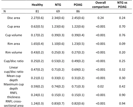
Optic nerve tomography was performed in all eyes using Heidelberg retina tomograph III (HRT; Heidelberg Engineering, Dossenheim, Germany). The optic disc was analysed using HRT version 3.0.3.0 software. The technique is described in detail elsewhere (130). Three high-quality images at 15x15-degree scanning angle were recorded per eye. Subsequent analysis was done on a mean image. All mean images included must have had a mean standard deviation of height measurement ≤ 40µm. The optic disc margin was manually marked at the inner edge of the Elschnig’s ring. The standard reference plane was used for calculations of optic disc topography with the relative and tilted coordinate system turned on.
The following variables were obtained: Disc area, Cup area and volume, rim area and volume, cup/disc overall and linear ratios, mean and maximum depth and retinal nerve fiber layer (RNFL) thickness and cross-sectional area.
IV. E) Blood pressure measurement
Blood pressure measurement was taken from the sitting’s subject’s right arm using an electronic sphygmomanometer (Omron, Schaumburg, IL 60173 U.S.A.). The device provided systolic and diastolic pressure readings, as well as peripheral pulse rate. The latter variable was used as a surrogate for the heart rate.
Other than systolic and diastolic pressures, calculations for the following parameters were performed off-line:
Blood pressure amplitude: Systolic BP – diastolic BP Mean arterial pressure: 2/3 diastolic+1/3 systolic BP Mean ocular perfusion pressure:
(2/3 diastolic+1/3 systolic BP)*2/3-Goldmann tonometry (131) IV. F) Visual acuity assessment
The Early treatment diabetic retinopathy study (ETDRS) chart was placed in the same location at the same distance (4 meters) from the patient under the same illumination for all subjects. The logmar scale was chosen to allow a direct statistical analysis, as no logarithmic transformation is needed.
56
IV. G) Visual field testing
Automated perimetry was performed using a Humphrey Field Analyzer, program 24-2, Sita standard strategy (Carl Zeiss, Oberkochen, Germany) or Octopus 301, program G1(30°), dynamic strategy (Interzeag AG, Schlieren, Switzerland). Unreliable visual fields (false positive, false negative or fixation loss values > 20%) were excluded. Visual field analysis was performed using Peridata software (PeriData Software GmbH, Huerth, Germany).
Mean defect (MD) values were recorded from these examinations. IV. H) SVP assessment
Optic disc observation was done under pharmacological dilation of the selected eye with tropicamide 0.5% (Tropicol®, Théa Pharma, Wetteren, Belgium). This was confirmed by two observers (LAP, EV), both of them masked to the subject’s diagnosis. In case of disagreement, a second observation by both researchers was made and a consensus was reached. The patient’s discs were observed continuously for at least one minute using a fundus camera for fundoscopy (Topcon TRC-50DX/EX fundus camera). The recording of visible venous pulsation over the disc was done using a binary code: + (present), - (absent).
A validation of the reproducibility of this SVP phenomenon assessment was later performed. As patients returned to either the cataract or Glaucoma clinic (for medical reasons only) during the
recruitment period, they were again observed using the same protocol for SVP detection.
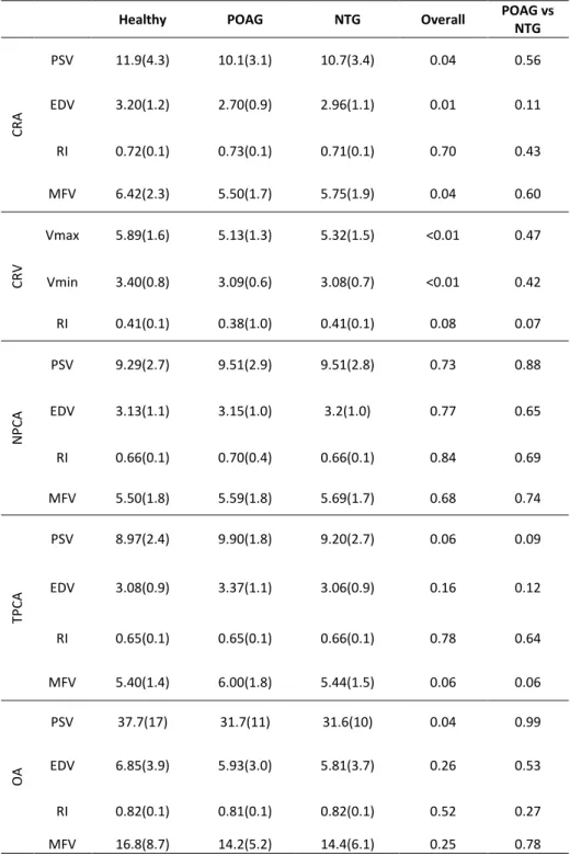
IV. I) Retrobulbar hemodynamics
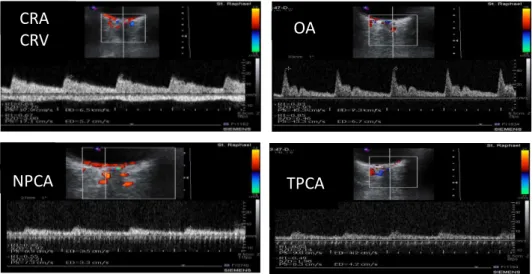
Hemodynamics of the retrobulbar vessels were studied using a probe capable of ultra-high frequency sound waves pulsed Doppler (7.5Mhz) from an ultrasound machine (Antares®, Siemens, Munich, Germany). The protocol of data acquisition was performed in accordance to the consensus of CDI technique for ocular blood flow studies (22) and whose reproducibility has been previously validated (132): The examination was performed on a supine patient after a minimum of 10 minutes of rest. The patient was instructed to keep legs uncrossed, arms along the body and to look straight while closing the eye. The examiner was behind the head of the subject, resting the base of his hand on the patient’s forehead to prevent exerting pressure on the globe. The examination started with a B-mode scan with the identification of the optic nerve as a landmark. Then, colour Doppler was applied to visualize the vessels (flow). The appropriate vessel was identified, the sample volume was placed in the center of the vessel, the angle was set parallel to the vessel and several seconds of Doppler waveform were recorded (angle was <60ο degrees in all measurements). The CRA and its corresponding vein were taken posterior to the lamina cribrosa. The nasal and temporal short posterior ciliary arteries (NPCA and TPCA were measured at a position that is close to the optic nerve and as anterior as possible. The OA was measured on the nasal side
58
of the optic nerve, immediately after it crossed the optic nerve (figure 1).
Figure 1. Example from a patients’ CDI examination of the retrobulbar vessels: central retinal artery and vein (CRA, CRV; respectively), ophthalmic artery (OA), nasal and temporal posterior ciliary arteries (NPCA, TPCA; respectively).The anatomic location of the Doppler signal and the characteristic patterns in the Doppler waveform allow for accurate labeling of these vessels.
The following data were retrieved from all the arteries’ Doppler waveform:
Peak Systolic Velocity (PSV) – highest point in waveform curve End-Diastolic Velocity (EDV) – lowest point in waveform curve Mean Flow Velocity (MFV) – mean time of the spectral outline Resistivity index (RI), calculated as RI = (PSV-EDV)/PSV.
N
OA CRA
CRV
In the CRV, the following variables were determined:
Maximal Velocity (Vmax) – highest point in venous waveform Minimal Velocity (Vmin) – lowest point in venous waveform Resistivity index (RI), calculated as RI = (Vmax-Vmin)/Vmax
60
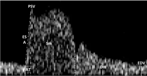
In the OA, the Doppler waveform was further studied for five other variables (figure2):
Early Systolic Acceleration (ESA) – fastest moving portion of systolic component
Acceleration Time (AT) – time period between the beginning and end of the fastest portion
Systolic mean Velocity (Sm) - mean time of the spectral outline from the beginning of the cycle and dicrotic notch
Diastolic mean Velocity (Dm) – mean time of the spectral outline from the dicrotic notch and the EDV
Ratio Sm/Dm was calculated offline
Figure 2. OA Doppler wavefront analyzed characteristics. ESA (early systolic acceleration) represents the fastest moving portion of the systolic component. Early systolic compliance peak (ESP), corresponds to the acute angle in the wavefront just after the first peak; AT, acceleration time; Sm, systolic mean blood flow velocity; Dm, diastolic mean flow velocity; PSV, peak-systolic velocity; EDV, end-diastolic velocity.
AT Sm Dm ES A PSV EDV
Additionally, the vascular cutpoints in glaucoma patients associated with the upper limit of the vessel regulation as previously published by our group (133) were also determined. Patients were labeled as being above or below the pre-determined value:
POAG patients: RI of CRA 0.77 NTG patients: RI of CRA 0.61 RI of OA 0.82
In the case of NTG patients, due to the existence of two cutpoints (for CRA and OA respectively), calculations were made to each artery separately and to the combination of both (CRA, OA and CRA+OA).
62
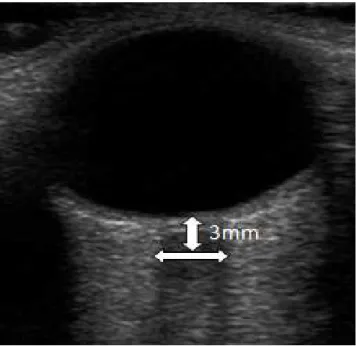
IV. J) Optic Nerve Sheath Diameter measurement
The same ultrasound device and probe as used for the Doppler studied was further used to assess the Optic Nerve Sheath Diameter (ONSD), using the previously published protocol (134). Using mode B, the insonation depth was set to 5–8 cm with the transducer placed over the upper eyelid in an axial plane. The ONSD was calculated perpendicular to the vertical axis of the scanning place 3 mm behind the globe (figure 3), where the optic nerve sheath structure is more prone to expansion due to increases in ICP (135).
Figure 3. Optic nerve sonography. The optic nerve complex is shown as a sharply-defined hypoechoic stripe in between the echogenic retrobulbar fat
V. Experimental design
Patients from both the Glaucoma Clinic and subjects from the general ophthalmology consultation were asked to participate in the study. Inclusion/exclusion criteria in both glaucoma patients and healthy subjects were screened by the head of the glaucoma clinic (IST). If conditions were met, each patient/subject was required to sign an informed consent statement before being enrolled into the study and prior to any study measurements being taken.
In this screening visit, best corrected visual acuity and prior medical history including topical medication intake was recorded. All subjects underwent a visual field as well as disc tomography on the day of the study visit. For the healthy individuals, randomization for the eye to be included in the study was made by a third person, blind to any of the subject characteristics or examination results.
64
Subjects from all groups were taken to a different observation room with another examiner (LAP). This examiner was masked to the subject diagnosis or any other clinical variable and whose only indication was a paper indicating which eye to perform the following examinations in a pre-determined order:
(1) OPA reading; (2) CCT measurement
(3) blood pressure and pulse rate measurement; (4) retrobulbar hemodynamic assessment; (5) ONSD measurements;
(6) SVP assessment.
Of note, 20 minutes were taken between step 5 and 6 in order to allow proper optic disc observation through adequate pupil dilation.
The recruitment of patients and the previously described methodology was performed during two periods, totalizing 4 months (January-February 2011, and September-October 2011).
VI. Statistical analysis
Chi-square tests (for 3x2 contingency tables) were used to analyze the present/absent spontaneous venous pulsation ratios. When 2x2 contingency tables were performed, a Fisher exact test was used. In all cases, the tests will be performed using absolute number of the variables. However, for graphical purposes, the figures will be designed as percentage of total.
Kruskal-Wallis tests were used to compare the three diagnostic groups on different variables. When statistical differences were detected, Mann-Whitney test was used in pairwise group comparisons.
Normal distribution of the data was verified with the D’Agostino and Pearson omnibus normality test. If the data were normal distributed, the existence of correlation between variables was tested using Pearson’s correlation. Otherwise, the Spearman correlation was used.
For practical purposes, acquisition of the data from a clinical setting could not match sample calculations based on previous publications from CDI technology alone for instance (136). In order to address this, the projected work aimed at 100 patients. Not only would it still be one of the largest works done in the subject of ocular blood flow in retinal venous studies, but also the division in three groups would still comply with the central limit theorem. This theorem implies that groups of independent variables that are larger than a certain number (usually above 30) tend to have a
66
normal distribution (137). Our group samples, by being at least twice these numbers, try to account for the practical limitations of clinical research in reaching sample calculations.
Statistical significance was considered when p<0.05. Values depicted as mean±SD unless otherwise indicated.
Analyses were performed using Graphpad Prism® ver. 5.0; (Graphpad Software Inc, La Jolla, CA).
Chapter 4. Results
Results will be presented as separate sets, according to the analyzed variables. This was done to allow a proper discussion of each variable/correlation.
As such, the following subchapters will be presented: I) Experimental group characteristics
II) SVP frequency
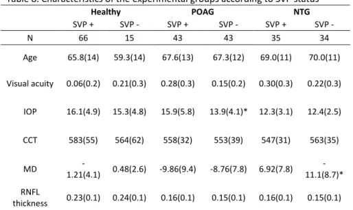
III) Patients’ overall characteristics by SVP status
IV) SVP phenomenon frequency and functional damage V) Optic Disc Tomography and SVP status
VI) IOP and SVP phenomenon
VII) OPA, ONSD and SVP phenomenon
VIII) Cardiovascular parameters and SVP phenomenon IX) Ocular blood flow and SVP phenomenon
I. Experimental groups’ characteristics
A thorough analysis of the characteristics of the different experimental groups is crucial to a correct interpretation of any further correlations. As such, in addition to the complete description of the variables in each group, the agreement of these results with the existing literature is discussed.
I. A) Overall characteristics
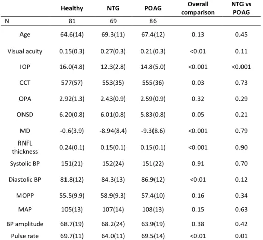
Table 1 summarizes the patients’ characteristics in the different diagnostic groups. Kruskal-Wallis test indicated no overall age differences between the studied groups (p=0.13). The best corrected visual acuity was significantly higher in the healthy group (p<0.01), although no difference was detected between the glaucoma groups (NTG vs POAG, p=0.11). Both CCT and IOP were also statistically different (p=0.03, p<0.001, respectively), with again the healthy individuals presenting higher IOP values as well as a higher pachymetry. In a pairwise comparison between NTG and POAG however, only IOP was detected to be different (p<0.001), with CCT presenting a significant overlap (p=0.73). OPA readings were not significantly different (p=0.32). Additionally, ONSD difference between the three groups was borderline significant (p=0.05). However, comparison between NTG and POAG group showed a greater similarity of ONSD values (p=0.21).
70
As expected, there was a significant difference in functional and structural parameters of glaucoma damage between the three groups (MD and RNFL thickness: p<0.001, respectively), with lesser damage values seen in healthy individuals. However, when comparing the two glaucoma groups, no difference was detected (MD: p=0.79; RNFL thickness: p=0.90).
Table 1. Patients characteristics
Healthy NTG POAG Overall comparison NTG vs POAG N 81 69 86 Age 64.6(14) 69.3(11) 67.4(12) 0.13 0.45 Visual acuity 0.15(0.3) 0.27(0.3) 0.21(0.3) <0.01 0.11 IOP 16.0(4.8) 12.3(2.8) 14.8(5.0) <0.001 <0.001 CCT 577(57) 553(35) 555(36) 0.03 0.73 OPA 2.92(1.3) 2.43(0.9) 2.59(0.9) 0.32 0.29 ONSD 6.20(0.8) 6.01(0.8) 5.83(0.8) 0.05 0.21 MD -0.6(3.9) -8.94(8.4) -9.3(8.6) <0.001 0.79 RNFL thickness 0.24(0.1) 0.15(0.1) 0.15(0.1) <0.001 0.90 Systolic BP 151(21) 152(24) 151(22) 0.91 0.70 Diastolic BP 81.8(12) 84.3(13) 86.9(12) <0.01 0.12 MOPP 55.5(9.9) 58.9(9.3) 57.4(10) 0.16 0.34 MAP 105(13) 107(14) 108(13) 0.15 0.63 BP amplitude 68.7(19) 68.2(24) 63.9(19) 0.38 0.42 Pulse rate 69.7(11) 64.0(11) 69.5(14) <0.01 0.01 Mean values (and SD) are depicted. SVP refers to number of patients who presented that phenomenon compared to the total number of patients. Kruskal-Wallis indicates P values of overall differences between the diagnostic groups. NTG vs POAG comparison was made using Mann-Whitney test.
The analysis of the cardiovascular parameters revealed systolic BP, median ocular perfusion pressure (MOPP), mean arterial pressure (MAP) and BP amplitude comparison not to be statistically different (p=0.91, p=0.16, p=15 and p=0.38; respectively). There were however differences in diastolic BP and pulse rate (p<0.01), with healthy individuals presenting lowest diastolic values and NTG patients presenting the lowest pulse rate. Pairwise comparison between NTG and POAG group demonstrated a significant difference in pulse rate, with NTG patients again having a lower value (p=0.01). The diastolic BP between these two later groups was not significant (p=0.12).
Topical medications in both glaucoma groups are summarized in table 2. The fixed combinations were documented according to the number of active drugs. Fisher exact-test detected no difference in the proportion of patients under beta-blockers, alpha-agonists and carbonic anhydrase inhibitors therapy between the two groups (p=0.19, p=1.00, p=0.21, respectively). A larger portion of the POAG group was taking prostaglandin analogs (p=0.01).
Table 2. Topical medications
Beta-blockers Alpha-agonists Carbonic anhydrase inhibitors Prostaglandin Analogs POAG 42(49) 8(9) 21(24) 53(62) NTG 26(38) 6(9) 24(35) 28(41)