Universidade de Trás-os-Montes e Alto Douro
Scaffold development for Rotator Cuff repair
Master Dissertation in Biomedical Engineering
Tânia Moura Peixoto
Advisor: Paula Luísa Nunes Braga da Silva Co-advisor: Maria Ascensão Ferreira Silva Lopes Co-advisor: João Manuel da Costa Ferreira Torres
Vila Real, 2017
i
Universidade de Trás-os-Montes e Alto Douro
Scaffold development for Rotator Cuff repair
Master Dissertation in Biomedical Engineering
Tânia Moura Peixoto
Advisor: Paula Luísa Nunes Braga da Silva Co-advisor: Maria Ascensão Ferreira Silva Lopes Co-advisor: João Manuel da Costa Ferreira Torres
Jury Composition:
President: Luís José Calçada Torres Pereira, PhD, assistant professor at Escola de Ciência e Tecnologia, Universidade de Trás-Os-Montes e Alto Douro.
Other members: Rui Jorge Sousa Costa de Miranda Guedes, PhD, associate professor at Faculdade de Engenharia da Universidade do Porto; Maria Ascensão Ferreira Silva Lopes, PhD, assistant professor at Faculdade de Engenharia da Universidade do Porto.
iii
Dissertation presented to the Universidade de Trás-os-Montes e Alto-Douro to obtain a Master's degree in Biomedical Engineering, carried out under the scientific orientation of Professor Paula Luísa Nunes Braga da Silva, Department of Mechanical Engineering, Escola de Ciências e Tecnologia, Universidade de Trás-os-Montes e Alto-Douro and under coorientation from Professor Maria Ascensão Ferreira Silva Lopes, from the Department of Metallurgic and Materials Engineering of the Faculdade de Engenharia da Universidade do Porto, and coorientation from Doctor João Manuel da Costa Ferreira Torres, from Hospital of S.João, Porto.
v
Abstract
The rotator cuff is a functional anatomical unit located in the upper extremity and it is defined, anatomically, as a group of muscles and their tendons that act to move and stabilize the shoulder. (Opsha, 2008) Rotator cuff tendon injuries are frequent and are responsible for substantial morbidity in athletes, and in working and elder population. As our society continues to age and remain active, these conditions not only have an impact on peoples' quality of life and activities of daily living, but also represent significant burden on social, economic and health terms. (Docheva, 2015)
Currently used therapies are mainly limited to pain control and/or tissue replacement, without fully restoring tissue functionality. (Morais, 2015) Although there was an increase in volume of rotator cuff surgeries during the last years, re-tear rates remain high, with structural repair failure remaining the main problem. (Docheva, 2015) Hence, there is a need for repair strategies that can augment the repair by mechanically reinforcing it, while at the same time biologically enhancing and optimizing the intrinsic healing potential of the tendon. Ultimately, tissue-engineering based alternatives, have been the target of extensive investigation and several biological, natural, and synthetic scaffolds have been proposed for rotator cuff tendon repair. (Richetti, 2012) However, to develop a structure that properly mimics the mechanical performance of the native tissue remains a challenge. (Morais, 2015)
Textile manufacturing techniques have been used in a wide range of engineering applications and have recently attracted great attention as potential biofabrication tools for engineering tissue constructs. The versatility of textile structures allows for tailoring their architecture, being possible to control the resulting physical properties and cellular behavior of the scaffolds. (Aibubu, 2016; Akbari, 2016)
In this work, four textile constructs, two three-dimensional (3D) and two planar warp-knitted structures were evaluated as potential scaffolds for rotator cuff tendon repair. Polyethylene terephthalate (PET) was the chosen base polymer for the structures because of its biocompatibility and excellent mechanical properties. Fourier-Transform Infrared Spectroscopy (FTIR), Scanning Electronic Microscopy (SEM), Porosity tests, Absorption tests, Tensile tests and Suture pull-through tests were performed to characterize the structures under study.
vi
All matrices presented regular patterns, with well oriented fibers. The estimated fiber diameter, mean pore size and porosity levels of all structures can be consider adequate for tissue-engineering applications. All structures demonstrated similar liquid absorptive capacity and wicking behavior. Regarding the mechanical characterization, structure D demonstrated failure load, stress at failure and elastic modulus values that can be considered suitable for rotator cuff tendon repair. Although the obtained stiffness and failure strain values were not ideal, pre-tensioning techniques were concluded to be adequate to increase stiffness and lower the overall elongation of the structures. Furthermore, this construct demonstrated a value for maximum suture retention load close to the required. Thus, it was concluded that among all constructs, structure D meets the minimum requirements to serve as a scaffold for rotator cuff tendon repair.
vii
Acknowledgments
The realization of this master's dissertation counted with important supports and incentives, without which it would not have become a reality and to which I will be eternally grateful.
To Professors Paula Braga and Maria Ascensão Lopes, I want to thank for the guidance, total support, availability, for the knowledge they transmitted, for the opinions and criticisms, total collaboration in solving the doubts and problems that arose throughout the realization of this work and for all the words of incentive. To Dr. João Torres, I want to thank for the collaboration, and for all the help provided. To professor Rui Guedes, I want to thank for the demonstrated availability, for the collaboration is solving the doubts and all the help provided. To CITEVE (Technological Center for Textiles and Clothing), specially to engineers José Morgado and Graça Bonifácio, I want to thank for the demonstrated availability and help, and for putting me in contact with the company LMA (Leandro Manuel Araújo, LDA), to which I must also thank for making available the textile structures for my work.
To UTAD (Universidade de Trás-Os-Montes e Alto Douro) and FEUP (Faculdade de Engenharia de Universidade do Porto), and all the staff that helped me, I want to thank for allowing me and facilitate the access to all the equipment that I needed for my work.
To my boyfriend, I want to thank for the incredible support and encouragement, for being there for me in the good and also the bad moments, and for all the love demonstrated since we are together. To my sister, I want to thank for all the fun moments to distract me, and words of support. Finally, to my parents, I want to thank for all the sacrifices they made so that I could study, and for their continuous support, understanding and patience. It is to them that I dedicate my dissertation, since I would not have been able to complete this stage without their love.
ix
Contents
Abstract ... v
Acknowledgments ... vii
List of Figures ... xiii
List of Tables ... xix
Chapter 1 ... 1
Introduction ... 1
1.1. Motivation ... 1
1.2. Objectives ... 2
1.3. Structure of the dissertation ... 2
Chapter 2 ... 3
The Rotator Cuff ... 3
2. 1. Subacromial space and rotator cuff anatomy ... 3
2.1.1. Anatomy of the Coracoacromial Arch and the Structures in the Subacromial Region 3 2.1.2. Anatomy of the rotator cuff ... 4
2.2. Rotator cuff biomechanics ... 6
2.2.1. Rotator cuff function in glenohumeral stability ... 7
2.2.2. Rotator cuff function in glenohumeral motion ... 7
2.3. Epidemiology of Rotator Cuff tears ... 8
2.4. Rotator cuff pathology ... 13
2.4.1. Extrinsic theory ... 13
2.4.2. Intrinsic theory ... 15
2.4.3. Demographic factors ... 19
2.5. Mechanobiology of tendon ... 20
2.5.1. Tendon molecular composition and structure ... 21
2.5.2. Tendon mechanical properties ... 23
2.5.3. Tendon healing ... 25
2.6. Rotator Cuff tendon injuries ... 28
2.6.1. Clinical assessment ... 28
2.6.2. Classification of rotator cuff tears ... 29
x
2.7. Clinical Relevance ... 32
Chapter 3 ... 37
Scaffold devices and current trends for Rotator Cuff Repair: a Tissue-Engineering approach ... 37
3.1. Scaffolding in tissue-engineering: general characteristics ... 37
3.1.1. Desirable attributes of tissue-engineered scaffolds ... 39
3.2. Current strategies for rotator cuff repair ... 40
3.2.1. Scaffolds for Tendon Regeneration ... 41
3.2.2. Commercially available scaffolds for Rotator Cuff repair ... 43
3.3. Scaffold development for Tendon Tissue-Engineering ... 45
3.3.1. Fabrication method for Tissue-engineering scaffolds ... 46
3.3.1.1. Polymer choice ... 46
3.3.1.2. Textile techniques ... 49
3.3.1.2.1. Manufacturing process of warp-knitted structures ... 50
3.3.1.3. Textile Technologies advantage and disadvantages ... 54
3.4. Tendon tissue-engineering enhancing strategies ... 55
3.4.1. Scaffolds Surface Modification ... 55
3.4.2. Cell seeding and delivery of growth factors ... 56
3.4.3. Mechanical stimulation ... 58
Chapter 4 ... 61
Materials and Test Methods ... 61
4.1. Morphological and Surface Characterization ... 62
4.1.1. Fourier-Transform Infrared Spectroscopy (FTIR) ... 62
4.1.2. Scanning Electronic Microscopy (SEM) ... 63
4.1.3. Porosity ... 63
4.2. Absorption tests ... 64
4.2.1. Liquid Absorptive capacity test ... 64
4.2.2. Liquid Wicking rate test ... 64
4.3. Mechanical Characterization ... 65
4.3.1. Tensile testing ... 65
4.3.1.1. Dry tests ... 65
4.3.1.2. Wet tests ... 66
xi
4.3.2. Testing suture pull-through ... 68
4.4. Statistical Analysis ... 68
Chapter 5 ... 69
Results and discussion ... 69
5.1. Morphological and Surface Characterization ... 69
5.1.1. Fourier-Transform Infrared Spectroscopy (FTIR) ... 69
5.1.2. Scanning Electron Microscopy (SEM) ... 70
5.1.3. Porosity ... 76
5.2. Absorption tests ... 77
5.2.1. Liquid Absorptive capacity test ... 77
5.2.2. Liquid Wicking rate test ... 79
5.3. Mechanical Characterization ... 81 5.3.1. Tensile testing ... 81 5.3.1.1. Dry tests ... 81 5.3.1.2. Wet tests ... 86 5.3.1.3. Cyclic testing ... 93 5.3.1.4. Final remarks ... 103
5.3.2. Testing suture pull-through ... 106
Chapter 6 ... 109
Conclusions ... 109
xiii
List of Figures
Figure 1. Rotator cuff anatomy: (a) Anterior view, (b) Posterior view, (c) Lateral view.
(Blum, 2009) ... 6
Figure 2. Proportion of population reporting pain, United States 2012. (Weinstein, 2014)
[Adapted] ... 9
Figure 3. Anatomic site for sprains/strains treated in Physician Offices, United States 2010.
(Pollak, 2014) ... 9
Figure 4. Work-related injuries involving days away from work by body part affected, United
States 2011. (Pollak, 2014) ... 10
Figure 5. Prevalence of rotator cuff tear on dominant limb in asymptomatic subjects that have
undergone ultrasound examination, in the various age groups, in a study conducted by Milgrom, 1995. (Gumina, 2017) ... 11
Figure 6. Prevalence of partial and full thickness tears in asymptomatic subjects that have
undergone MRI examination, in the various age groups, in a study conducted by Sher, 1995. (Gumina, 2017) ... 11
Figure 7. (a) Percentages of partial and full-thickness tears with unilateral tear (199 patients)
(Yamaguchi, 2006); (b) Percentages of partial tears, full-thickness tears and partial associated to full-thickness tears, in the group of patients with bilateral damage (177 patients) (Yamaguchi, 2006). (Gumina, 2017) ... 11
Figure 8. Bigliani's classification of acromion undersurface with corresponding supraspinatus
outlet view radiograph. (Pandey, 2015) ... 14
Figure 9. Rotator cuff degeneration secondary to inflammation and oxidative stress. (Nho,
2008) [Adapted] ... 17
Figure 10. Summary of extrinsic and intrinsic pathways of rotator cuff tear (ECM:
extracellular matrix; MMP-1:matrix metalloproteinase-1; ROS: reactive oxygen species). (Pandey, 2015) ... 18
Figure 11. Rotator cuff tears: from impingement syndrome or overuse tendinitis to tear.
(Mayo clinic, 2016) ... 18
Figure 12. A schematic drawing of basic tendon structure. (Towler, 2006) ... 23 Figure 13. Tendon stress-strain curve. (Wang, 2006) ... 24
xiv
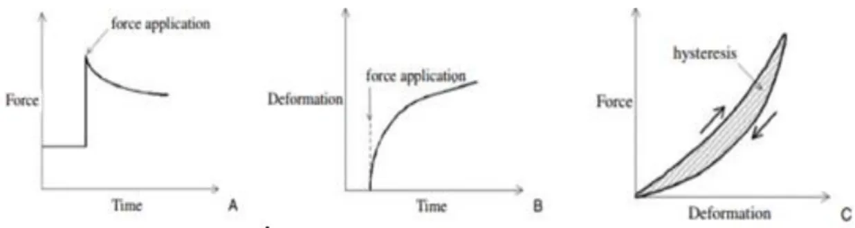
Figure 14. (A) Typical force-relaxation curve in a tendon. - the force required to cause a
given deformation decreases over time. (B) Typical creep curve in a tendon - the deformation caused by a given force increases over time. (C) Typical mechanical hysteresis in a tendon - the arrows indicate loading and unloading directions during a test with a tensile load within the elastic limit of the tendon. The area of the loop between the loading and unloading curves relative to that underneath the loading curve represents the fraction of strain energy lost as
heat by the tendon viscous damping. (Maganaris, 2005) ... 25
Figure 15. Human tendon repair process. The healing of ruptured tendons passes through three main phases containing distinctive cell and molecular cascades. These phases overlap and their duration depends upon the location and severity of the tendon injury. (Docheva, 2015) ... 26
Figure 16. Distribution of pre-operative costs. (Yeranosian, 2013) ... 34
Figure 17. The role of scaffolds in tissue engineering strategies. (BMP - bone morphogenetic protein; FGF-2, fibroblast growth factor 2; IGF, insulin-like growth factor; MSC, mesenchymal stem cell; PRP, platelet-rich plasma; TGF-β; transforming growth factor β. (Smith, 2015) ... 38
Figure 18. Engineering process of fibrous architectures for tissue-engineering (TE) applications. (Aibubu, 2016) [Adapted] ... 46
Figure 19. Two-Dimensional woven, nonwoven, knit and braid structures. (Textile study Center) ... 49
Figure 20. Warp-knitting. (Anand, 2000) [Adapted] ... 51
Figure 21. Guide bar movement. (Kothari, 2011) ... 52
Figure 22. Warp-Knitting machine. (Kothari, 2011) ... 52
Figure 23. 3D Warp knitted fabric structure. (Ma, 2017) [Adapted] ... 53
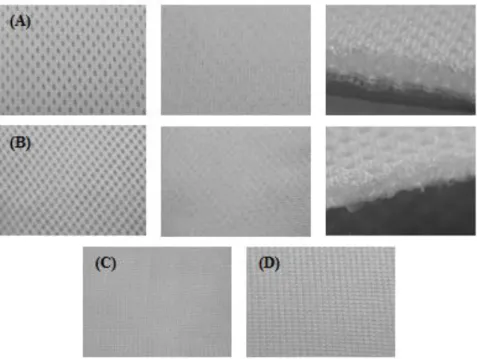
Figure 24. (A) and (B) represent the two 3D warp-knitted structures and (C) and (D) the two conventional warp-knitted structures, tested in this work. Notice that the structure (A) and (B) have two different faces. ... 62
Figure 25. Wicking apparatus. ... 65
Figure 26. Shimadzu EZ-LX Long-Stroke Model tensile testing machine used. ... 66
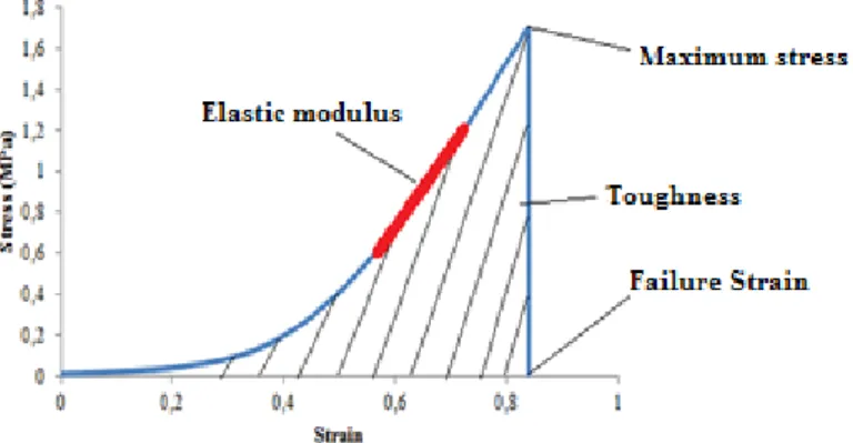
Figure 27. Illustration of the determination of the tensile parameters under study. ... 66
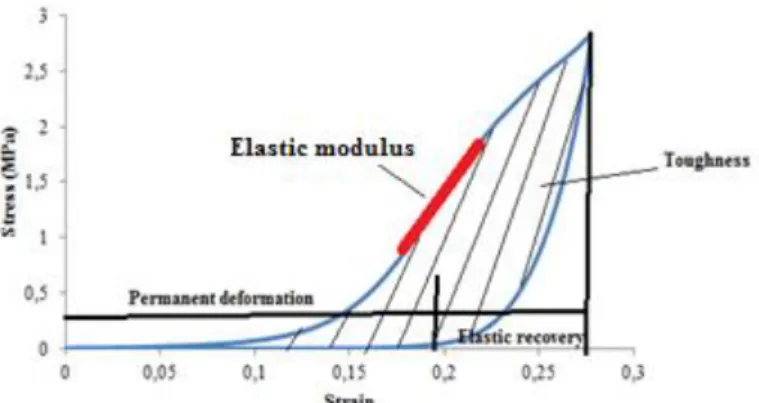
Figure 28. Illustration of the determination of the cyclic tensile parameters under study. ... 68
Figure 29. ATR-FTIR analysis of the polyester scaffold. ... 69
xv
Figure 31. SEM image obtained of the 3D warp-knitted structure A. The upper images
correspond to one face, and the lower images to the other. ... 71
Figure 32. SEM image obtained of the 3D warp-knitted structure B. The upper images
correspond to one face, and the lower images to the other. ... 72
Figure 33. SEM image obtained of the planar warp-knitted structure C. ... 72 Figure 34. SEM image obtained of the planar warp-knitted structure D. ... 72 Figure 35. Relationship between the weight change of the two 3D warp-knitted structures A
and B and the two planar warp-knitted structures C and D and the immersion time in water. 77
Figure 36. Relationship between the wicking behavior of the two 3D warp-knitted structures
A and B and the two planar warp-knitted structures C and D over time. ... 79
Figure 37. (a) Force (N) / Elongation (mm) curve of the 3D warp-knitted structure A in the
longitudinal direction; (b) Force (N) / Elongation (mm) curve of the 3D warp-knitted structure A in the transversal direction. ... 82
Figure 38. (a) Force (N) / Elongation (mm) curve of the 3D warp-knitted structure B in the
longitudinal direction; (b) Force (N) / Elongation (mm) curve of the 3D warp-knitted structure B in the transversal direction. ... 82
Figure 39. (a) Force (N) / Elongation (mm) curve of the 2D warp-knitted structure C in the
longitudinal direction; (b) Force (N) / Elongation (mm) curve of the 2D warp-knitted structure C in the transversal direction. ... 82
Figure 40. (a) Force (N) / Elongation (mm) curve of the 2D warp-knitted structure D in the
longitudinal direction; (b) Force (N) / Elongation (mm) curve of the 2D warp-knitted structure D in the transversal direction. ... 83
Figure 41. Maximum stress (MPa) values of the different structures, in both directions
(longitudinal and transversal). ... 83
Figure 42. Young’s modulus (MPa) values of the different structures, in both directions
(longitudinal and transversal). ... 84
Figure 43. Failure strain values of the different structures, in both directions (longitudinal and
transversal). ... 84
Figure 44. Stiffness (N/mm) values of the different structures, in both directions (longitudinal
and transversal). ... 85
Figure 45. Toughness (J/mm3) values of the different structures, as measured by failure in
xvi
Figure 46. (a) Force (N) / Elongation (mm) curve of the wet 3D warp-knitted structure A. (b)
Force (N) / Elongation (mm) curves of the wet 3D warp-knitted structures B; (c) Force (N) / Elongation (mm) curve of the wet 2D warp-knitted structure C; (d) Force (N) / / Elongation (mm) curve of the wet 2D warp-knitted structure D. ... 86
Figure 47. Maximum stress (MPa) values of the different structures, under wet conditions. 87 Figure 48. Young’s modulus (MPa) values of the different structures, under wet conditions.
... 87
Figure 49. Failure strain values of the different structures, under wet conditions. ... 88 Figure 50. Stiffness (N/mm) values of the different structures, under wet conditions. ... 88 Figure 51. Toughness (J/mm3) values of the different structures, as measured by failure in
tension, under wet conditions. ... 89
Figure 52. Longitudinal (y-axis) and transverse (x-axis) directions of the knitted matrix.
(Almeida, 2013) ... 90
Figure 53. (a) Relationship between Maximum Stress (MPa) and thickness (mm); (b)
Relationship between Elastic Modulus (MPa) and Thickness (mm). ... 90
Figure 54. (a) Relationship between Maximum Stress (MPa) and Porosity (%); (b)
Relationship between Elastic Modulus (MPa) and Porosity (%). ... 91
Figure 55. Typical load-extension characteristic of a knitted fabric tested in the wale
direction. Step 1: stretching of the curved yarns up to the critical stretch state (yarns are straightened and not elongated). Step 2: elongation of the straightened yarns up to the breaking point. (Camino, 2007) ... 92
Figure 56. Force (N) / Elongation (mm) curves obtained from the cyclic tests for 3D
warp-knitted structure A, in which in (a), as it is observable, the test was continuous, while in (b) the specimen length was readjusted at each cycle to maintain the initial length of 40 mm. .... 93
Figure 57. Force (N) / Elongation (mm) curves obtained from the cyclic tests for 3D
warp-knitted structure B, in which in (a), as it is observable, the test was continuous, while in (b) the specimen length was readjusted at each cycle to maintain the initial length of 40 mm. .... 94
Figure 58. Force (N) / Elongation (mm) curves obtained from the cyclic tests for 2D
warp-knitted structure C, in which in (a), as it is observable, the test was continuous, while in (b) the specimen length was readjusted at each cycle to maintain the initial length of 40 mm. .... 95
Figure 59. Force (N) / Elongation (mm) curves obtained from the cyclic tests for 2D
warp-knitted structure D, in which in (a), as it is observable, the test was continuous, while in (b) the specimen length was readjusted at each cycle to maintain the initial length of 40 mm. .... 95
xvii
Figure 60. Force (N) / Elongation (mm) curves obtained for 3D warp- knitted structure A and
their overlap, obtained for each test condition, in which the blue curve represents the test condition in which it was maintained the initial specimen length for all loads, the red curve represents the continuous test, and the green curve represents the overlap. Graphic (a) represents the curves obtained for 20N, (b) for 40N, (c) for 60N, (d) for 80N, (e) for 100 (N) and (f) for 120N. ... 96
Figure 61. Force (N) / Elongation (mm) curves obtained for 3D warp- knitted structure B,
and their overlap, obtained for each test condition, in which the blue curve represents the test condition in which it was maintained the initial specimen length for all loads, the red curve represents the continuous test, and the green curve represents the overlap. Graphic (a) represents the curves obtained for 10N, (b) for 20N and (c) for 30N. ... 97
Figure 62. Force (N) / Elongation (mm) curves obtained for 2D warp- knitted structure C,
and their overlap, obtained for each test condition, in which the blue curve represents the test condition in which it was maintained the initial specimen length for all loads, the red curve represents the continuous test, and the green curve represents the overlap. Graphic (a) represents the curves obtained for 20N, (b) for 40N and (c) for 60N. ... 97
Figure 63. Force (N) / Elongation (mm) curves obtained for 2D warp- knitted structure D,
and their overlap, obtained for each test condition, in which the blue curve represents the test condition in which it was maintained the initial specimen length for all loads, the red curve represents the continuous test, and the green curve represents the overlap. Graphic (a) represents the curves obtained for 40N, (b) for 80N, (c) for 120N, (d) for 160N and (e) for 200N. ... 98
Figure 64. Relationship between load (N) and elongation (mm) for all the structures under
study demonstrated a directly proportional relationship between the increase in load and the deformation suffered. The laxity of the structures increased throughout the test. ... 100
Figure 65. (a) The elastic modulus (MPa) values increased throughout the cycles (with
increasing load) for all structures. (b) The stiffness values (N/mm) increased throughout the cycles (with increasing load) for all structures. ... 101
Figure 66. For all structures, the initial behavior of the loop structure decreased throughout
the cycles, as the mechanical response was basically taken upon the yarns, and as the structures became stiffer, they resisted deformation, resulting in smaller elongations with increasing loads. ... 102
xviii
Figure 67. A comparison of the toughness (J/mm3) of the different structures, as measured by
xix
List of Tables
Table 1. Origins, Insertions, Actions, and Nerve Supplies of the Rotator Cuff muscles and
tendons. ... 8
Table 2. Studies in literature that investigated the prevalence of RCT in cadavers,
asymptomatic and symptomatic patients. (Tashjian, 2012 ;Wani, 2016; Gumina, 2017) ... 10
Table 3. Classification of rotator cuff tears. (Vollans, 2016; Wani, 2016) ... 30 Table 4. Commercially available devices for rotator cuff repair. (Ricchetti, 2012; Morais,
2015) ... 44
Table 5. Effect of different growth factors on tendon healing. (Docheva, 2015; Morais, 2015)
... 58
Table 6. Information obtained through the analysis of the SEM images, of the four structures
under study. ... 71
Table 7. Physical properties of the different warp-knitted structures. ... 76 Table 8. Measurements obtained from the tensile tests of the four warp-knitted structures
under study. ... 81
Table 9. Measurements obtained from the wet tensile tests of the four warp-knitted structures
under study. ... 86
Table 10. Measurements obtained from the cyclic tests of the four warp-knitted structures
under study. ... 99
Table 11. Measurements obtained from the cyclic tests of the four warp-knitted structures
under study, regarding the structures deformation under different loads. ... 99
Table 12. Failure load (N) obtained from the tensile tests performed on the three types of
braids developed. ... 106
1
Chapter 1
Introduction
1.1. Motivation
Of the 30 million musculoskeletal injuries reported worldwide, over half are thought to involve tendons and ligaments. (Walden, 2017) Tendon injuries often result in pain, substantial tissue morbidity and disability, affecting athletes, active workers and elder population. (Wani, 2016) Existing studies have shown a high prevalence of rotator cuff tears in the general population, with some reporting up to a 60% lifetime incidence, affecting between 30% and 50% of people older than 50 years, reaching 80% in patients aged more than 80. (Minagawa, 2013; Varkey, 2016) Multiple etiologies, involving extrinsic and intrinsic factors, have been implicated in the pathogenesis of rotator cuff tears. (Gumina, 2017) In general, rotator cuff tearing is thought to occur, to some extent, as a normal degenerative process, that increases with aging (Tashjian, 2012). It is expected that the total number of individuals with rotator cuff dysfunction will increase in the future, as a result of a progressively older population that tend to maintain their level of activity and are less tolerant of functional limitations.
Due to the limited tendon ability for self-healing, conservative treatment requires long treatment periods, with potential partial function loss and recurrent injury, failing in many cases. So, when this approach does not result or is not appropriate attending to the extension of the lesion, surgical intervention is used. (Morais, 2015) Despite the high rate at which these surgeries are performed and regardless of advances in technology and surgical techniques, re-tear after rotator cuff surgery remains a common problem with current approaches. (Patel, 2016) Current surgical treatments involve suturing the injured ends together or reattaching the tendon to bone, securely with high-strength suture anchors, or the use of tendon grafts, when the defect is large. However, tendon detachment remains the primary cause of surgical failure in most cases, and grafts often do not provide adequate mechanical strength during the remodeling process. (Longo, 2010; Morais, 2015)
Tissue-engineering strategies for rotator cuff repair are emerging as important tools in the treatment of large, massive or chronic degenerative tears. The main challenge when
2
designing scaffolds for tendon tissue-engineering is to develop a long-term functional structure that properly recreates the mechanical and physical-chemical performance of the native tissue. (Ávila, 2015) Textile manufacturing techniques have recently attracted great attention as potential biofabrication tools for engineering tissue constructs. The versatility of textile manufacturing processes allows for tailoring the architecture and features of the textile structures, thus, it is possible to control the physical properties and cellular behavior of the engineered constructs, essential for tissue regeneration. (Akbari, 2016)
1.2. Objectives
This work aims to study four textile structures, namely four different warp-knitted structures, to determine their potential as scaffolds for rotator cuff tendon repair. The choice of these textile structures rested on the fact that these type of textiles can be manipulated by the surgeon, i.e., can be cut and adapted in size without unravel, enabling the surgeon to adapt the scaffold to the patient needs.
Several tests were performed in order to understand the impact of different architectures in the overall performance of the structures, to determine if they could be used for rotator cuff tendon augmentation and how important parameters could be tailored to further improve their properties in order to develop an optimal scaffold for rotator cuff tendon repair.
1.3. Structure of the dissertation
This dissertation is composed by 6 chapters. After this introductory section, Chapter 2 addresses the anatomical and physiological characterization of the rotator cuff tendons, the involving pathology and the overall disease characterization and treatment options. Current treatments for rotator cuff repair, regarding the application of biomaterials are discussed in Chapter 3, along with the new role of textile technologies in the field of tendon/ligament tissue-engineering. Chapter 4 presents the textile structures under study and the experimental methodology applied to characterize the structures, and Chapter 5 the main results and discussion. Finally, Chapter 6 gives a global perspective of the developed work, presenting relevant conclusions and final remarks. It also provides guidelines and questions to be explored in the future.
3
Chapter 2
The Rotator Cuff
2. 1. Subacromial space and rotator cuff anatomy
The rotator cuff is a functional anatomical unit located in the upper extremity and it is defined, anatomically, as a group of muscles and their tendons that act to move and stabilize the shoulder. This group of muscles and tendons performs multiple functions and is often stressed during various activities. A rotator cuff tear is a common cause of pain and disability among adults. (Opsha, 2008) Although many factors influence the treatment of rotator cuff tears, understanding the anatomy and how it relates to function is an important one (the anatomy and physiology of the rotator cuff is complex and interconnected to other muscle groups in the shoulder). During the past decade, advances in basic science and surgical technology have improved the knowledge of this anatomy. (Opsha, 2008; DeFranco, 2009)
2.1.1. Anatomy of the Coracoacromial Arch and the Structures in the Subacromial Region
The subacromial space is delimited above by the coracoacromial arch (formed by the anterior-inferior margin of the acromion, coracoacromial ligament, apex and distal third of the posterior surface of the coracoid) and below by the humeral head, by the tendons of the rotator cuff and of the long head of the biceps. (Gumina, 2017) The humeral head articulates with the glenoid cavity of the scapula to form the glenohumeral joint. (Opsha, 2008)
The coracoacromial arch is intimately related to the structures below it. The acromion is flat in shape and in it, it is distinguishable an upper surface, in close contact with the skin; an inferior concavity, which forms the tip of the glenohumeral joint; a lateral margin, the bundles from which the deltoid muscle originates and a medial margin where the surface of the acromiovascular joint is. The coracoacromial ligament is a strong structure stretching from the coracoid process (located anteriorly to the glenoid and laterally to the scapular notch) to insert into the inner border of the acromion just in front of the acromioclavicular joint. The rotator cuff, particularly its supraspinatus portion, is in close proximity to the under surface of
4
the coracoacromial ligament, the acromioclavicular joint and the under surface of the acromion. Between the cuff and the arch, lies the subacromial bursa whose floor is adherent to the greater tuberosity of the humerus and its roof to the acromion and the coracoacromial ligament. The subacromial bursa and the areolar tissue in this region provide the gliding mechanism. (Brand, 2008; Gumina, 2017)
The superior surface of the cuff is often termed the bursal surface. The inferior or more caudal surface of the cuff lies adjacent to the capsule of the glenohumeral joint, and is termed the articular surface of the cuff. (Opsha, 2008)
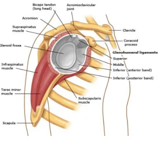
2.1.2. Anatomy of the rotator cuff
The rotator cuff consists of four muscles: the subscapularis, the supraspinatus, the infraspinatus and the teres minor muscles. These muscles end in short, flat, broad tendons which fuse intimately with the articular capsule of the humerus, to form the musculotendinous cuff. (Brand, 2008)
The subscapularis muscle is a large, flat structure that originates from the subscapular fossa along the anterior aspect of the scapula. It is innervated by the upper and lower subscapular nerves (C5, C6 and C7) and supplied by the subscapular artery. Most of the fibers of the subscapularis tendon insert onto the lesser tuberosity, however, the more superficial fibers extend to the greater tuberosity of the humerus and together with the transverse humeral ligament, form the roof of the bicipital groove (deep groove on the humerus that separates the greater tubercle from the lesser tubercle). (Brand, 2008; Opsha, 2008; Gumina, 2017)
The teres minor muscle takes origin from the axillary border of the scapula, it is innervated by the axillary nerve (C5, C6) and supplied by the subscapular and circumflex scapular arteries. Much of the teres minor tendon inserts into the inferior facet of the greater tuberosity while a small portion is inserted directly below this area. (Brand, 2008; Opsha, 2008; Gumina, 2017)
The infraspinatus muscle takes origin from the infraspinous fossa on the posterior aspect of the scapula, it is intimately associated both structurally and functionally with the teres minor muscle, it is innervated by the suprascapular nerve and supplied by the suprascapular and circumflex scapular arteries. The tendon of the infraspinatus inserts onto the middle facet of the greater tuberosity of the humerus. (Brand, 2008; Opsha, 2008)
5
The supraspinatus muscle consists of the anterior and posterior bellies: the anterior supraspinatus belly is a larger, more fusiform structure which originates entirely from the supraspinatus fossa and becomes a thick tubular tendon comprising the anterior 40% of the supraspinatus tendon, and the posterior supraspinatus belly is smaller, unipennate structure which originates mostly from the scapular spine and glenoid neck and becomes a thin, flat tendon comprising the posterior 60% of the supraspinatus tendon. It is supplied by the suprascapular artery and innervated by the suprascapular nerve (C5 and C6). The tendon of the supraspinatus inserts on the superior facet of the greater tuberosity, just posterior to the bicipital groove, and it is bordered by the acromion and subacromial bursa superiorly, and by the joint capsule inferiorly. Anteriorly, the supraspinatus tendon blends with the coracohumeral ligament and posteriorly it merges with the infraspinatus tendon. The region just medial to the convergence of the posterior fibers of the supraspinatus and the anterior fibers of infraspinatus has been referred to as the posterior rotator interval. (Brand, 2008; Opsha, 2008) The anterior rotator cuff interval separates the supraspinatus and subscapularis tendons. Within the gap between the tendons run the coracohumeral ligament, the superior glenohumeral ligament, and the long head of the biceps tendon, which passes from the bicipital groove through the glenohumeral joint before inserting on the superior glenoid. (Opsha, 2008)
At this point it should be noted that the supraspinatus, infraspinatus and teres minor muscles at their points of insertion cannot be separated into anatomic units.
Details on the anatomy of the rotator cuff are depicted in Figure 1.
(a) (b)
6 (c)
Figure 1. Rotator cuff anatomy: (a) Anterior view, (b) Posterior view, (c) Lateral view. (Blum, 2009)
2.2. Rotator cuff biomechanics
Normal glenohumeral motion consists of a roll-gliding combination that keeps the humeral head centered on the glenoid. (Huijbregts, 2015) The rotator cuff provides dynamic stability and is critical to normal shoulder function: allows the humerus to change position with respect to the scapula and stabilizes the humeral head in the glenoid fossa. The two functions cooperate: when a rotator cuff muscle is working, at the same time the antagonist must remain immobile. Forces generated by the rotator cuff facilitate the motions involved in activities of daily living and the more demanding movements of athletics and manual labor. Any alteration of this mechanism will lead to a reduction in the range of motion and in the stability of the shoulder. (Karas, 2011; Gumina, 2017)
Considering the movement, stability and biomechanical properties of the cuff, it is possible to understand how a small rotator cuff tear will lead to the alteration of the factors reported and a redistribution of the load to the remaining tendons, thus determining the progression of the tear. (Karas, 2011; Gumina, 2017)Injury and pathology of the rotator cuff are common and the unique anatomical and biomechanical characteristics of the cuff contribute to the etiology of its injury thus, understanding the biomechanics and function of the rotator cuff is important when considering the surgical options during repair to optimize the outcomes for a given individual. (Karas, 2011; Park, 2012)
7
2.2.1. Rotator cuff function in glenohumeral stability
Glenohumeral joint stability is the result of a delicate balance between the static stabilizers of the shoulder (e.g., bony anatomy, negative intra-articular pressure, the glenoid labrum, and the glenohumeral ligaments along with the joint capsule) and the dynamic stabilizers of the shoulder, including rotator cuff muscles and tendons.
The static and dynamic stabilizers react to the forces applied through the glenohumeral joint to provide stability at different positions during the motion arc. At the end range of shoulder motion, where forces acting on the glenohumeral joint are increased (abduction and maximum external rotation), the capsuloligamentous structures such as the inferior glenohumeral ligament contribute to shoulder stability, by restraining the translation of the glenohumeral joint. In the mid-range of shoulder motion, because the capsuloligamentous structures are lax, shoulder stability is mainly provided by the glenoid concavity and the compressive force generated by the rotator cuff muscles - dynamic compression of the humeral head against the glenoid cavity, termed “concavity compression,” is the primary mechanism by which the rotator cuff dynamically stabilize the glenohumeral joint (the contraction of the rotator cuff muscles anteriorly and posteriorly provides antagonistic forces that compress the humeral head into the concavity of the glenoid). (Lugo, 2008; Opsha, 2008; Karas, 2011; Yamamoto, 2015) Decentering of the humeral head on the glenoid in any direction will cause excessive tensile, compressive, and shear forces in active and passive structures predisposing the patient to eventual pathology. (Huijbregts, 2015)
The coracoacromial arch has a fundamental role in stabilizing the glenohumeral joint as well, also by static restraining the superior translation of the humeral head, as it limits anterior and superior motion of the humeral head and overlying tendons. Anything that decreases the space within the coracoacromial arch could lead to impingement symptoms and subsequently, to rotator cuff tears. (Opsha, 2008)
2.2.2. Rotator cuff function in glenohumeral motion
The supraspinatus is relevant for flexion/abduction functions – the posterior supraspinatus acts as external rotator during abduction and the anterior supraspinatus as internal rotator during flexion of the arm, cooperating with the deltoid and other cuff muscles. (Ackland, 2011) However, its main function is as initiator of abduction, having functions throughout the range of abduction in synergy with the deltoid muscle (the supraspinatus is
8
believed to be more important in initiating elevation and the middle portion of the deltoid more important for elevation of the arm at higher angles of abduction). (Opsha, 2008; Gumina, 2017)
In literature, the infraspinatus and teres minor are often considered together, especially as far as biomechanics is concerned, being considered external rotators of the shoulder. The infraspinatus primarily acts with the arm in neutral position while the teres minor is more active with external rotation in 90◦ of abduction. (Opsha, 2008; Ackland, 2011; Gumina, 2017)
The subscapularis is considered the main internal rotator of the shoulder. While internal rotation remains its main function, its secondary roles change depending on the position of the humeral head with respect to the scapula. Consequently, it is also an abductor and a flexor (with significantly greater muscle activity in the lower subscapularis in comparison to the upper subscapularis during these activities). Because of the shared insertion of the supraspinatus and subscapularis, it is possible that in certain positions of the arm, the subscapularis may work as external rotator. (Opsha, 2008; Wickham, 2014; Gumina, 2017)
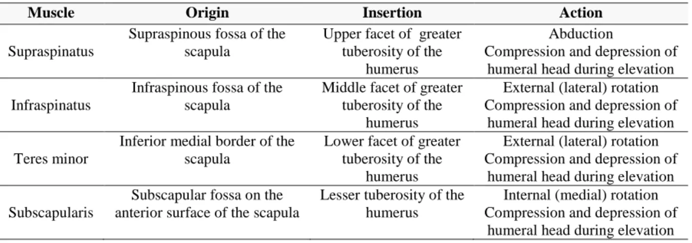
Table 1 summarizes the main functions of the rotator cuff, in glenohumeral motion and stability.
Table 1. Origins, Insertions, Actions, and Nerve Supplies of the Rotator Cuff muscles and tendons.
Muscle Origin Insertion Action
Supraspinatus
Supraspinous fossa of the scapula
Upper facet of greater tuberosity of the
humerus
Abduction
Compression and depression of humeral head during elevation Infraspinatus
Infraspinous fossa of the scapula
Middle facet of greater tuberosity of the
humerus
External (lateral) rotation Compression and depression of
humeral head during elevation Teres minor
Inferior medial border of the scapula
Lower facet of greater tuberosity of the
humerus
External (lateral) rotation Compression and depression of
humeral head during elevation Subscapularis
Subscapular fossa on the anterior surface of the scapula
Lesser tuberosity of the humerus
Internal (medial) rotation Compression and depression of
humeral head during elevation
2.3. Epidemiology of Rotator Cuff tears
Rotator cuff tears are a common cause of pain and disability of the shoulder and are a source of significant morbidity for patients, with potential long-term implications to the health system and economy. When affecting the younger, working population, these injuries can
9
cause significant lost wages or time lost from work. (Mall, 2013; Liem, 2014) Some epidemiologic studies have been carried to determine the prevalence of rotator cuff tears. However, the available data is limited, even though it could provide important information about rotator cuff tears natural history. (Liem, 2014; Gumina, 2017)
Worldwide, of the 30 million musculoskeletal injuries reported, over half are thought to involve tendons and ligaments. (Walden, 2017) Shoulder pain is a common musculoskeletal complaint (Figure 2), with rotator cuff disease representing a common source of shoulder pain (Urwin, 1998; Longo, 2012; Weinstein, 2014), and being one of the most common disorders presenting to clinics worldwide (Figure 3). (Ensor, 2013; Pollak, 2014) Injuries to the upper extremities have the highest prevalence in the working population (Figure 4) (Pollak, 2014), with rotator cuff tears considered one of the usual causes of disability related to the shoulder. (Wani, 2016)
Figure 2. Proportion of population reporting pain, United States 2012. (Weinstein, 2014) [Adapted]
10
Figure 4. Work-related injuries involving days away from work by body part affected, United States 2011. (Pollak, 2014)
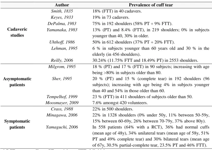
Many authors have focused on epidemiologic studies, performed on cadavers, asymptomatic and symptomatic patients, using medical imaging techniques, to determine the prevalence and incidence of rotator cuff tears, the average age of the patients affected, the work and creative activities related to the lesion and a possible sex-linked predisposition. Table 2 resumes some of these studies. Figures 5, 6 and 7 illustrate some of the findings.
Table 2. Studies in literature that investigated the prevalence of RCT in cadavers, asymptomatic and symptomatic patients. (Tashjian, 2012 ;Wani, 2016; Gumina, 2017)
Author Prevalence of cuff tear
Cadaveric studies
Smith, 1835 18% (FTT) in 40 cadavers.
Keyes, 1933 19% in 73 cadavers.
DePalma, 1983 75% in 192 shoulders (58% PT + 9% FTT).
Yamanaka, 1983 13% (PT) and 8.4% (FTT), in 219 shoulders; 0% in subjects younger than 40, 30% in older.
Uhthoff, 1986 50% in 612 shoulders (37% PT + 20% FTT).
Lehman, 1995 Reilly, 2006
6 % in subjects younger than 60 years old and 30 % in the elderly (in 456 shoulders).
30.24% (11.75% FTT and 18.49% PT) in 2553 shoulders. Asymptomatic patients Milgrom, 1995 Sher, 1995 Tempelhof, 1999 Moosmayer, 2009
18 % (PT) and 17 % (FTT) in 90 subjects; increasing with age being >80% in subjects older than 80.
20 % (PT) and 15 % (complete tear) in 192 shoulders (96 subjects); increasing with age being 4% in subjects younger than 40 and 54% in those older than 60.
23 % (FTT) in 411 shoulders of subjects older than 50. 7.6% amongst 420 volunteers. Symptomatic patients Crass, 1988 Minagawa, 2006 Yamaguchi, 2006 22% in 500 shoulders.
22% in 1328 shoulders (0% under 50y, 11% between 50-59y, 15% between 60-69y, 26% between 70-79y, 37% above 80y). In 558 patients (64% with a RCT), 36% had normal cuffs (mean age of 48y), 34% unilateral tears (mean age of 58y, 51% PT and 49% complete tear) and 30% bilateral tears (mean age of 67y, 30.5% partial-complete tear, 23.5% PT and 46% FTT).
11
Figure 5. Prevalence of rotator cuff tear on dominant limb in asymptomatic subjects that have undergone ultrasound examination, in the various age groups, in a study conducted by Milgrom, 1995. (Gumina, 2017)
Figure 6. Prevalence of partial and full thickness tears in asymptomatic subjects that have undergone MRI examination, in the various age groups, in a study conducted by Sher, 1995. (Gumina, 2017)
(a) (b)
Figure 7. (a) Percentages of partial and full-thickness tears with unilateral tear (199 patients) (Yamaguchi, 2006); (b) Percentages of partial tears, full-thickness tears and partial associated to full-thickness tears, in the group of
12
Existing studies have shown a prevalence of rotator cuff tears that varies extensively. Limitations of cadaveric research include the lack of background information of patients, oftentimes regarding demographic factors such as age, habits, work and activities, among others, or the fact that the criteria established by hospitals or law for postmortem procedures may involuntarily select a population more predisposed to have lesions (patients with chronic diseases, metabolic disorders). (Yamamoto, 2010; Gumina, 2017) Patients who have rotator cuff tears can present with symptoms ranging from minimal discomfort without functional deficits to severe pain, weakness and marked disability, so there is a high rate of asymptomatic patients that frequently remain undiagnosed (Ensor, 2013), but on the other hand, many reports have focused on the patients with symptoms, which may too be misleading with regard to the entire clinical picture of a rotator cuff tear, making difficult to accurately interpret its incidence. (Wani, 2016)
Population based studies, regardless of the presence or absence of symptoms, are thus the best way to study the natural history of rotator cuff tears, however, they are scarce. Studies conducted by Yamamoto (Yamamoto, 2010), Minagawa (Minagawa, 2013) and Gumina (Gumina, 2017) demonstrated a high prevalence of rotator cuff tears in the general population (>20%), increasing amongst those in their fourth compared with those in their seventh-decade, reaching 80% in patients aged more than 80, highlighting the increasing association of rotator cuff tears with age. Asymptomatic tears were twice as common as symptomatic tears, which corroborates with the previous idea that there is a high rate of undiagnosed patients. (Minagawa, 2013) In all studies, side and dominant arm, occupation and activities resulting in the overuse of the shoulder joint and a history of trauma, were significantly associated with the risk of rotator cuff tear.
In conclusion, since twenty decades ago (Milgrom, 1995) to recent studies (Gumina, 2017), the results suport the theory that rotator cuff tearing occurs, to some extent, as a normal degenerative process, that increases with aging (Tashjian, 2012), being most commonly associated with elderly patients, males, affected in the dominant arm and engaged in heavy labor. (Yamamoto, 2010; Gumina, 2017) On the other hand, disease was also present in patients only in the non-dominant shoulder, and, furthermore, some authors (Harryman, 2003; Yamaguchi, 2006) also found a small percentage of subjects with bilateral tears and tears in sedentary individuals who did light work only. (Yamamoto, 2010; Gumina, 2017) In some studies, authors also found that females are at a greater risk of cuff tears, while others found no correlation between tears and gender, which suggests that there isn’t enough
13
evidence to prove that gender has an influence on the development of this pathology. (Oh, 2007) Thus, these risk factors might have a role in contributing to cuff disease, but they are likely to be only one of several factors.
2.4. Rotator cuff pathology
Multiple etiologies have been implicated in the pathogenesis of rotator cuff tears. The rotator cuff is weakened by both extrinsic and intrinsic factors, leading to gradual failure of tendon, which finally results in tear. (Pandey, 2015) Extrinsic factors reflect anatomic variables that compress the tendon by bony impingement or direct pressure from the surrounding soft tissue. Intrinsic factors are where the pathologic changes lie predominantly within the rotator cuff tendon itself (alterations in material properties or matrix composition), because of tensile overload and repetitive stress, aging, microvascular supply, traumatisms, or degeneration. In many patients, it is likely their pathologic abnormality is a byproduct of the interaction between both intrinsic and extrinsic factors, but it is still unclear whether the origin of the tear is related to tendon degeneration itself or induced by several morphologic changes. Demographic factors, such as shoulder overuse, smoking, and any medical condition that impairs the inflammatory and healing response, such as diabetes mellitus, may also contribute to rotator cuff tear. (Torrens, 2007; Nho, 2008; Maffulli, 2011)
2.4.1. Extrinsic theory
Rotator cuff tears may be initiated, or at least propagated, by factors extrinsic to the cuff itself. Shoulder impingement syndrome is caused by an anatomic narrowing of the subacromial space by the structures that form the coracoacromial arch (acromion, the coracoacromial ligament, and the coracoid process) leading to progressive bursitis, tendinitis, and partial and/or complete rotator cuff tears.
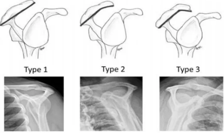
Neer's classic work advocated extrinsic factors for rotator cuff tendon failure in which repetitive translation of the cuff under the acromion led to partial tears that in turn, led to full-thickness tears (Neer, 1987). Neer's theory got a boost when Bigliani described acromial morphologic characteristics with 3 classifications of acromions: type I, or flat acromions, were seen in 17%, type II, or curved acromions, in 43% and type III, or hooked acromions, in 39%, of rotator cuff tears (Figure 8), and concluded that hooked, curved, and laterally sloping
14
acromions were associated with cuff tears and may contribute by causing tractional damage to the tendon. (Bigliani, 1991) The development of these different acromial shapes is likely both congenital and acquired (in response to traction forces applied). (Wang, 1997) Several authors confirmed close relationship between hooked acromion and impingement and/or higher incidence of rotator cuff tears. (Epstein, 1993; Farley, 1994; MacGillivray, 1998; Wang, 2000; Gill, 2002; Worland, 2003). Other factors such as the presence of acromial spurs (bone projections in response for stress found at the attachment of a tendon or ligament, or in joint spaces) (Ogawa, 2005; Natsis, 2007; Oh, 2010; Hamid, 2012), acromial index (large lateral extension of the acromion) (Nyffeler, 2006; Torrens, 2007; Balke, 2013), acromion slope (decreased lateral acromion angle) (Banas, 1995), os acromiale (unfused accessory center of ossification of the acromion that leads to additional motion segment) (Mudge, 1984; Hutchinson, 1993; Park, 1994; Warner, 1998), acromioclavicular joint spur (Petersson, 1983; Gohlke, 1993), coracoacromial ligament morphology (Fremerey, 2000; Kesmezacar, 2008), coracoid morphology (Richards, 2005; Schulz, 2005), internal impingement between the glenoid and the humeral head (Edelson, 2000; Heyworth, 2009) and posture (Gumina, 2008; Sambandam, 2015; Yamamoto, 2015) may also contribute towards extrinsic compression and therefore, for impingement and rotator cuff tears.
Figure 8. Bigliani's classification of acromion undersurface with corresponding supraspinatus outlet view radiograph. (Pandey, 2015)
Even though research studies support the association between these anatomical features and rotator cuff tears, there is evidence disputing it that argues that these features are the result, rather than the cause, of the lesion, that is, an adaptation to an already damaged, poorly balanced rotator cuff that is creating increasing stress on the coracoacromial arch, what suggests a primary intrinsic initiating factor. In general, there is a lack of in-depth data on this condition, likely due to its multifactorial, insidious, and often asymptomatic presentation.
15
More studies are needed to determine whether variation in the morphology causes impingement or is a function of it. (Nho, 2008; Yadav, 2009; DeFranco, 2009; Pandey, 2015; Gumina 2017).
2.4.2. Intrinsic theory
Instrinsic factors encompass the range of mechanisms that occur within the rotator cuff itself. The degenerative-microtrauma theory proposes that age-related degeneration compounded with repetitive microtrauma leads to a partial tear that gradually converts into a full-thickness tear. It is believed that repetitive microtrauma is associated with deterioration and disorganization of collagen constituents through the release of specific angiogenic and inflammatory markers. (Nho, 2008; Pandey, 2015; Wani, 2016)
(i) Degeneration-Microtrauma theory
As previously described, epidemiological studies support a relationship between age and rotator cuff tear prevalence, which suggests that rotator cuff tears could be seen within the framework of “normal” aging rather than an endpoint of an overtly pathological process. Hashimoto (Hashimoto, 2003) reported that the rate of tear increases from 13% to 51% from the age of 50 to 80, suggesting that it could be a normal attrition process and that with advancing age, the cuff undergoes several internal changes, such as collagen disorganization and thinning, myxoid and hyaline degeneration, fatty infiltration, chondroid metaplasia, and calcification. The authors supposed that these features represent primarily degenerative changes contributing to a reduced tensile capacity. These findings are in agreement with earlier studies (Kannus, 1991), that concluded that tendon degeneration is characterized by loss of cellularity, vascularity, tissue architecture and fibrocartilaginous mass, resulting in a mechanically inferior tendon.
Other studies have emphasized the role of altered collagen fiber quality and composition as an important mediator of cuff degeneration, defending that advanced age leads to collagen fiber disorganization and structural changes, namely the increase in type III collagen, that forms smaller and less organized fibrils, along with decrease in type I and II collagen, that confer stiffness and strength to the matrix and fibrocartilage junctions in the tendon-to-bone interface, respectively, decreasing capability of the tendon to withstand load, predisposing it to tear. (Kumagai, 1994; Plate, 2014; Gumina, 2017) Some authors (Lindblom, 1939; Nakajima, 1994) confirmed through histological and biomechanical studies
16
that articular side fibers have a smaller cross-sectional area and a greater vulnerability to tensile load, than the bursal side fibers. These findings are consistent with the early studies of Codman (Codman, 1934) that demonstrated that partial tears typically began on the articular side of the tendon, because the load capacity is lower than that of the bursal side, making it more prone to damage. The heterogeneity of tendon and the difference in its mechanical stress shear, may therefore contribute to tendon failure.
This is then compounded by repetitive microtrauma – mechanical loading of degenerative tendon leading to several small tears that only partially heal until the tendon is so weakened that a full-thickness tear develops. This theory suggests that repetitive stress is responsible of a vicious circle that predisposes the tendon to rupture, in which small repetitive injuries have insufficient time to heal before further trauma presents, and the remaining fibers endure increased load, becoming susceptible to further microrupture. (Nho, 2008; Yadav, 2008; Pandey, 2015) Typically after the deep fibers tear, they retract because they remain under tension, even with the arm at rest, which results in an inhibition of healing. This results in an increased load on the remaining fibers, increasing the likelihood of further rupture. (Premdas, 2001)
(ii) Inflammation and oxidative stress
As a result of repetitive microtrauma in the setting of a degenerative rotator cuff tendon, inflammatory mediators alter the local environment and oxidative stress induces tenocyte apoptosis causing further degeneration (Figure 9). (Yadav, 2008)
Soslowsky (Soslowsky, 2000) demonstrated in a rat model an increase in angiogenic and inflammatory markers in response to overuse, associated with concomitant declines in normal collagen constituents and architecture, resulting in lower load-to-failure. Tsuzaki (Tsuzaki, 2003) investigated the biochemical cascade of interleukin 1 (Il-1) in vitro, on human tendon cells subjected to progressive cyclic loading, on the basis that it may be a pro-inflammatory mediator. He found increases in mRNA levels of COX-2, with increase in tissue concentrations of prostaglandin E2 (PGE2), and increased expression of matrix metalloproteases (MMP-1, MMP-3 and MMP-13). This study supposes that the painful symptoms of rotator cuff disease are mediated via COX-2 and PGE2, while the loss of tissue architecture is mediated by the range of MMPs released by the activated tendons, as it is believed that certain MMPs are involved in the turnover of the extracellular matrix of
17
tendons, probably through collagen structure alteration, affecting normal matrix remodelling. Other authors supported these ideas. (Riley, 2002; Li, 2004; Koshima, 2007)
Another theory reflects on oxidative stress in the local environment. Yuan (Yuan, 2002) noted an increased proportion of apoptotic cells at the edge of a rotator cuff tear compared to controls. The oxidative stress is produced due to repetitive injury followed by reparative processes, and induces tenocyte apoptosis due to excess levels of reactive oxygen species (ROS), contributing to tendon degeneration, predisposing it to rupture. Possible mediators for these apoptotic pathways include: (i) matrix metalloproteinase-1 (MMP-1) within the extracellular matrix, found in normal tendon at very low concentrations to effect the natural turnover of collagen, and increased in damaged tendon, contributing to disorganized tissue architecture, reduced collagen synthesis and weakened tendon biomechanics; and (ii) c-Jun N-terminal protein kinase (JNK) within the intracellular environment, induced in tendon by interleukins (like the pro-inflammatory cytokine IL-1) and cyclic mechanical stretch, that when phosphorylated, activates a number of downstream transcription factors linked to the apoptotic pathway (Han, 2001; Yuan, 2004; Wang, 2007). (Nho, 2008; Yadav, 2008; Pandey, 2015)
Figure 9. Rotator cuff degeneration secondary to inflammation and oxidative stress. (Nho, 2008) [Adapted]
(iii) Cuff vascularity
It has been taught that a “critical” or hypovascular zone exists proximal to the insertion of the tendon, but these assertions have become an area of controversy. Several authors concluded that no significantly hypovascular areas exist or that hypovascularity was a minimal contributor, if at all, to cuff tears. (Moseley, 1963; Fukuda, 1990; Swiontkowski, 1990; Brooks, 1992; Uhthoff, 1992)
18
Lindblom, (Lindblom 1939) and Benjamin (Benjamin, 1986) asserted that histological transition from tendon to calcified fibrocartilage during tendon-to-bone insertion leads to hypovascular zones in tendon, responsible for ruptures and poor healing of tendon after repair. Rudzki (Rudzki, 2008) demonstrated that cuff vascularity is significantly less after the age of 40 as compared with those under 40, especially on the articular side. This corroborates with Codman’s (Codman, 1934) theory that lesion starts on the articular side of the cuff, also due to decreasing vascularity in ageing tendons. Some authors (Rathbun, 1970; Maffulli, 2011) also demonstrated that perfusion within the rotator cuff is a dynamic phenomenon, with markedly reduced perfusion when the arm is in full adduction, so in situations with supraspinatus compressed at the humeral head, it is possible that hypovascularity may be a contributory factor to tear. (Nho, 2008; Yadav, 2008; Pandey, 2015)
Figure 10 summarizes the extrinsic and intrinsic theories for rotator cuff tear and Figure 11 illustrates some of the mentioned causes for tear.
Figure 10. Summary of extrinsic and intrinsic pathways of rotator cuff tear (ECM: extracellular matrix; MMP-1:matrix metalloproteinase-1; ROS: reactive oxygen species). (Pandey, 2015)
19
2.4.3. Demographic factors
Demographic factors, such as shoulder overuse, smoking, and any medical condition that impairs the inflammatory and healing response such as diabetes mellitus, may also contribute to rotator cuff tear, playing an additive role to the underlying influence of age-related degeneration. (Tashjian, 2012) However, the relationship between epidemiology and cuff pathology is a challenging one due to the lack of quality data available. Several risk factors have been identified for rotator cuff disease, and can be grouped into non-mechanical and mechanical factors. (Nho, 2008; Yadav, 2008; Maffulli, 2011)
(i) Mechanical factors
As described above, mechanical overuse is involved in the development of rotator cuff tears. Position of lateral decubitus during sleep, in which the shoulder is repetitively subject to the weight of the body itself, leading to compression of the supraspinatus tendon against the coracoacromial arch, generating inflammation and tendon degeneration (Cabuk, 2015), use of wheelchair, sports (such as tennis, volleyball, baseball, weightlifting and badminton) and occupations characterized by a repetitive arm activity and history of trauma, can be also included in this group. (Nho, 2008; Yadav, 2008; Maffulli, 2011)
(ii) Non-mechanical factors
Cigarette smoking plays a significant role in the development, progression and healing of rotator cuff tears following surgery. The negative effects of tobacco arise from the vasoconstrictive properties of nicotine, which decrease the delivery of oxygen to tissues, and to the capability of carbon monoxide to decrease cellular oxygen tension levels. Since rotator cuff tears are thought to occur in a hypovascular zone, it is likely that an already hypovascular region is further compromised in smokers. (Determe, 1995; Mallon, 2004; Dave, 2006; Baumgarten, 2010; Carbone, 2012; Gumina, 2017) Furthermore, since the accumulation of collagen, important for the tensile strength of a wound, is dependent on adequate oxygenation and perfusion, due to the described effect of nicotine, it is safe to say that it can be responsible for persistent inflammation, reduced collagen concentrations and so, reduced mechanical properties. Thus, postoperative results of smokers are also less favorable than those of nonsmokers, (Jorgensen, 1998; Mallon, 2004; Galatz, 2006) and smokers are at an increased risk of repeat tear. (Maffulli, 2011 ;Tashjian, 2012; Wani, 2016)
20
Diabetes mellitus is also an important risk factor. Collagen is naturally glycosylated, that is, the collagen protein molecules have sugar molecules covalently bonded to them. In a diabetic patient, this results in an increasing amount of intermolecular collagen cross-links, causing a reduction in the solubility of collagen. The collagen that remains is stiffer, however, has lost its elasticity and is prone to tearing. (Abate, 2010; Bedi, 2010; Dhar, 2013) Failure rate, after repair, is also significantly higher in patients with diabetes mellitus since they have a higher rate of complications, including infections. (Clement, 2010; Chung, 2011; Fermont, 2014; Wani, 2016)
Arterial hypertension is a cause of peripheral hypovascularity; therefore, patients with arterial hypertension could conceivably have a more frequent prevalence of rotator cuff tears. (Gumina, 2013)
Deposition of cholesterol by-products has also been implicated in increasing the risk for tendon rupture because like nicotine, they impair tendon microcirculation, increasing the likelihood of tear and prolonging healing. (Abboud, 2010; Gumina, 2017) High levels of cholesterol also result in the formation of tendon xanthomas (increased deposition of lipids within the connective tissue, that increases with age) which alters the tendon’s mechanical properties, contributing to tears. (Tashjian, 2012; Beason, 2014; Wani, 2016) Obesity is also considered a risk factor for rotator cuff tears through its known associations with elevated cholesterol level, diabetes or hypertension. (Wendelboe, 2004; Gumina, 2014; Gumina, 2017)
Studies have revealed that genetic predisposition has a role in the development and progression of tears. (Harvie, 2004; Yamaguchi, 2006; Gwilym, 2009; Tashjian, 2009; Longo, 2012; Dominique, 2017) As the development of these tears is seen as multifactorial, no single gene has been directly implicated. (Tashjian, 2012; Wani, 2016)
2.5. Mechanobiology of tendon
Tendons are mechanically responsible for transmitting muscle forces to bone, and in doing so, permit locomotion and enhance joint stability. Moreover, tendons are a living tissue, able to store elastic energy and withstand the high tensile forces upon which locomotion is entirely dependent, responding to mechanical forces by changing their structure, composition and mechanical properties - a process called tissue mechanical adaptation. (Wang, 2006)
Structurally, tendons are composed of tenoblasts and tenocytes lying within a network of extracellular matrix (ECM), composed of collagen and a smaller fraction of elastin,
![Figure 9. Rotator cuff degeneration secondary to inflammation and oxidative stress. (Nho, 2008) [Adapted]](https://thumb-eu.123doks.com/thumbv2/123dok_br/16075731.1106631/39.892.226.670.673.883/figure-rotator-degeneration-secondary-inflammation-oxidative-stress-adapted.webp)