Outubro de 2011
Ana Catarina Correia Leirós
Evaluation of the interactions between
human cells and silicone rubber surfaces
modified by Atom Transfer Radical
Polymerization
UMinho|20
11
Ana Catarina Corr
eia Leir ós Ev aluation of t he interactions be tw
een human cells and silicone rubber sur
faces modified b
y Atom T
ransfer Radical P
ol
Dissertação de Mestrado
Mestrado Integrado em Engenharia Biomédica
Ramo de Engenharia Clínica
Trabalho realizado sob a orientação da
Doutora Lígia Rodrigues
e do
Doutor Theo van Kooten
Outubro de 2011
Universidade do Minho
Escola de Engenharia
Ana Catarina Correia Leirós
Evaluation of the interactions between
human cells and silicone rubber surfaces
modified by Atom Transfer Radical
COMPROMETE;
Universidade do Minho, ___/___/______
iii I would like to thank Dr. Lígia Rodrigues for being always present. I am very grateful for all the patience, comprehension and help; without it I am sure I wouldn’t be able to do the best work I could. Every word, every advice, every incentive were more than I would ever expect from a supervisor.
From Dr. Theo van Kooten I got the best orientation during my research work development. I am thankful for the patience and the teaching, for having someone that gave me orientation, that wanted me to learn how to solve problems, that was there to listen to my ideas and that always had time to help me.
To Dr. Miguel Gama I would like to thank the wise words that made me move on and not giving up the opportunity of working abroad and learn new working methods, cultures and traditions.
I would like to thank the Biomedical Engineering Department colleagues, which made me feel home and always made me smile. To Joram, Thomas, Joana and Das I express special thanks, because they were more than colleagues, they were friends.
Finally, a really heartfelt gratitude to my family, which supported me during the last 5 years and never gave up on me, to Diana, the best friend and roommate, and to my fiancé Steffen, the most patient and sweet partner one can have.
v Poly(dimethylsiloxane) or PDMS is one of the materials of choice to be used in implants due to some of its properties, such as low-cost, versatility, elasticity, chemical inertness, biocompatibility, non-toxicity, among others. However, PDMS also presents some drawbacks. The major limitation of PDMS is its hydrophobic nature, which makes the transferring and spreading of aqueous solutions difficult and may lead to complications in cell culture, as well as its low adhesiveness for cell attachment.
The PDMS used in the current work was modified by atom transfer radical polymerization (ATRP), which is a technique used to carry out a controlled/living radical polymerization that is easy to apply and makes possible the use of different monomers and reaction media. The surface was coated with poly(ethylene glycol) methyl methacrylate (PEGMA), a polymer known for its ability to reduce biomolecules adsorption. With this coating, anti-fouling properties are expected, as well as a more hydrophilic surface. Contact angles were measured showing that the PEGMA coating turned the surface more hydrophilic. Moreover, by atomic force microscopy the surface topography was assessed. It was possible to observe that the coating possessed a high roughness, thus suggesting that ATRP is a suitable technique to create brushes in the PDMS surface. However, the contact angle variability that was found in the modified PDMS samples suggests that the technique is not reproducible.
The interaction of the modified PDMS with human skin fibroblasts (HSkF) and human umbilical vein endothelial cells (HUVECs) was assessed in order to determine the biocompatibility of the surface. To observe these interactions, an immunocytochemistry assay was used to stain the cell nuclei and the vinculin and fibronectin complexes. The stained structures were visualized with confocal microscopy. The staining of the cell nuclei made possible the estimate of the number of cells in the surface, and the formed fibronectin was also quantified. Some colorimetric assays were also performed (MTT and CV) to quantify the metabolic activity per cell, giving some insight about cell viability and adhesion.
It was possible to conclude that the modified surfaces decrease cell adhesion, which is expected due to the anti-fouling properties of PEGMA. Then, if an increase in cell adhesion is desired, PDMS coated with PEGMA is not the most suitable material for vascular implants. However, due to the anti-fouling properties, a better hemocompatibility can be achieved.
vii O poli(dimetilsiloxano) ou PDMS é um dos materiais de eleição no desenvolvimento de implantes devido a algumas das suas propriedades, tais como baixo-custo, versatilidade, elasticidade, inércia química, biocompatibilidade, não toxicidade, entre outras. No entanto, o PDMS apresenta algumas desvantagens. A maior limitação do PDMS é a sua natureza hidrofóbica, a qual pode levar a complicações na cultura de células, bem como a sua reduzida aderência para a adesão celular.
O PDMS usado neste trabalho foi previamente modificado por polimerização radicalar controlada por transferência de átomo (ATRP), que consiste numa técnica usada para levar a cabo uma polymerização radicalar controlada fácil de aplicar e que torna possível o uso de diferentes monómeros e meios de reacção. A superfície foi revestida com poli(etileno glicol) metil metacrilato (PEGMA), um polímero conhecido pela sua capacidade de reduzir a adsorção de biomoléculas. Com este revestimento, esperava-se a obtenção de uma superfície anti-adesiva e mais hidrofílica. Os ângulos de contacto foram medidos confirmando que o revestimento tornou a superfície mais hidrofílica. Adicionalmente, a topografia da superfície foi avaliada por microscopia de força atómica (AFM). Observou-se que o revestimento apresenta uma elevada rogusidade, mostrando que o ATRP é uma técnica apropriada para criar “escovas” na superfície do PDMS. No entanto, as grande variabilidade dos ângulos de contacto medidos no PDMS modificado indicam que a técnica não é reprodutível.
A interacção do PDMS modificado com fibroblastos da pele humana (HSkF) e células endoteliais da veia do cordão umbilical humano (HUVECs) foi avaliada de modo a determinar a biocompatibiltidade da superfície do material em estudo. De modo a observar as interacções, utilizou-se técnicas de imunocitoquímica que permitiram marcar o núcleo celular e complexos de vinculina e fibronectina com fluorescência. As estruturas marcadas foram visualizadas com micorscopia confocal. A marcação do núcleo celular permitiu estimar o número de células na superfície, e a fibronectina formada foi também quantificada. Alguns estudos colorimétricos foram também utilizados (MTT e CV) com o intuíto de quantificar a actividade metabólica por células, fornecendo informação acerca da viabilidade e da adesão celular.
Foi possível concluir que a superfície modificada diminui a adesão celular, o que era expectável devido às propriedades anti-adesivas do PEGMA. Assim sendo, se um aumento na adesão celular for desejado, o PDMS revestido com PEGMA não é o
viii
material mais apropriado para implantes vasculares. No entanto, devido às propriedades anti-adesivas, uma melhor hemocompatibilidade pode ser alcançada.
ix Acknowledgments iii Abstract v Resumo vii List of Contents ix List of Figures xi List of Tables xv
List of Abbreviations xvii
Framework xix Chapter 1. Introduction 1 1. Vascular implants 2 2. Silicone Biomaterials
3 2.1. Properties 4 2.2. Poly(dimethylsiloxne) (PDMS) 5 2.3. Applications 6 3. Surface Modification 8 3.1. Physical Treatments 8 Plasma Treatment 9 3.2. Chemical Treatments 9 Surface Grafting 10
Atom Transfer Radical Polymrization (ATRP) 11
3.3. Coating Materials 12
3.4. Vascular Grafts Modification 13
4. Body-Biomaterial Interactions 14
4.1. Cell-Biomaterial Interaction 15
4.1.1. Extracellular Matrix (ECM) 16
4.1.2. Adhesion Molecules – Integrins 18
x
4.3. Host Immune Response 20
Chapter 2. Materials & Methods 23
1. Surface Modification 24
1.1. Fibronectin Coating 24
1.2. Gelatin Layer 24
2. Surface Characterization 24
2.1. Contact Angles 24
2.2. Atomic Force Microscopy (AFM) 25
3. Surface Cytotoxicity 26
3.1. MTT Assay 28
3.2. Crystal Violet (CV) Assay 28
4. Cell-Biomaterial Interaction 28
3.1. Cell Culture 28
3.1.1. Specific conditions 30
3.2. Cell Interaction with Modified and Unmodified PDMS 30
3.2.1. Immunocytochemistry Assay 32
3.2.2. Confocal Microscopy 33
5. Statistics 34
Chapter 3. Results & Discussion 35
1. Surface Characterization 35 1.1. Contact Angles 35 1.2. AFM 36 2. Surface Cytotoxicity 39 3. Cell-Biomaterial Interaction 44 Chapter 4. Conclusions 55
Chapter 5. Recommendations and future work 57
xi
Figure 1. Repeat unit of siloxane and of PDMS. [Taken from (1)] (4)
Figure 2. Surface grafting of a generic surface. On the top figure it is possible to
observe a representation of the grafting on approach. In the bottom the grafting from approach is represented. [Taken from (33)] (10)
Figure 3. Schematic representation of the ATRP process. After the initiator (X) covers
the polymer surface (R), the transition metal complex (Met) will remove the halides from the polymer surface. The monomer (M) will then interact with the polymer surface and the propagation of the monomers begins, culminating with the formation of polymeric chains in the polymer surface. [Taken from (33)] (12)
Figure 4. Progression of the mammalian cell adhesion. (A) Initial contact of the cell
with the material covered by proteins. (B) Formation of bonds between the cell surface receptors and the cell adhesion ligands in the proteins. (C) Cytoskeletal reorganization with a progressive spreading of the cell in the surface of the implant, increasing the attachment strength. [Taken from (1)] (15)
Figure 5. Cell proteins involved in cell adhesion on a biomaterial. [Taken from (50)] (16)
Figure 6. Surface placed in a protein mixture. In a matter of seconds the surface is
covered by a layer of adsorbed proteins. Initially, the “red” and the “green” proteins adsorb in a higher concentration. With time, these proteins are displaced by the “blue” protein. This can happen because the concentration of proteins in the adsorbed layer is usually different from the concentration in the solution, being able to change in time.
[Taken from (47)] (19)
Figure 7. The top scheme shows a protein denaturing with increasing adsorption time.
The bottom scheme shows a proteins adsorbing to the surface in different orientations. This is possible because the conformation and the orientation of adsorbed proteins depend on adsorption conditions and surface properties. [Taken from (47)] (20)
Figure 8. Reaction of the host to the implanted biomaterial. (1) The biomaterial is
implanted in the surgical site by the surgeon. (2) A layer of proteins quickly adsorbs to the implant surface. (3) The neutrophils and the macrophages examine and attack the biomaterial. However the implant is too large to be ingested. (4) The macrophages find that they cannot digest the implant, so they fuse into giant cells to engulf the implant.
xii
The giant cells send out cytokines to attract other cells. (5) The fibroblasts arrive and start synthesizing collagen. (6) The implant is completely entrapped in an acellular, avascular collagen bag. [Taken from (47)] (21)
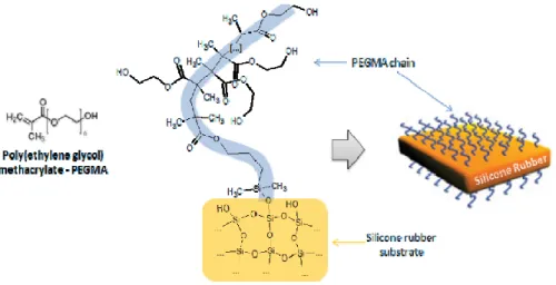
Figure 9. Representation of poly(ethylene glycol) methyl methacrylate (PEGMA)
polymer brushes formed in the PDMS surface due to the ATRP modification. [Taken
from (60)] (23)
Figure 10. Water drops formed in the surface. Contact angle between the water drop
and the surface depends on the surface hydrophobic/hydrophilic character. [Taken
from (61)]. (25)
Figure 11. Scheme of the cytotoxicity assessment experiment. First, samples were cut
into 4 small squares (1 cm2). One of the samples was washed and the other was only sterilized. The 4 squares of each sample were placed in a 12 well-plate and the cells
were seeded. (26)
Figure 12. Schematic representation of the 96 well-plate used to seed the cells. Cells
were seeded in a concentration of 5 x 10⁴ cells/ml. After 24 h the medium was changed. Row A: medium incubated with the washed modified PDMS in the first 12 well-plate. Row B: medium incubated with the non-washed modified PDMS in the first 12 well-plate. Row C: medium incubated with the washed modified PDMS in the second 12 well-plate. Row D: medium incubated with the non-washed modified PDMS in the second 12 well-plate. Row E: medium incubated with the washed modified PDMS in the third 12 well-plate. Row F: medium incubated with the non-washed modified PDMS in the third 12 well-plate. Row G: control with fresh medium. (27)
Figure 13. Distribution of PDMS samples in the 12 well-plate, for the experiments with
HSkF. The first 4 wells contain unmodified PDMS surfaces, the next 4 contain modified PDMS surfaces and the last 4 wells contain modified PDMS with a fibronectin coating. The 12 well-plate was prepared in duplicate, one to be incubated for 48 h and the other
for 120 h. (31)
Figure 14. Distribution of PDMS samples in the 12 well-plate, for the experiment with
HUVECs. In the left plate, the first 4 wells contain unmodified PDMS surfaces, the next 4 contain modified PDMS surfaces and the last 4 wells contain modified PDMS with a fibronectin coating. In the second plate, the first 4 wells contain modified PDMS with a gelatin coating. Both the 12 well-plate were prepared in duplicate, one to be incubated for 48 h and the other for 120 h. (31)
xiii surfaces with adsorbed fibronectin, the next 4 contain modified PDMS surfaces with adsorbed fibronectin and the last 4 wells only contain a fibronectin coating. In the second plate, the first 4 wells have no additional surface or coating. Both the 12 well-plate were prepared in triplicate, one to be incubated for 3 days, the other for 4 days and the last for 5 days. (32)
Figure 16. Scheme representing the image processing for cell number quantification.
(A) image stack (B) overlay image (C) selection of DAPI staining fluorescence (D) particle analysis image. [Taken from (60)] (33)
Figure 17. Images obtained by atomic force microscopy (AFM) from the modified
PDMS without (left) and with (right) a fibronectin coating. Both the pictures correspond to an area of 5 x 5 μm. Surface roughness is represented between 0 and 200 μm.
(37)
Figure 18. Images obtained by atomic force microscopy (AFM) from the modified
PDMS with a fibronectin coating after cell seeding. The picture on the left has a normal resolution and corresponds to an area of 5 x 5 μm. The picture on the right is a 3D picture from a high resolution picture taken from the surface. (38)
Figure 19. Ratio between the MTT values and CV values for each dilution of the
medium in contact with washed and non-washed modified PDMS surfaces. Control consists of the fibroblasts seeded in fresh medium. Results correspond to the average of 3 independent assays ± standard deviation. (39)
Figure 20. Ratio between the MTT values and CV values for each dilution of the
medium in contact with washed and non-washed modified PDMS surfaces. Control consists of the HUVECs seeded in fresh medium. Results correspond to the average of 3 independent assays ± standard deviation. (41)
Figure 21. Ratio between the MTT values and CV values for each dilution of the
medium in contact with washed and non-washed modified PDMS surfaces. Fibroblasts were seeded with medium that was previously incubated with modified PDMS and diluted afterwards. Control consists of the fibroblasts seeded in fresh medium. Results correspond to the average of 3 independent assays ± standard deviation. (42)
Figure 22. Ratio between the MTT values and CV values for each dilution of the
xiv
were seeded with medium that was previously incubated with modified PDMS and diluted afterwards. Control consists of the fibroblasts seeded in fresh medium. Results correspond to the average of 3 independent assays ± standard deviation. (43)
Figure 23. Images obtained by confocal microscopy of the immunochemistry assay
conducted after exposing fibroblasts to the modified PDMS for 48 and 120 h. A and B - fibroblasts adhered to unmodified PDMS, after 48 and 120 h, respectively. C and D - fibroblasts adhered to modified PDMS, after 48 and 120 h, respectively. E and F - fibroblasts adhered to modified PDMS with a fibronectin coating, after 48 and 120 h, respectively. (45)
Figure 24. Average number of nuclei counted after 48 and 120 h of cells exposure to
unmodified PDMS, modified PDMS and modified PDMS with a fibronectin coating. Results correspond to the average of 6 counts ± standard deviation. (47)
Figure 25. Estimate of the fibronectin percentage present after 48 and 120 h using
unmodified PDMS, modified PDMS and modified PDMS coated with fibronectin. Results correspond to the average of 6 experiments ± standard deviation. (48)
Figure 26. Images obtained by confocal microscopy of the immunochemistry assay
conducted after exposing HUVECs to the modified PDMS for 48 and 120 h. A and B - HUVECs adhered to unmodified PDMS, after 48 and 120 h, respectively. C - HUVECs adhered to modified PDMS, after 48 h. D - HUVECs adhered to modified PDMS coated with fibronectin after 48 h. E and F - HUVECs adhered to modified PDMS coated with gelatin, after 48 and 120 h, respectively. (49)
Figure 27. Images obtained by confocal microscopy of the immunochemistry assay
conducted after seeding the HUVECs in different surfaces for 3, 4 and 5 days. A and B - HUVECs adhered to the 12 well-plate wells with no gelatin, after 3 and 5 days, respectively. C and D - HUVECs adhered to the 12 well-plate wells with gelatin, after 3 and 5 days, respectively. E and F - HUVECs adhered to modified PDMS coated with gelatin after 3 and 5 days, respectively. G and H - HUVECs adhered to unmodified PDMS coated with gelatin, after 3 and 5 days, respectively. Pictures from day 4 are not represented, because they didn’t represent any additional information. (52)
xv
Table 1. Key milestones in the development of silicone. [Adapted from (1)] (4)
Table 2. Applications of silicone biomaterials in the biomedical field. [Adapted from (3)]
(7)
Table 3. Contact angles measured for the modified and unmodified PDMS. Results
represent the average of 5 (modified PDMS) and 2 (unmodified PDMS) independent measures ± standard deviation. (35)
xvii GABGs- Coronary Artery Bypass Grafts
VAGs- Vascular Access Graft
PET- Polyethylene Terephthalate
ePTFE- Expanded Polytetrafluoroethylene
PU- Polyurethane
PEtUs- Poly(ether)urethanes
PDMS- Poly(dimethyl siloxane)
UV- Ultraviolet
ATRP- Atom Transfer Radical Polymerization
PEGMA- Polyethylene Glycol Methyl Methacrylate
PHEMA- Poly(2-hydroxyethyl methacrylate)
PEO- Poly(ethylene oxide)
PEG- Poly(ethylene glycol)
ECM- Extracellular Matrix
GAGs- Glycosaminoglycans
RGD- Arg-Gly-Asp
VEGF- Vascular Endothelial Cell Growth Factor
FGF- Fibroblast Growth Factor
EGF- Epithelial Cell Growth Factor
FAK- Focal Adhesion Kinase
HSkF- Human Skin Fibroblasts
HUVECs- Human Umbilical Veins Endothelial Cells
RPMI- Roswell Park Memorial Institute
PBS- Phosphate Buffer Saline
EDTA- Ethylenediamine Tetraacetic Acid)
PFA- Paraformaldehyde
xviii
DAPI- 4',6-diamidino-2-phenylindole
MTT- “3‐(4,5‐dimethylthiazol‐2‐yl)‐2,5‐diphenyl‐tetrazolium bromide) CV- Crystal Violet
xix The cardiovascular diseases are becoming more and more common mostly due to the increasing use of blood-contacting devices. One example of cardiovascular interventions is the replacement of obstructed parts of arteries, which involves the use of vascular grafts. The most common materials used in the synthetic vascular grafts are the polyethylene terephthalate (PET), expanded polytetrafluoroethylene (ePTFE), but they present a thrombogenic nature that can cause serious complications. Thus, some alternatives have been studied, like the poly(dimethyl siloxanes) (PDMSs) that has been used for vascular vessels and as a coating, presenting some promising results. However, this material is hydrophobic, which will repel proteins from serum and consequently will decrease cell adhesion to the surface. Hence, some coating techniques like the ATRP can be used in order to modify the surface by covering it with hydrophilic brushes.
The aim of this work is to evaluate the biocompatibility of modified silicone rubber for vascular applications. Specifically, this work aims to assess if the PDMS surfaces modified with polyethylene glycol methacrylate (PEGMA) brushes by atom transfer radical polymerization (ATRP) are suitable for vascular grafts applications. These modified surfaces were kindly provided by the Department of Biological Engineering, University of Minho, Portugal. The hydrophobicity of the modified surfaces will be determined, since the goal of the modification was to turn it more hydrophilic thus increasing the interaction of the biomaterial with endothelium cells. Cell-biomaterial interactions will be studied and to enhance cell adhesion the surfaces will also be coated with fibronectin and gelatin.
1
Chapter 1. Introduction
Biomaterials, that were unknown 50 years ago, are now widely used in medicine, dentistry and biotechnology. Back on that time, medical devices manufacturers, regulatory approval processes and the biocompatibility concepts did not exist. (1) The development of biomaterials was not based in any scientific criteria. On the contrary, when biomaterials were used to create new devices, these were tested on a trial-and-error basis using animals and even humans.
However, this scenario has changed dramatically due to the needs of the continuous and ever-growing practice of medicine, and currently there are thousands of biomedical devices. Besides the biomedical devices, diagnostic products, pharmaceutical preparations and disposable devices, now the list of biomaterials includes intelligent drug delivery, tissue culture, tissue engineering and hybrid organs. (2)
The biomaterials can be divided into four major classes: polymers, metals, ceramics and natural materials. The polymers represent the major class of biomaterials and are used in several biomedical applications in the orthopedic, dental, soft tissue and cardiovascular areas. (1)
Implants made of biomaterials can range from temporary implants, as venous catheters, to long-term implants, as artificial joints. It is highly probable that most individuals in developed countries will use biomaterials and medical devices at some point in their lives. (3) However, some implants and extracorporeal devices can have adverse interactions with the patient, which constitute a device failure, and thus can cause injury or death of the patient. These complications arise largely from the tissue-biomaterial interaction and infection. (1)
Vascular implants (or grafts) are one type of implants that have been widely used in order to enhance the insufficient functioning of blood vessels. There are several types of vascular implants, which can be grouped in three main classes: coronary artery bypass grafts (CABGs); small-diameter grafts; and large-diameter grafts. The CABGs are used in aortocoronary bypass procedures and have a diameter between 1–4 mm. The small-diameter grafts (4 –6 mm) are used peripherally to relieve lower-extremity ischemia and as vascular access graft (VAG) to treat endstage renal
Chapter 1
Introduction
2
diseases. Regarding the large-diameter grafts (> 7 mm) are used in the thoracic and abdominal cavities, typically for the replacement of aortic sections. (3)
1.
Vascular implants
The medicine practice has always involved the use of blood-contacting devices, such as the simple blood bags and catheters. Nowadays, these devices are even more present in our reality and in the so-called modern medicine, being more complex and intimately connected to cardiovascular diseases. Cardiovascular diseases, which include any disorder that affects both the heart and the blood vessels, are one of the main causes of death in the World. (4) According to the World Health Organization (WHO), in 2002 there were 393.000 deaths caused by cardiovascular diseases in Portugal, and around 167 million deaths worldwide. The use of the blood-contacting devices is deeply connected to the cardiovascular diseases, thus being its major application in the biomedical field.
The vascular grafts are included in the group of blood-contacting devices, being widely used to restore the blood flow in patients with different cardiovascular problems. The replacement of obstructed parts of arteries is becoming a very common medical procedure, so the use of vascular grafts is increasing.
The ideal features of a vascular graft include an easy handling, mimicking the native blood vessels, being non thrombogenic, immunologically inert, resistant to infection and puncture trauma, being able to retain tensile strength, and being manufactured at reasonable costs. There are two main groups of vascular substitutes, the biological grafts and synthetic grafts. (5)
The biological grafts or autografts consist in taking tissue from one site on a patient and transplant it to another site on the same patient. Usually it consists in an autogenous vessel, like the saphenous or the arm veins. Currently, the autografts are the vascular grafts with the best performance. However, due to the unavailability, poor quality or the failure (thrombosis, emboli production, intimal hyperplasia) of autologous conduits, the use of prosthetic grafts is often required. (2; 6)
The prosthetic grafts are manufactured from synthetic materials, like polyester and expanded polytetrafluoroethylene. (6) Regardless of being extremely useful and necessary these devices are not perfect, presenting some biocompatibility issues and some undesirable side effects, thus requiring the search for more suitable materials and alternative coating techniques. Though a large variety of polymeric materials are
3 used (e.g. polyethylene terephthalate (PET), expanded polytetrafluoroethylene (ePTFE) (7)), they present a thrombogenic nature that can cause serious complications, and consequently the failure of the devices. It is recognized that these materials have excellent mechanical and physical properties. Nevertheless, they are also characterized by some hemo-incompatibility, thus activating the humoral and cellular defense mechanisms of the body when contacting with blood. (4; 8) To overcome this biocompatibility problem, the patients are usually exposed to systemic anticoagulation regimens, which in a long-term situation can increase the probability of hemorrhage, even if the levels of anticoagulants are controlled. Indeed thrombosis is, for example, the cause of 80% of vascular graft dysfunction. In addition to this problem, there is a risk of damaging the endothelium lining of blood vessels during implantation of some devices like vascular grafts, which can lead to proliferation of smooth muscle cells or vascular stenosis (decrease in vessel diameter). (9)
For that reason, searching for more suitable materials and coating techniques is highly recommended. Alternative polymers that have been studied are the polyurethane (PU) elastomers, which are thermoplastic polymers with outstanding physical properties. A group of the PU, the aromatic poly(ether)urethanes (PEtUs), due to their flex life, tensile strength, elongation and good blood compatibility, have been used as chorinc implants (e.g. ventricular assist devices, intra-aortic balloons). However, these polymers have been presenting variable clinical results, showing some thrombogenicity and tendency to degrade uncontrollably. (7) Furthermore, the poly(dimethyl siloxanes) (PDMSs) constitute another example of materials that have been used for vascular vessels and as a coatings, presenting some promising results (8). The PDMS presents better blood contact properties when compared to the PU. (7)
2.
Silicone Biomaterials
Silicones, or poly(diorganosiloxanes), are a class of synthetic polymeric biomaterials with high chemical stability that were not explored before 1940. Six years later this polymer was referred for the first time as suitable for biomedical applications, and nowadays is one of the materials that found several applications into the biomedical field being widely used as an implant or any other invasive device. (1; 3; 10) The key milestones in the development of silicone are represented in the Table 1.
Chapter 1
Introduction
4
Table 1. Key milestones in the development of silicone. [Adapted from (1)]
Year Milestone
1824 Silicon is discovered by Berzelius. It is obtained through the reduction of potassium fluorosilicate with potassium: 4K + K2SiF6 Si + 6KF.
1863 Tetraethylsilane: the first silicon organic compound is synthesized by Friedel and Craft.
1940 After Hide of Dow Corning demonstrates the thermal stability and the high electrical resistance of silicone resins, and Rochow of General Electric finds a method to separate silicone form silicon and methylchloride, silicones become commercial materials.
2.1. Properties
The silicone backbone consists in repeating silicon atoms bonded to oxygen atoms, with R groups (organic groups) attached to the silicon atom (Figure 1). (1; 11) Usually the organic groups are methyl groups, which gave origin to the name silicone. This happened because Kipping (1904) believed that there was a structural similarity with ketones, which was proved to be incorrect. However the name was kept. (1; 12)
Figure 1. Repeat unit of siloxane and of PDMS. [Taken from (1)]
There are several features that make this material suitable for biomedical applications, such as its good chemical and physical properties, its blood compatibility, low toxicity, good thermal and oxidative stability, low modulus and anti-adhesive properties. (1; 13; 14) It is the presence of organic groups attached to an inorganic backbone that provides excellent properties to the silicone, enabling this material to be used as a fluid, an emulsion, a compound, a resin or an elastomer. (1) Silicone finds many applications due to its hydrophobic nature, which makes it suitable to water
5 repellent applications. On the other hand, its low surface energy makes it suitable to applications where a material with non-stick characteristics is required. Furthermore, there are also many applications in the electronic field, due to the excellent dielectric properties exhibited by silicone. (12)
However, silicone also presents some biocompatibility issues, due to the presence of siloxane oligomers or some catalyst residues that can diffuse out from the material and cause inflammation, and presents a surface that is difficult to modify due to its hydrophobic character. Also, silicone favors protein adsorption, which can cause problems like blood coagulation and biofilm formation. (10; 14)
2.2. Poly(dimethylsiloxane) (PDMS)
The most common poly(diorganosiloxane) structure is the poly(dimethylsiloxane) or PDMS (-Me2SiO-) (Figure 1), which was first used for external
prostheses in 1960, and from then on has been one of the materials of choice to be used in implants. (3; 12; 15) PDMS is a linear silicone that is in the fluid state at room temperature and is soluble in organic solvents. Its physical properties depend widely from its molar mass. A low molar mass it’s in the origin of materials with low viscosity, and high molar mass materials have a viscoelastic behavior and are gum-like. (12) PDMS has a low surface tension (20.4 mN/m) and can wet most surfaces. Because of its methyl groups that point to the outside, PDMS gives rise to very hydrophobic films and presents a surface with good releasing properties. This material is widely used in many applications due to the characteristics mentioned above and due to its lack of toxicity. (1)
The introduction of chemical crosslinks in this material enables the formation of silicone elastomers or rubbers. These materials present a high reversible extensibility, which is called rubberlike elasticity. Then, due to the resulting three-dimensional network, the silicone rubber only swells in organic solvents, thus it doesn’t dissolve anymore. The silicone resins, which present low extensibility and low degrees of swelling in the presence of organic solvents, are formed when there is a high crosslinking degree. Both the silicone fluids, elastomers and resins are highly stable. Their thermal stability is so high that they can support temperatures up to 250 °C in air and up to 350 °C in vacuum. (12)
PDMS shows several characteristics that makes it attractive for biomedical applications. It has a low-cost and presents versatility, elasticity, chemical inertness,
Chapter 1
Introduction
6
biocompatibility, non-toxicity, excellent optical properties, gas permeability, lack of autofluorescence, transparence down to 280 nm, reversible deformation. Moreover, PDMS makes it possible to reproduce features in the micron scale with high fidelity and is easy to process. (16; 17; 18; 19; 20; 21; 22; 23; 24)
Despite all the interesting properties, PDMS presents also some drawbacks. The major limitations of PDMS are its hydrophobic nature, which makes the transferring and spreading of aqueous solutions difficult and may lead to complications in cell culture, as well as its low adhesiveness for cell attachment. (19; 20; 24) Moreover, PDMS may present large residual deformations (18), which can difficult its application in the micromachining field. Although being widely used as an implant material, PDMS leads to intermittent local and systemic adverse immunological effects, which includes the formation of a fibrotic capsule around the implant that can result in contraction causing severe pain and local tissue damage. (21)
2.3. Applications
Silicone biomaterials are used in several areas, due to its attractive properties. Some of the applications (Table 2) where silicone can be used are vascular grafts, urinary and intravenous catheters, heart valves, artificial joints, breast implants, contact lenses, voice prostheses, oxygenators, fingerjoints, artificial skin, among others. (3; 13; 10)
7 Table 2. Applications of silicone biomaterials in the biomedical field. [Adapted from (3)]
Application Examples
Implants Retinal tamponade;
Gel swelling agents.
Lubricants Silicone valves;
Syringes; Needles; Condoms.
Soft tissue implant fillers Brest implants;
Testicular implants.
Membranes Blood oxygenators;
Dialysis.
Encapsulants Cochlear implants;
Pacemakers/Defibrillators.
Plastic surgery Soft tissue implant envelopes;
Tissue expanders; Maxillofacial implants.
Ophthalmology Punctum plugs;
Intraocular lenses.
Urology Penile implants;
Incontinence devices.
Cardiology Artificial heart valves.
PDMS itself found its way in several applications, like numerous active and passive implantation devices, microfluidic devices (separations systems, micromixers, micropumps), microreactors, hydrophobic vent valves, microdevices for cell-culture, cell-based assays, DNA hybridization assays, among others. (16; 17; 18; 19; 20; 21; 25; 26)
Chapter 1
Introduction
8
3.
Surface Modification
Polymer surfaces are the boundaries between the bulk polymer and the surrounding environment. It is the surface that is responsible for the interaction of the polymer with the outer environment. Polymer surfaces are usually hydrophobic and chemically inert, which can lead to undesirable protein adsorption and cell adhesion. In order to overcome this problem, a great amount of research has been carried out in order to develop proper surface modifications of polymeric materials. (27)
As previously mentioned, PDMS has been widely used in biomedical applications although this polymer also presents some drawbacks. (28) Hydrophobicity is usually the main drawback, which can compromise its use in several medical devices (catheters, vascular grafts). The hydrophobic character of PDMS surface enhances the adsorption of numerous proteins, which can lead to microbial adhesion and, lately, to biofilm formation, and causes a lack of interaction between the device and the tissues. (28; 29)
To overcome the problems caused by the hydrophobic character of PDMS surface, the surface energy has to be increased in order to increase the hydrophilicity, and consequently PDMS surface functionality. Increasing the hydrophilic character of the surface, with no influence in the bulk properties of PDMS, can be achieved with surface modification techniques. (28) For the PDMS surface modification different treatments may be used, namely chemical, physical and a combination of both. (27)
3.1. Physical Treatments
The physical modification of a polymer surface can be achieved in two different ways, one that involves the chemical modification of the surface layer and the other that involves the deposition of an external layer on top of the material surface. In the first case the physical techniques require the generation of high-energy species on the surface, like radicals, ions or molecules in the excited electronic state. In the second case it requires the deposition of atoms or atomic clusters coatings on the polymer surface. (28)
There are several physical techniques, including sputtering and flame, corona, cold and hot plasma, ultraviolet (UV), laser, electron-beam and ion-beam treatments. Some of these techniques were already used to modify the PDMS surface, but the most used ones are corona, plasma and laser treatments. (28)
9
Plasma Treatment
Plasma treatment is the most common technique used to modify the hydrophobic character of a surface. This simple treatment leads to modifications that depend on the characteristics of the created plasma and on the system used. (29)
When a surface is modified with a plasma technique, depending on the gas that is used, the treatment can introduce groups in the surface or can activate the surface, which will make possible the linkage of water soluble polymers afterwards. (29)
There are two types of gases that can be used in the plasma treatment, reactive gases and inert gases. When a reactive gas is used, the reaction that occurs between the polymer surface and the activated gas will lead to the surface modification. On the other hand, if an inert gas is used, free radicals are formed in the polymer surface. (30)
Owen et al, in 1994, modified the PDMS surface with a plasma treatment. This group reported an increase in the wettability of PDMS surface. Several gases like argon, helium, oxygen and nitrogen were used, and a thin silica like layer was produced in the PDMS surface with all the gases. However, the surface modification is reversible. The surface rapidly recovers its hydrophobic character, which can be explained with the reorientation of surface silanol groups into the bulk polymer making possible the movement of free PDMS chains to the surface. (23; 29; 31)
3.2. Chemical Treatments
The chemical modification of a polymeric surface can be done in two different ways, a modification achieved by the direct chemical reaction with a solution (wet treatment) or a modification by covalent binding of macromolecular chains to the surface (grafting). Among different techniques the one most widely used is the surface grafting.
Besides the surface grafting, other techniques can be used to achieve a chemical modification of the polymer surface. Some of these techniques include surface oxidation, etching, hydrolysis and functionalization. (28)
Chapter 1
Introduction
10
Surface Grafting
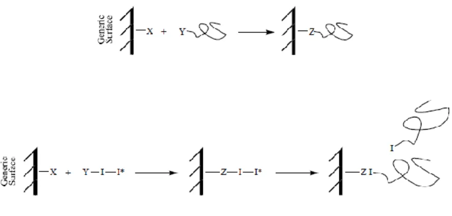
Depending on the polymer, different grafting techniques can be chosen to modify its surface (Figure 2). If the polymer surface presents suitable functional groups, an option is to attach large macromolecules to the appropriate surface (grafting on). The other option is to exploit these functional groups as possible sites to start the polymerization of a monomer (grafting from). (28; 32)
Figure 2. Surface grafting of a generic surface. On the top figure it is possible to observe a representation of the grafting on approach. In the bottom the grafting from approach is represented. [Taken from (33)]
The grafting from approach (Figure 2: bottom) presents several advantages when compared to the grafting on (Figure 2: top). Although the grafting on approach offers better define and characterized structures in the surface (the structures can be isolated and purified before being grafted), the grafting from approach has reduced preparative steps (the macromolecular material to be grafted doesn’t need to be prepared and isolated) and the surface density is not influenced by the dimension and mutual sterical constraints of the grafting material (the surface density depends on the density of the initiation groups). (32) Nevertheless, the formation of islands or mushrooms on the grafted on surface is often observed. (33)
Additionally, if the polymer has no chemically-reactive functional groups in its surface that allows the grafting initiation, it is possible to create radicals in the surface by chemical treatments followed by an initiation reaction or by irradiation. The irradiation can be carried out in the presence of a monomer (simultaneous irradiation) or the monomer can be added after the irradiation (pre-irradiation). (28)
Grafting modification that is induced by radiation is an extremely promising method for surface modification and can be used in most of the polymers that are
11 known. This technique enables the development of materials with new properties, since it allows to change the nature, morphology and structure of the modifying polymer and to control the thickness of the grafted layer. (28)
Several radiation sources have been used, with corona, plasma or glow-discharge being the most common. When the radiation source is chosen, there are some factors to be considered, as the availability, the impact on the modification process and the penetration depth. (28)
Among the grafting from techniques, atom transfer radical polymerization (ATRP) has been widely used and discussed, being a promising technique. It can be performed in mild conditions (room temperature in aqueous solution) and can be used in a wide range of polymers. In addition, this technique has negligible transfer reactions, due to the presence of monomers only in the end of the growing chains. (32)
Atom Transfer Radical Polymerization (ATRP)
Atom transfer radical polymerization (ATRP), which consists in a catalyzed reversible redox process, is one of the techniques used to carry out a controlled/living radical polymerization. (34; 35) This technique is easy to apply and makes possible the use of different monomers and reaction media (aqueous or organic) (36; 37) The monomers that may be used are styrenes, methacrylates (e.g. polyethylene glycol methyl methacrylate (PEGMA) and poly(2-hydroxyethyl methacrylate) (PHEMA)), methacrilamides, methacrylic acids and 4-vinylpyridine. (35) Furthermore, this technique allows to obtain polymer chains with controlled molecular weights and low poly-dispersity. (34) ATRP is also a suitable technique for growing polymeric brushes on surfaces. (37)
The name of the technique is based in the fact that both the activation and the deactivation of the radicals involve an atom transfer reaction. Being a radical polymerization process, this technique has four main steps: initiation, propagation, transfer and termination.
The initiation step consists in the formation of a reactive site, thus initiating the polymerization. After the initiation, the propagation step begins, with the monomers being added one by one to the active chain end. During the reaction, the active site can be transferred to the monomer, the initiator, the polymer or the solvent itself, in the transfer step. This step may result in the terminated molecule or in some new active site where the propagation may occur. The final step is the termination, which results in
Chapter 1
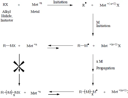
Introduction
12
inert macromolecules. This may occur by coupling reactions of two active centers or active transfer between active chains. (33)
All of these steps may be found in the ATRP process, as represented in Figure 3. First the initiator covers the polymer surface. Many commercially available initiators (e.g. alkyl halides) can be used, as long as they present a weak halogen-heteroatom bond. The initiator will provide the polymer a surface with simple halogen as end groups, which is easily converted into useful functionalities. The transition metal complexes used (Ru, Cu, Fe, Ni, among others) are responsible for the conversation into useful functionalities, removing the halides from the polymer surface. The surface is then ready to a polymerization. (35)
Figure 3. Schematic representation of the ATRP process. After the initiator (X) covers the polymer surface (R), the transition metal complex (Met) will remove the halides from the polymer surface. The monomer (M) will then interact with the polymer surface and the propagation of the monomers begins, culminating with the formation of polymeric chains in the polymer surface. [Taken from (33)]
In order to achieve a controlled polymerization of the monomers, the initiation process has to be fast enough to provide a constant concentration of growing polymer chains, and there has to be a dynamic equilibrium between the inactive chains and the growing radicals. (38)
Xiao et al (2002) used ATRP to modify the PDMS surface. They used polyacrylamide chains to increase the hydrophilicity of PDMS. The amount of polyacrylamide reached a maximum after 15 min. However, after 30 min of exposure
13 visual inspection revealed PDMS damage. It was also observed that the treatment could be performed optimally before the occurrence of bulk damage. The surface became hydrophilic and, unlike the plasma treatment, the hydrophilic character of the modified PDMS lasted for at least one month. (39)
3.3. Coating Materials
Several materials have been used in order to increase PDMS surface hydrophilicity. Among these materials it is possible to find polymers and biomolecules. (28; 29; 37; 39; 40) Some polymers that are widely used in order to increase the hydrophilicity are the poly(ethylene oxide) (PEO), the poly(ethylene glycol) (PEG), the polyacrylamides (PAAm), among others.
PEG and its derivates have been used to change the hydrophilicity and the biomolecule-repelling properties of PDMS surfaces. This polymer is frequently used due to its low toxicity, low immunogenicity, and its ability to prevent protein, cell, and bacteria adhesion. However, PEG has only two hydroxyl groups available per chain, because of its linear structure. This can lead to some limitations in the surface modification. (37; 41)
PEO is structurally similar to PEG, but has a higher molecular weight. The surface coating with this polymer usually results in the reduction of biomolecules adsorption (e.g. proteins and bacteria). PEGMA, which is also used as a coating material, has a similar structure and similar properties. (37)
Other solution may be the use of the hydrophobic nature of PDMS and immobilize blood proteins on its surface. Because of the hydrophobic interaction, the proteins would bond and stay on the surface and remain part of the fluid that is washed off. (42) Some extracellular matrix (ECM) proteins (collagen, laminin, fibronectin) may also be used as a coating, promoting cell adhesion and migration. (40)
3.4. Vascular Grafts Modification
Vascular grafts have drawn high interest in the medical society, with the increased need of regeneration of vascular tissues. One of the most common materials utilized in vascular grafts is ePTFE. (43) Although this material presents interesting properties, some graft failure has been observed, due to thrombosis and intimal occlusion of the vessel. In order to avoid these limitations, an increased effort to seed the vascular grafts has been performed by several researchers. (44)
Chapter 1
Introduction
14
Cezeaux and co-workers (1998) modified the surface of ePTFE using vacuum ultraviolet, with the purpose of increasing the endothelial cell adhesion. The modified surfaces could not increase endothelial cell adhesion, however the surfaces yielded an increased cell proliferation. These results suggest that the vacuum ultraviolet surface modification can be used to obtain more suitable surfaces for endothelial cell colonization of ePTFE vascular grafts. (44)
Adali et al (2010) reported a new approach for the cell seeding of grafts, namely the in situ endothelialization of implanted grafts inside the body. For that purpose, the endothelial progenitor cells (EPCs), which consist in a small population of CD34+ circulating mononuclear cells capable of attaining endothelial cell characteristics in
vitro, are the best candidates. The limitation of using such cells for in situ
endothelialization of grafts is due to the fact that these cells are mainly located in the bone marrow and only small amounts of EPCs circulate in the blood. Thus, these cells need to be mobilized to the implant site and seed their surface. The proposed strategy is coating the graft with capture molecules that attract the circulating EPCs and increase their adhesion to the surface. (4)
Furthermore, Larsen et al (2006) synthesized a novel peptide fluoro-surfactant polymer modification that facilitates the adhesion and growth of endothelial cells on ePTFE vascular grafts. The peptide fluoro-surfactant polymer consists of a poly(vinyl amine) backbone with RGD sequences and perfluorocarbon pendant branches. Endothelial cells showed a specific adhesion to the RGD sequences and retained an hemostatic function. (45)
4.
Body-Biomaterial Interactions
Replacing injured tissues with biomedical devices is currently the main approach in biomaterials science, because substitutes of biological origin are recognized by the immune system. This happens due to the presence of biological motifs in this substitutes that are immunologically recognizable. (2)
Once the biomaterials are intended to contact directly with living tissues and biological fluids, they are targets for the protective mechanisms within the body (protein adsorption, hemostasis, inflammation, foreign body response). During the past decade it was recognized that all implantable biomaterials invoke an almost identical inflammatory and foreign body response, despite the biomaterial nature. Currently
15 obstacles related to the design of biomaterials involve the interaction of biomaterials with the body and the reaction of the body to biomaterials. (46)
Therefore, understanding and predicting the interaction between tissues or body fluids with biomaterials is crucial to all kinds of medical technologies. The reconstructive medical implants require a perfect integration of the biomaterial with the surrounding tissues to restore adequate function, with no release of harmful products. (2)
4.1. Cell-Biomaterial Interaction
Cell adhesion to biomaterials surface is a critical step in the integration of implants. Therefore, the interaction of cells with biomaterials is an important feature of
in vitro biocompatibility and cytotoxicity studies. This adhesion is mediated by adsorbed
proteins, like immunoglobulins, vitronectin, fibrinogen and fibronectin. (47; 48)
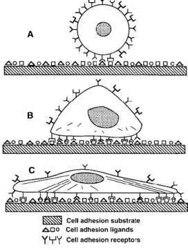
The main parameters of cell-biomaterial interactions are the cell adhesion and the cell spreading. Cell adhesion and spreading are the consequence of a series of molecular events (Figure 4) that occur in and around the cells, which are mediated by the trans-membrane receptors present in the extracellular matrix (ECM). (49)
Figure 4. Progression of the mammalian cell adhesion. (A) Initial contact of the cell with the material covered by proteins. (B) Formation of bonds between the cell surface receptors and the cell adhesion ligands in the proteins. (C) Cytoskeletal reorganization with a progressive spreading of the cell in the surface of the implant, increasing the attachment strength. [Taken from (1)]
Chapter 1
Introduction
16
4.1.1. Extracellular Matrix (ECM)
The ECM is secreted by the cells that populate a tissue or organ. Its composition will be determined by factors like the mechanical forces, the oxygen requirements, the gene expression patterns. This matrix plays a crucial role in the mammalian development and physiology, and both the amino acid sequence and the quaternary components of the ECM are greatly preserved across species lines. (49)
The composition of the ECM is a mixture of functional and structural proteins (collagen, fibronectin), glycosaminoglycans (GAGs), glycoproteins and small molecules (figure 5). All this components are arranged in a unique three-dimensional architecture. (49)
Figure 5. Cell proteins involved in cell adhesion on a biomaterial. [Taken from (50)]
Collagen
Collagens represent more than 90% of the dry weight of the ECM, being the most abundant proteins in the mammalian ECM. More than 20 different types of collagen where identified and the type I collagen is the major structural protein in tissues. Type I collagen is abundant in tendinous and ligamentous structures, providing the necessary strength that these tissues require. Other types of collagens can be found in the ECM in much lower amounts than the type I collagen, although providing different mechanical and physical properties to the ECM. One example is the type IV
17 collagen, which is present within the basement membrane of most vascular structures and tissues with an epithelial cell component. (49)
Fibronectin
Fibronectin is the second most abundant protein in the ECM. It is a large dimeric glycoprotein that exists either in the soluble state or as a tissue isoform. This protein is a mediator of mammalian cells adhesion, because it possesses ligands for adhesion of several cell types. Fibronectin is rich in the tripeptide Arg-Gly-Asp (RGD), which is recognized by the cell surface receptors, the integrins, thus being extremely important in cell adhesion. When bound via integrins, these proteins trigger a number of signal transduction pathways that activate events like cell spreading, proliferation, differentiation and migration. This protein is critical for normal biologic development, especially the development of vascular structures. (49; 51)
Laminin
Laminin is an adhesion protein present in ECM that exists in numerous forms, depending in the specific mixture of the several peptide chains. It is determinant in the formation and maintenance of vascular structures. This protein is one of the most critical ECM factors in the process of cell and tissue differentiation. (49)
Glycosaminoglycans
It is possible to find several mixtures of GAGs in the ECM, depending on the tissue location, the age of the host and the microenvironment. The GAGs have various functions, like binding growth factors and cytokines, promoting water retention and contributing to gel properties of the matrix. Heparin and hyaluronic acid are two GAGs present in the ECM. (49)
Growth Factors
Growth factors and cytokines, although in small amounts, are also present in the ECM. However, they act as potent modulators of cell behavior. There is an extensive list of growth factors present in the ECM, including the vascular endothelial cell growth factor (VEGF), the fibroblast growth factor (FGF) family, and the epithelial cell growth factor (EGF), among others. These factors can be found in different isoforms, each with a specific biologic activity. (49)
Chapter 1
Introduction
18
4.1.2. Adhesion Molecules - Integrins
The adhesion molecules include four main classes – selectins, immunoglobulin super family, adhesins and integrins- and are capable of interacting with specific ligands situated on the membrane of neighbor cells or on the ECM. (50)
Among the four classes of adhesion molecules, the integrins are the main cell surface receptors for proteins within the ECM. (52) The integrin family is composed of 22 heterodimers with two types of subunits, α and β, that are non-covalently associated. There have been discovered 16 α subunits and 8 β subunits, that combined in different ways can origin a diversity of structures with various ligand-binding possibilities. In the integrin structure, each subunit has a large extracellular domain, a transmembrane domain and a short cytoplasmic domain. (48; 50; 52; 53; 54; 55)
Integrins can bind to specific amino acid sequences, such as the RGD motif that is present in many ECM proteins. (49; 53; 54) Besides the ECM proteins, integrins can interact with components of the cytoskeleton and signaling molecules through their intracellular domain. Being an interface between the extracellular and the intracellular environment, integrins can translate the attachment of external ligands to internal information, inducing adhesion, spreading, cell migration and cell growth and differentiation. (50)
After binding specifically to a ligand, the integrins cluster together into focal adhesions. Focal adhesions consist of additional cytoskeletal proteins, adapter molecules and kinases, being an area of close contact between the cell and the ECM. The integrins are present in these areas in higher amount than their normal membrane distribution. Focal adhesions are barely formed on hydrophobic surfaces, and well developed in surfaces that sustain cell adhesion. (53; 54)
When clustered, the recruitment of tensin and focal adhesion kinase (FAK), as well as their phophorylation start, resulting in the recruitment of talin, vinculin and α-actinin, which are responsible to link the F-actin fibers to the plasma membrane. The rearrangement of F-actin fibers induces changes in the organization of the cytoskeleton (and consequently in the cell shape), affecting cell adhesion and mobility. (53; 54)
19
4.2. Adhesion Proteins
When a biomaterial is implanted, in a question of seconds to minutes, a layer of proteins rapidly adsorbs and covers its surface. (47; 56) Therefore, instead of the original surface of the implanted material, the cells will recognize this protein layer. It is possible to say that the adhesion proteins convert the biomaterials into a biologically recognizable material. The adsorption of these adhesion proteins is the basis for all the reactions that may occur in the body. (1)
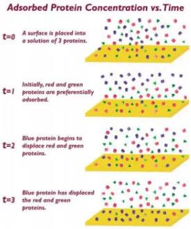
Figure 6. Surface placed in a protein mixture. In a matter of seconds the surface is covered by a layer of adsorbed proteins. Initially, the “red” and the “green” proteins adsorb in a higher concentration. With time, these proteins are displaced by the “blue” protein. This can happen because the concentration of proteins in the adsorbed layer is usually different from the concentration in the solution, being able to change in time. [Taken from (47)]
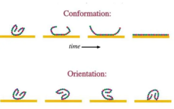
The surface properties of the biomaterials (chemistry and hydrophobicity) determine the type, amount and conformation of the adsorbed proteins. The composition of this protein layer can be different, depending of the fluid composition and adsorption time. In figure 6 it is possible to observe that initially the “red” and the “green” proteins are present in a higher concentration, but with time these proteins are displaced by the “blue” proteins. Besides the composition of the protein layer, the conformation and the orientation of the protein can also change with time, as represented in figure 7 (44,54).
Chapter 1
Introduction
20
Figure 7. The top scheme shows a protein denaturing with increasing adsorption time. The bottom scheme shows a proteins adsorbing to the surface in different orientations. This is possible because the conformation and the orientation of adsorbed proteins depend on adsorption conditions and surface properties. [Taken from (47)]
The layer of proteins that is formed will increase the cell adhesiveness, since the cells have receptors in the cell membrane, which bind specifically to the adhesion proteins. This layer of adhesion proteins also increases the cell spreading in the surface.
4.3. Host Immune Response
Nowadays it is known that there are no inert biomaterials. The medical implants, being foreign to the host body, will trigger tissue responses during the healing process that are dependent on the nature of the biomaterial and the implant site. (57) Indeed, the host-biomaterial interaction is a very complex process that will control the biological performance of the medical implants. (56)
When the biomaterial is implanted in the body a wound is created and a series of events, initially similar to the ones in normal wound healing, will occur. The process of implantation disturbs the homeostatic mechanism in the body, thus activating the healing process. (58)
The way the implant is accepted by the host and how well the host will heal depends largely on the way the complex wound healing around the device will occur. The wound healing process includes four phases, namely the inflammation, the foreign body reaction, the fibrous encapsulation and the matrix formation and remodeling. (57; 58)
The host body reacts similarly to nearly all the biomaterials, as represented in figure 8. After one month of implantation, all the biomaterials are found to heal basically in the same way.
21 Figure 8. Reaction of the host to the implanted biomaterial. (1) The biomaterial is implanted in the surgical site by the surgeon. (2) A layer of proteins quickly adsorbs to the implant surface. (3) The neutrophils and the macrophages examine and attack the biomaterial. However the implant is too large to be ingested. (4) The macrophages find that they cannot digest the implant, so they fuse into giant cells to engulf the implant. The giant cells send out cytokines to attract other cells. (5) The fibroblasts arrive and start synthesizing collagen. (6) The implant is completely entrapped in an acellular, avascular collagen bag. [Taken from (47)]
The inflammation process can be trigged by surgery trauma or the presence of a foreign body in the host. In the 24 hours after the surgery cellular and non cellular responses will take place. The first event is a non cellular response, the vasodilatation of the local vessels, which culminates with the increased permeability of the vascular endothelium and the edema formation. In parallel the complement cascade is activated by the membrane damage. Then the cellular response also takes place with cell recruitment. (59) Several molecular signals will act as chemoattractants at the implant site, recruiting the inflammatory cells. (58)
The first cells migrating to the injury site are the neutrophils, which are responsible for the phagocytosis, the engulfing and the degradation of the foreign body. Once neutrophils start their function, the monocytes circulating in the blood enter the tissue and become macrophages, which are also responsible for the phagocytosis and release several biochemical factors that can activate other cells. (59) The activated macrophages adhere to the material and spread on its surface, trying to phagocyte it. Because they cannot digest or engulf the implant, they fuse together and origin a foreign body giant cell, which can phagocytize larger particles. It is the presence of the
![Table 1. Key milestones in the development of silicone. [Adapted from (1)]](https://thumb-eu.123doks.com/thumbv2/123dok_br/17934664.852202/26.893.239.640.782.880/table-key-milestones-development-silicone-adapted.webp)
![Table 2. Applications of silicone biomaterials in the biomedical field. [Adapted from (3)]](https://thumb-eu.123doks.com/thumbv2/123dok_br/17934664.852202/29.893.118.773.148.872/table-applications-silicone-biomaterials-biomedical-field-adapted.webp)
![Figure 5. Cell proteins involved in cell adhesion on a biomaterial. [Taken from (50)]](https://thumb-eu.123doks.com/thumbv2/123dok_br/17934664.852202/38.893.183.716.469.817/figure-cell-proteins-involved-cell-adhesion-biomaterial-taken.webp)