UNIVERSIDADE DE LISBOA
FACULDADE DE CIÊNCIAS
DEPARTAMENTO DE QUÍMICA E BIOQUÍMICA
Modulation of Neuronal Maturation:
Influence of Neurotrophic Factors
and Neuromodulators
Ana Filipa Ferreira da Cunha Ribeiro
MESTRADO
EM
BIOQUÍMICA
UNIVERSIDADE DE LISBOA
FACULDADE DE CIÊNCIAS
DEPARTAMENTO DE QUÍMICA E BIOQUÍMICA
Modulation of Neuronal Maturation:
Influence of Neurotrophic Factors and
Neuromodulators
Ana Filipa Ferreira da Cunha Ribeiro
Dissertação orientada por:
Professora Doutora Ana M. Sebastião
Doutor Pedro A. Lima
MESTRADO EM BIOQUÍMICA
O trabalho experimental descrito nesta tese foi realizado no Instituto de Farmacologia e Neurociências, Faculdade de Medicina de Lisboa e Unidade de Neurociências, Instituto de Medicina Molecular.
R
ESUMO
O cérebro é uma estrutura complexa formada por inúmeras células com diversas formas e funções, nomeadamente neurónios e células da glia, que incluem astrócitos, oligodendrócitos e microglia. No caso dos neurónios, estes divergem num grande número de subtipos, com base em características moleculares, morfológicas e electrofisiológicas. São células especializadas na transmissão e integração da informação, sendo que para tal, a sua morfologia é essencial. Para além do corpo celular, são normalmente formados por dois tipos diferentes de processos especializados, inúmeras dendrites, que recebem e levam a informação para o corpo celular, e um único axónio, que leva a informação para fora do corpo celular. Deste modo, o desenvolvimento de uma morfologia especializada e a expressão de moléculas específicas durante a diferenciação e maturação neuronal, constituem eventos essenciais à formação de neurónios maduros funcionais. O ambiente neural influencia grandemente a diferenciação. Os neurónios em diferenciação e maturação estão sujeitos à acção do meio envolvente, de moléculas sinalizadoras, as chamadas “pistas de orientação” (do inglês, signalling cues), que são libertadas pelas células vizinhas, induzindo alterações morfológicas nos primeiros. A existência de estruturas especializadas nas extremidades dos processos em desenvolvimento, os cones de crescimento, a partir dos quais os processos crescem, é fundamental para conduzir os processos em crescimento para um local específico, em resposta às pistas de orientação no meio extracelular, de modo a estabelecer contactos com outros neurónios. Deste modo, a maior alteração nas células nervosas durante o seu desenvolvimento é o crescimento extensivo de axónios e dendrites, bem como a proliferação de sinapses. Um exemplo conhecido de uma molécula sinalizadora que influencia a diferenciação e maturação neuronal é o factor neurotrófico derivado do cérebro, BDNF (do inglês, brain-derived neurotrophic factor). As suas acções estendem-se também à sobrevivência neuronal, bem como à modulação da transmissão e plasticidade sináptica. Para executar estas acções, o BDNF liga-se a dois tipos de receptores, nomeadamente aos receptores de alta afinidade de tirosina cinase, TrkB (do inglês, tropomyosin-related kinase B), e aos receptores de baixa afinidade, pertencentes à superfamília dos receptores do factor de necrose tumoral, p75NTR (do inglês, p75 neurotrophin receptor). A nível da maturação
neuronal, o BDNF dá origem a alterações estruturais na célula, tais como a indução de dendrites primárias e arborização axonal e dendrítica, uma acção que envolve activação de cascatas de sinalização, como as vias das cinases PI3K (do inglês, phosphatidylinositol
3-kinase), a MAPK (do inglês, mitogen-activated protein kinase) e, com menor importância, a
PLC (do inglês, phospholipase C) A adenosina é um neuromodulador do sistema nervoso central, partilhando com o BDNF funções moduladoras da actividade sináptica. É o caso da facilitação das acções sinápticas do BDNF através da co-activação dos receptores A2A da
adenosina, os quais estão acoplados à formação de cAMP, com consequente activação da proteína cinase A (PKA, do inglês, protein kinase A). Dada esta interacção entre ambos receptores, foi colocada a hipótese de que a adenosina, por activação dos receptores A2A,
influencia as acções tróficas do BDNF na maturação neuronal.
O objectivo do trabalho descrito nesta tese foi avaliar se os receptores A2A da adenosina
modulam as acções tróficas do BDNF na maturação neuronal. Para além disso, também foi estudada a importância da adenosina endógena na activação dos receptores A2A e TrkB.
Finalmente, foram exploradas as potenciais vias de sinalização associadas às acções tróficas dos receptores A2A na maturação neuronal. Para tal, recorreu-se à utilização de um software
de medição de características morfológicas, HCA-Vision, o qual, a partir de uma imagem de um neurónio obtida por microscopia de fluorescência, cria um neurónio virtual, através do qual é possível realizar medições morfométricas, tais como o comprimento total das neurites e da neurite máxima, bem como o número de neurites primárias e de pontos de ramificação.
Ao terceiro dia de cultura, neurónios corticais foram pré-incubados na presença ou ausência do agonista selectivo dos receptores A2A da adenosina, CGS 21680 (10nM), e/ou do
antagonista desse receptor, ZM 241385 (50nM), sendo 20 minutos depois incubados com BDNF (20ng/mL). As culturas ficaram a incubar a 37˚C numa atmosfera humidificada com 5% CO2, com os ligandos anteriormente descritos até serem fixadas ao sétimo dia. Através da
técnica de imunocitoquímica, os neurónios foram marcados para a proteína de ligação aos microtúbulos MAP2 (do inglês, microtubule-associated protein 2), com um fluoróforo conjugado com um anticorpo anti-MAP2, e o núcleo celular, com o marcador nuclear DAPI, que se liga ao DNA (do inglês, deoxyribonucleic acid). Observou-se que, tanto o BDNF como a activação dos receptores A2A da adenosina modulam a morfogénese neuronal, no entanto
com efeitos distintos. O BDNF induz a formação de dendrites primárias, enquanto que a activação dos receptores da adenosina promovem um crescimento da neurite máxima. Para além disso, ambos induziram um aumento do número de pontos de ramificação. No caso da formação de dendrites primárias, estas mostraram ser independentes da activação dos receptores A2A da adenosina, contudo, os resultados obtidos levam a sugerir uma possível
modulação deste receptor nos efeitos do BDNF na arborização dendrítica. Para além disso, a utilização de um antagonista selectivo para o receptor A2A permitiu observar uma diminuição
do tamanho da neurite máxima e número de pontos de ramificação na ausência de ligandos activadores dos receptores em estudo, sugerindo uma actividade basal dos receptores A2A
com consequências para a maturação neuronal.
Para além disso, dado que no meio extracelular existem sempre quantidades consideráveis de adenosina, que é libertada tanto por neurónios como por células da glia, ou mesmo sintetizada extracelularmente por acção de ectoenzimas, fui explorar se a adenosina endógena, presente no meio de cultura, seria importante nos efeitos tónicos e basais dos receptores A2A e TrkB. Para tal, neurónios corticais em cultura foram tratados com adenosina
desaminase (ADA, do inglês adenosine deaminase) 1h antes da adição dos ligandos dos receptores TrkB e A2A. Estes ficaram a incubar até serem fixados ao sétimo dia de cultura,
sendo de seguida marcados para o MAP2 e núcleo. A remoção da adenosina endógena do meio extracelular pela utilização de ADA, não afectou a acção dos receptores A2A, no entanto
atenuou o efeito do BDNF no aumento do número de pontos de ramificação e de neurites primárias. No primeiro caso, apesar de se já ter observado uma activação tónica dos receptores A2A (resultados com o antagonista A2A), a ausência de efeito aquando da remoção
da adenosina endógena, sugere a presença de um outro receptor de adenosina com uma função oposta à dos A2A. No caso do BDNF, a atenuação dos seus efeitos na maturação
neuronal pela remoção da adenosina extracelular, mas não pelo antagonista A2A, sugere
novamente o envolvimento de um outro receptor de adenosina.
Finalmente, foi-se explorar as vias de sinalização activadas aquando das acções tróficas dos receptores A2A da adenosina na maturação neuronal. Para tal, experiências com inibidores
farmacológicos foram realizadas ao terceiro dia de cultura. As vias de sinalização PI3K, MAPK, PLC e PKA foram bloqueadas através da incubação de neurónios corticais com,
respectivamente, os inibidores LY294002 (50μM), U0126 (10μM), U73122 (5μM) e Rp-cAMPS (100μM), os quais foram adicionados 30 minutos antes da adição do agonista dos receptores A2A, permanecendo durante mais 5h em contacto com as células. Observou-se que o
crescimento da neurite máxima, promovido pela activação dos receptores A2A, foi
completamente prevenido pelo bloqueio das vias de sinalização PI3K e MAPK e PLC; por outro lado, o aumento do número de pontos de ramificação por activação dos receptores A2A
foi prevenido pelo bloqueio da PI3K, MAPK e PKA.
Em conclusão, os resultados deste trabalho demonstram que tanto a activação dos receptores TrkB e A2A têm efeitos na maturação neuronal. Enquanto o primeiro promove a
formação de dendrites primárias, o segundo promove o crescimento da neurite máxima, sendo que ambos apresentam uma acção coincidente na arborização dendrítica. Em estádios mais precoces da diferenciação neuronal, parece haver uma independência entre as acções tróficas do BDNF e as dos receptores A2A da adenosina, pois o aumento do número de
dendrites primárias induzido pelo BDNF não é modulado pela activação, nem pela inibição, dos receptores A2A da adenosina. No entanto, ao longo do processo de maturação neuronal,
as funções do BDNF parecem começar a ser moduladas pelo receptor A2A, como é o caso da
arborização dendrítica. Para além disso, apesar da formação de neurites primárias não ser modulada pelo receptor A2A, parece ser modulada por um outro receptor de adenosina, tal
como os restantes parâmetros morfológicos estudados. Finalmente, a acção trófica da adenosina, via activação dos receptores A2A, no crescimento da neurite máxima e na
arborização dendrítica, mostrou ser dependente das vias de sinalização PI3K, MAPK, sendo a primeira também dependente da PLC e a segunda da PKA.
A
BSTRACT
BDNF is known to induce the formation of primary dendrites in cortical neurons via PI3K and MAPK pathways. Adenosine, through A2A receptors (A2ARs), facilitates BDNF synaptic
actions, but it is not known if this extends to BDNF influence upon neuronal maturation. Since the effects of BDNF are dependent on cAMP formation and A2ARs are coupled to the AC/cAMP
transducing system, I tested the hypothesis that adenosine, through A2AR activation, was able
to modulate BDNF actions upon neuronal maturation.
E18 forebrain neurons from Sprague-Dawley rats were seeded at 104cells/cm2, and treated, at DIC 3, with CGS21680 (10-100nM) and/or ZM241385 (50nM) 20 minutes before BDNF (20ng/mL), remaining there until DIC 7. Cells stained with anti-MAP2 and DAPI were analysed by HCA-Vision software in terms of maximal neurite length and number of roots and branch points. The A2AR agonist, CGS21680, increased maximal neurite length, and the
number of branch points in a concentration-dependent manner, with maximal effect at 30nM. BDNF increased the number of roots and branch points. The effect of BDNF upon neuronal branching was modified by CGS21680 or by ZM241385. The effects of BDNF upon the number of roots were not influenced by CGS21680, but it was attenuated by endogenous adenosine removal with adenosine deaminase. To address the signalling pathways activated by A2AR
upon neuronal maturation, neurons were treated, at DIC 3, with inhibitors of PI3K, MAPK, PLC and PKA signalling pathways 30 minutes before the addition of CGS21680, which remained in the medium for 5h until fixation. The effects of the A2AR agonist were totally
prevented by blocking PI3K and MAPK pathways; blocking PLC prevented the influence of the A2AR agonist upon maximal neurite elongation, whereas PKA blockade prevented its influence
upon dendritic branching.
These results suggest that A2ARs are involved in neurite elongation, while BDNF
influences new primary dendrites formation, being that both receptors operate independently at early stages of neuronal maturation. However, they seem to crosstalk at later stages. Endogenous adenosine seems to influence BDNF-induced formation of roots, a process that might involve other adenosine receptors. Finally, the signalling pathways involved in the A2AR
actions upon neural maturation require PI3K, MAPK and PLC signalling pathways for maximal neurite elongation and PI3K, MAPK and PKA for branching.
KEYWORDS:
I
NDEX
1 INTRODUCTION ... 5
1.1 Corticogenesis ... 5
1.2 Neuronal Interaction with its surrounding Environment ... 6
1.3 Neurotrophins ... 9
1.3.1 BDNF ...12
1.4 Adenosine ...14
1.5 Crosstalk between TrkB and Adenosine A2A Receptors ...17
2 OBJECTIVES ...19
3 METHODS ...21
3.1 Neuronal Cortical Cultures ...21
3.2 Immunocytochemistry ...23
3.3 Morphometric Analysis ...24
3.4 Electrophoresis and Immunoblotting ...28
3.5 Drugs ...31
3.6 Statistics ...31
4 RESULTS ...33
4.1 Expression levels of Adenosine A2A and BDNF TrkB Receptors during Neuronal Differentiation in Cortical Cultures ...33
4.2 Function of BDNF TrkB and Adenosine A2A Receptors in Neuronal Maturation ...37
4.2.1 Specific role of Adenosine A2A Receptor activation in Neuronal Maturation ...40
4.2.2 Specific role of BDNF action in Neuronal Maturation ...41
4.2.3 Modulatory action of Adenosine A2A Receptor upon BDNF effects in Neuronal Maturation ...42
4.3 Concentration dependency of the effects of the A2A Receptors agonist ... 45
4.4 Endogenous influence of Adenosine in Neuronal Maturation ... 47
4.4.1 Endogenous influence of Adenosine upon A2A Receptor activation ... 48
4.4.2 Endogenous influence of Adenosine upon BDNF actions ... 50
4.5 Signalling Pathways ... 51
4.5.1 Validation of the tools used by evaluating their influence upon BDNF actions ... ... 52
4.5.2 Signalling Pathways activated by Adenosine A2A Receptors in Neuronal Maturation ... 55
5 DISCUSSION ... 59
5.1 Roles of TrkB and A2AR activation in Neuronal Maturation ... 59
5.2 Modulation of BDNF actions by A2A Receptor activation ... 60
5.3 Endogenous influence of Adenosine ... 63
5.4 Signalling Pathways activated by the Adenosine A2A Receptor ... 66
6 CONCLUSION ... 71
7 ACKNOWLEDGEMENTS ... 73
A
BBREVIATION
L
IST
A2AR – adenosine A2A receptor
ADA – adenosine deaminase
AMP – adenosine 5’-monophosphate cAMP – 3’,5’-cyclic AMP
ATP – adenosine 5’-triphosphate
BDNF – brain-derived neurotrophic factor BSA – bovine serum albumin
CGS 21680 – 4-[2-[[6-Amino-9-(N-ethyl-b-D-ribofuranuronamidosyl)-9H-purin-2-yl]amino] ethyl]-benzene propanoic acid hydrochloride
CNS – central nervous system
CREB – cAMP-response element binding protein DAPI – 4’, 6’-diamidino-2-phenylindole
DIC – days in culture
ECL – enhanced chemiluminescencent
ERK – extracellular signal-regulated protein kinases GAPDH – glyceraldehyde-3-phosphate dehydrogenase GPCR – G-protein coupled receptor
HBSS – Hank’s balanced salt solution HRP – horseradish peroxidase
LY294002 – 2-(4-Morpholinyl)-8-phenyl-1(4H)-benzopyran-4-one MAP – microtubule-associated protein
mRNA – messenger ribonucleic acid NGF – nerve growth factor
P75NTR – p75 neurotrophin receptor PBS – phosphate-buffered saline PFA – paraformaldehyde
PI3K – phosphatidylinositol 3-kinase PKA – protein kinase A
PKC – protein kinase C PLC – phospholipase C
PVDF – polyvinylidene fluoride RGC – radial glial cell
Rp-cAMPS – Rp-Adenosine 3’,5’-cyclic monophosphorothioate triethylammonium salt hydrate SDS – sodium dodecyl sulphate
TBS – tris-buffered saline Tris – tris-hydroxymethyl-aminomethane Trk – Tropomyosin-related kinase U0126 – 1,4-Diamino-2,3-dicyano-1,4-bis(2-aminophenylthio)butadiene U73122 –1-[6-[[(17b)-3-Methoxyestra-1,3,5(10)-trien-17-yl]amino]hexyl]-1H-pyrrole-2,5-dione ZM 241385 – 4-(2-[7-Amino-2-(2-furyl)[1,2,4]triazolo[2,3-a][1,3,5]triazin-5-ylamino]ethyl) phenol
1 I
NTRODUCTION
The mammalian neocortex is a remarkably complex structure. In the case of the human, the neocortex exists as its most complex and evolved state, as it is the region of the brain that is responsible for higher functions such as sensation, action, cognition, thoughts, memories, language and consciousness. Thus, it corresponds to the part of the brain that most distinguishes humans from the rest of the species. Neocortex is composed by two major neuronal cell types, excitatory pyramidal neurons and inhibitory interneurons, and glial cells such as astrocytes, oligodendrocytes, and microglial cells, establishing rich local and external connections, i.e. synapses, with other regions of the central nervous system. Both neuronal subtypes diverge in a large number of subtypes, each of which possessing distinct molecular, morphological, and electrophysiological characteristics. Nonetheless, despite such cell type heterogeneity, cortical neurons display an extraordinary laminar organization since their cell bodies are distributed into six distinct layers along the radial axis of neocortex. This orderly cortical laminar organization provides key advantages for the processing and storage of neural information in the cerebral cortex.
The formation of a functional neuronal network in the cerebral cortex, as we know in the adult brain, is dependent on the coordination of two high organized cellular processes, such as neuronal migration and neuronal maturation. Therefore, the unravelling of the early development of this complex structure that is the cerebral cortex might provide the key to understand the mechanisms underlying the pathogenesis of many cognitive disorders.
1.1 Corticogenesis
During embryonic development, the vast majority of neurons of the cerebral cortex are generated from precursor cells that are located in a pseudostratified epithelial cell layer which surrounds the early ventricular lumen. This neuroepithelial cell layer is composed by two distinct germinal zones: the ventricular zone and the subventricular zone. The ventricular zone corresponds to the innermost neuroepithelial cell layer of progenitor cells that adjacently surrounds the lumen of the neural tube that lines the lateral ventricles, and the subventricular
zone lies superficially on the ventricular zone (see Diaz and Gleeson, 2009). The neuroepithelial progenitor cells in the germinal zones have a high mitotic activity in order to maintain its progenitor population and produce radial glial cells (RGC), which retain contact with both ventrical and pial surfaces. Cortical neurons are generated from asymmetric division of RGSs (Anthony et al., 2004) to produce one proliferative glial cell that stays in germinal zone and one neuron that migrates towards the pial surface to the cortical plate, through a migratory process guided by RGSs (see Huang, 2009). Within the cortical plate, neurons become organized into well-defined layers, being known six layers in the mammalian neocortex. The layering of neurons in the cerebral cortex is established in an inside-first, outside-last manner. In other words, the cells that migrate from the ventricular zone and leave the cell cycle at early stages give rise to neurons that form the deepest layers of the cortex, while cells that leave the ventricular zone and exit the cell cycle at progressively later stages migrate over longer distances, pass early-born neurons, and settle in more superficial layers of the cortex, forming the more superficial layers of the neocortex (Sidman and Rakic, 1973; Rakic, 1988; Marin-Padilla, 1998; Molnar et al., 2006; Abdel-Mannan et al., 2008). Nevertheless, besides the majority of neurons originate in the local neuroepithelium, recent studies have revealed that some neurons, such as Cajal-Retzius cells (Bielle et al., 2005), cortical interneurons and cortical predecessor neurons, have different origins and complex migratory routes (Bystron et al., 2006; Bystron et al., 2008).
1.2 Neuronal Interaction with its surrounding Environment
Neurons are polarized cells, formed by two distinct types of processes, typically a single axon and multiple dendrites, which allow them to receive, process and transmit information in a single direction. During the period of migration, from the germinal zone to the cortical plate, neurons have just a simple bipolar form, which is very different from their more complex adult morphology. Many specialized biochemical properties, such as neurotransmitters, crucial for a mature neuron to participate in neuronal circuits, are not yet expressed in the cells. So, besides neurons may be in the correct place, when they arrive to the cortical plate, the dendritic trees are hardly formed, as well as many axons have yet to find their correct
destination. As a result, it is in the cortical plate that cortical neurons begin their final differentiation into a functional mature form, through the formation of a distinct axon and a dendritic arborization, together with the expression of specific molecules important to the establishment of functional synapses.
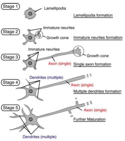
Experiments using cultured embryonic neurons have revealed that neurons initially generate several equivalent neurites and begin to polarize when one neurite becomes an axon, with the other neurites, lately, becoming dendrites. Dotti et al. (1988), in cultured hippocampal neurons, observed this differentiation process and divided the morphological events into five stages (Figure 1.1). Dotti and co-workers demonstrated that shortly after plating, neuronal development starts with round spheres that spread lamellipodia around the cell body (stage 1). Then, these protrusions develop into several short immature neurites, which contain dynamic growth cones at their tips (12-24h; stage 2). Neurites, at this early developmental stage, alternate phases of elongation and retraction and are approximately equal in length, being, consequently, unpolarized cells. Neuronal polarization occurs when one of these equally long neurites starts to grow rapidly to become the axon (24-48h; stage 3). A few days after the formation of the axon, the remaining neurites slowly elongate to become dendrites (> 3-4 days; stage 4). The axon and dendrites continue to mature, expressing specific proteins very important to the formation of synaptic contacts and establishment of a neuronal network with the formation of dendritic spines and filopodia (> 1 week in culture; stage 5) (Dotti et al., 1988).
Figure 1.1 Processes of neuronal differentiation in cultured hippocampal neurons. Cultured hippocampal
neurons from rodent hippocampus acquire their characteristic polarized morphology in five well-defined stages (From Yoshimura et al., 2006).
However, in vivo, neuronal differentiation and maturation is more complicated than in cultures, as neurons are involved by the complex environment existent in the brain, i.e. neurons have to extend axons over great distances bypassing billions of potential synaptic targets before reaching the correct area and recognizing the appropriate targets. Though, these pathways along which axons grow provide a large number of diverse molecular cues that are crucial to guide axons to their targets, as axons possess specific receptors to recognize and interpret these cues. By sensing the extracellular cues, signalling cascades are activated and the growth cone at the tip of the axon steers towards (attractants) or away (repellents) from them, through changes in cytoskeleton organization. Given the great distance that can separate growth cones from their cell bodies, axon pathfinding presents a challenge to
neurons in ensuring that growth cones respond properly and rapidly to complex extracellular stimuli encountered along the way. Classically, it has been thought that the proteins required for directional steering originate in cell bodies, where they are synthesized and subsequently transported to growth cones to mediate responses to extracellular stimuli. However, recent evidence suggests that the functional autonomy seen in axonal growth cones is mediated by local protein synthesis. According to this view, subsets of neuronal mRNAs are trafficked into axons and growth cones, and different guidance cues elicit translation of a distinct set of proteins in a spatially restricted manner, being locally translated independently of cell bodies. In the case of dendrites, local translation of mRNAs is now relatively well characterized and is critically important for synaptic plasticity and memory. Some of the axonal mRNAs known in vertebrate central nervous system (CNS) include cytoskeletal proteins, such as actin, β-tubulin, microtubule-associated protein 1b (MAP1b), tau, and neurofilament, as well as proteins involved in cytoskeleton remodelling, as actin depolymerizing factor ADF/cofilin, a protein involved in cytoskeleton disassembly. Other mRNAs present encode other functional proteins that include cell signalling molecules, transcription factors like cAMP-response element binding (CREB) protein and transmembrane receptors. This suggests that axons, and in a similar way dendrites, contain a diverse repertoire of mRNAs that can be potentially translated, enabling a neuron to rapidly respond to the extracellular environment, by translating specific subsets of proteins that will induce cytoskeletal reorganization (for a review see Tahirovic and Bradke, 2009).
All these processes of neurogenesis, neuronal migration and maturation, during the development of the cerebral cortex, are enabled by the dynamic regulation of the cytoskeletal machinery in response to extracellular guidance or positional cues, factors that are released from the surrounding cells, including neighbouring and distant neurons and glial cells. An example of extracellular guidance cues are neurotrophins.
1.3 Neurotrophins
During the process of the embryonic development of the nervous system there is an overproduction of neurons, and glial cells, followed by the programmed death of superfluous
cells, which is now known to occur in almost all regions of the central and peripheral nervous systems. It was Hamburger and Levi-Montalcini who developed the neurotrophic factor
hypothesis, the idea that the target cells of developing neurons have a critical role in
regulating the number of surviving neurons, by producing a limited amount of an essential nutrient or trophic factor that is taken up by the nerve terminals. Based on this hypothesis neurons extend axons to the vicinity of target cells which secrete limited amounts of neurotrophic factors that bind to specific receptors localized on the surface of nerve terminals. Neurons that do not receive adequate amounts of neurotrophic factor die by apoptosis, while the others survive. Following this hypothesis, Levi-Montalcini and Stanley Cohen isolated nerve growth factor (NGF), the first neurotrophic factor to be identified by isolating it from snake venom, which provided the first direct support for the neurotrophic hypothesis (Cohen et al., 1954; Cohen and Levi-Montalcini, 1956). NGF is one of a large array of secreted factors that have the ability to promote the survival of neurons. Apart from NGF, there are three more major neurotrophins that have been isolated from mammals which are brain-derived neurotrophic factor (BDNF), neurotrophin 3 (NT3), and neurotrophin 4/5 (NT4,5) (see Huang and Reichardt, 2001; Chao, 2003).
These neurotrophins interact with two types of transmembranar cell surface receptors: the high affinity tropomyosin-related kinase receptor (Trk), a receptor that belongs to the tyrosine kinase family of receptors; and the pan-neurotrophin receptor p75NTR, which belongs to the tumor necrosis factor receptor family. Neurotrophic factors were originally believed to promote the survival of neural cells by stimulating their metabolism in beneficial ways. Instead, it now appears that such factors act predominantly by suppressing a latent biochemical pathway present in all cells of the body. This biochemical pathway is a suicide program, induced by the activation of p75NTR. Once activated it kills cells by apoptosis. The binding of neurotrophic factors to their tyrosine kinase receptors is thought to promote anti-apoptotic Bcl-2-like activities or inhibit caspase activity (see Kaplan and Miller, 2000). Apart from that, neurotrophin-mediated activation of Trk receptors leads to a variety of biological responses, which includes proliferation, axonal and dendritic growth, guidance and remodelling, assembly and remodelling of cytoskeleton, membrane-trafficking, and modulation of synaptic formation, transmission and plasticity. In the case of p75NTR, it
activates a distinct set of signalling pathways within cells that are in some instances synergistic and in other instances antagonistic to those activated by Trk receptors (Figure 1.2) (Huang and Reichardt, 2003).
Figure 1.2 Neurotrophin receptors, Trk and p75NTR, and correspondent signal transducing pathways. Binding
of neurotrophins to Trk triggers the activation of signalling cascadas, such as Ras, phosphatidylinositol 3-kinase (PI3K), and phospholipase C (PLC) pathways, resulting in gene expression, neuronal survival and neurite outgrowth (From Chao, 2003).
Trk family of tyrosine kinases are the major signal-transducing receptors and consist of three membrane tyrosine kinases named TrkA, TrkB and TrkC. NGF interacts selectively with TrkA, whereas BDNF and neurotrophin 4/5 interact primarily with TrkB. Neurotrophin 3 activates TrkC and, to a lesser extent, TrkB and TrkA (Chao, 2003). On the other hand, neurotrophins also bind to p75NTR with a similar low affinity, although proneurotrophins, the precursors of the above mature neurotrophins, bind with high affinity to this class of receptors (Lee et al., 2001).
Regarding neuronal differentiation and maturation, neurotrophins were reported to increase the dendritic complexity of pyramidal neurons by increasing total dendrite length, the number of branch points, and/or the number of primary dendrites (McAllister et al., 1995;
Baker et al., 1998; Niblock et al., 2000). Of the major signalling pathways activated by Trk receptors, the mitogen-activated protein kinase (MAPK) and phosphatidylinositol 3-kinase (PI3K) pathways have been implicated in neurite and dendrite filopodia formation in both neuronal cell lines and primary neurons (Posern et al., 2000; Wu et al., 2001).
1.3.1 BDNF
BDNF is a small, basic protein, with 252 amino acid residues, that was purified from the mammalian (pig) brain (Barde et al., 1982), being its actions the best characterized of the four neurotrophins in nervous system. BDNF levels are dynamically regulated in postnatal development, in part by activity-dependent mechanisms. BDNF is not synthesized as a mature protein, but as a precursor protein (preproBDNF) in endoplasmic reticulum. Upon cleavage of the signal peptide, proBDNF is formed, which is transported to the Golgi for sorting into either constitutive or regulated secretory vesicles. proBDNF can have functions by itself, being highly expressed, together with p75NTR, during a perinatal window when axonal outgrowth and synapse maturation is robust and having a more localized expression during adolescence/adulthood. ProBDNF may be converted into mature BDNF (mBDNF) intracellularly in the trans-Golgi or in the immature secretory granules (Chao and Bothwell, 2002; Greenberg et al., 2009).
This neurotrophin has several functions, such as promoting neuronal survival (Thoenen et al., 1987), modulating synaptic plasticity in learning and memory (Yamada and Nabeshima, 2003), facilitating neuromuscular activity (Boulanger and Poo, 1999; Pousinha et al., 2006), promoting neurogenesis during cortical development (Bartkowska et al., 2007), as well as regulating the growth and branching of dendrites and inducing the formation of new primary dendrites (Bartrup et al., 1997; Dijkhuizen and Ghosh, 2005). These actions of BDNF are possible through the activation of BDNF the above mentioned specific membrane receptor TrkB, to which BDNF binds with high affinity, inducing receptor dimerization and autophosphorylation in tyrosine residues in the cytoplasmic domain. These phosphorylated tyrosines promote the activation of multiple intracellular signalling pathways, by the recruitment of various adapter proteins, such as Src homologous and collagen-like (Shc)
adaptor protein, or phospholipase C (PLC). Shc adaptor protein links the activated Trk receptor to two separate intracellular signalling cascades, namely PI3K and Ras pathway that leads to activation of Akt and MAPK. In addition, PLC binds to activated Trk receptors and initiates an intracellular signalling cascade, resulting in the release of inositol phosphates, with consequent release of intracellular calcium (Ca2+), and activation of protein kinase C (PKC) (Figure 1.2) (Chao, 2003; Huang and Reichardt, 2003). The Ras and MAPK pathways have been more related to primarily stimulate processes responsible for neuronal differentiation, and PI3K pathway for cell survival, whereas PLC, is more associated with fast actions of BDNF, as it happens in synaptic activity. In the case of cultured cortical neurons, BDNF was reported to induce primary dendrite formation via activation of the PI3K and MAPK pathways with a minor role of PLC (Dijkhuizen and Ghosh, 2005).
BDNF and TrkB receptors are highly expressed in the developing CNS during times of neuronal differentiation (McAllister et al., 1999). Depending on the alternative splicing, TrkB receptors can lead to the formation of truncated receptors, which lack most of the intracellular domain (Chao, 2003). In neocortex, full-length TrkB receptor is the major trkB form during early development, while truncated TrkB predominates in all forebrain regions of late postnatal and adult rats (Allendoerfer et al., 1994; Fryer et al., 1996). In cortical neurons, TrkB was shown to be present both in excitatory and inhibitory neurons, being that in excitatory ones, TrkB was found in all of the major neuronal compartments, namely in the cell body, dendrites and axons both at 4 and 10 days in culture, as well as in the surface of axonal growth cones and dendritic filopodia before synapse formation, which showed an increasingly localization to synapses through the development (Gomes et al., 2006). The presence of TrkB on the surface of growth cones and dendritic filopodia allows BDNF to activate local signalling pathways, playing thus a role in the BDNF-induced chemotropic responses. Guidance cues, such as BDNF, require local protein synthesis, to induce rapid cytoskeletal modifications, making it possible for growth cones to respond to chemo-attractants or repellents, by changing their pathways. An example of a protein recently reported, which is locally translated in response to BDNF signalling, is -actin, through the phosphorylation of zipcode-binding protein (ZBP) 1 by Src family kinases (SFKs) (Yao et al., 2006; Sasaki et al., 2010). In addition, activation of the PI3K activation was demonstrated to be involved in the modulation of
protein translation by regulating the phosphorylation of eukaryotic translation initiation factor (eIF) 4E, as well as MAPKs, such as p42/p44 MAPK which is involved in many aspects of cellular responses, including cell growth and differentiation.
In neurons, the reorganization of the cytoskeleton by BDNF is a fundamental mechanism not only to initial stages of neuronal differentiation, such as regulate axon guidance, but also final stages regarding maturation and synaptic plasticity. Fast changes in synaptic efficacy may be translated into structural alterations when the synapses are exposed to BDNF for a longer period of time. These include axonal branching (Cohen-Cory and Fraser, 1995; Gallo and Letourneau, 1998), dendritic growth and complexity (McAllister et al., 1995; McAllister et al., 1997; McAllister et al., 1999), and activity-dependent refinement of synapses and regulation of dendritic spine formation (Shimada et al., 1998).
Because dendritic morphology determines the number, pattern, and types of synapses received by a neuron, regulation of cortical dendritic growth and branching by BDNF is likely to play a major role for the proper functioning of the brain, and especially the cerebral cortex.
1.4 Adenosine
Adenosine is a ubiquitous endogenous purine nucleoside that is released from most cells, including neurons and glia, and modulates many physiologic and biochemical processes in the brain. Due to its extremely short half-life, adenosine acts in a paracrine way, acting in the same cell or in the cells adjacent to the adenosine-releasing cell (Abbracchio and Cattabeni, 1999). Adenosine can be produced intracellularly and released through an equilibrative nucleoside transporter (ENT) (Thorn and Jarvis, 1996), or extracellularly by the hydrolysis of released adenine nucleotides, namely adenosine 5’-triphosphate (ATP), through the action of a cascade of ectoenzymes. Inactivation of extracellular adenosine predominantly occurs through reuptake, although deamination of extracellular adenosine to inosine or phosphorylation to 5’-monophosphate (5’-AMP) can also occur (Meijer et al., 2008).
Adenosine is known to modulate neuronal activity, namely neurotransmitter release and its postsynaptic actions, as well as by nonsynaptically hyperpolarizing or depolarizing
neurons, thus regulating post-synaptic excitability. This nucleotide is also known to modulate the response of others receptors and neuromodulators (see Sebastião and Ribeiro, 2009). These actions mediated by adenosine are possible through the activation of four specific metabotropic G-protein coupled receptors (GPCRs), namely the A1, A2A, A2B and A3 subtypes.
As A1 and A2A subtypes are considered to be high affinity receptors for adenosine, being that
in basal conditions these subtype receptors are probably tonically activated, having physiological importance. On the other hand, the low affinity A2B and A3 receptors have more
relevance in pathophysiological conditions, when the concentration of adenosine is higher, although in humans A3 subtype is a high affinity receptor (Ribeiro et al., 2003). GPCRs are
seven-transmembrane domain receptors that are linked to a variety of transduction mechanisms. The main intracellular signalling pathways involve the formation of 3’,5’-cyclic AMP (cAMP), being A1 receptors mainly coupled to adenylate cyclase inhibitory G-proteins (Gi
or Go), while A2A and A2B receptors to activation of adenylate cyclase stimulatory G-proteins
(Gs), though coupling to PLC excitatory G-protein (Gq) can also occur. In the case of A3
receptors, they are coupled to Gq and Gi (Fredholm et al., 2001a).
Adenosine receptors are widely distributed through the brain, however depending on the brain area, age and cell sublocalization, there are differences in receptor expression. In the adult brain, the A1 receptor is highly expressed in cerebral cortex, cerebellum and
hippocampus (Reppert et al., 1991; Rivkees et al., 1995; Svenningsson et al., 1997), while the others adenosine receptors are expressed in a more restricted way. A2A receptor is present in
several brain areas, with a higher expression in the olfactory bulb and striatum (Jarvis and Williams, 1989), and at lower levels in the cerebral cortex (neocortex) and hippocampus (Cunha et al., 1994a; Kirk and Richardson, 1995; Svenningsson et al., 1997). Regarding A2B and
A3 adenosine receptors, these have been demonstrated to be fairly dispersed through the
brain, including the neocortex, although at very weak levels (Dixon et al., 1996; Fredholm et al., 2001a). Thus, due to its high expression in certain brain areas and to its high affinity, the neuromodulatory role of adenosine in the brain is mainly mediated by the balance between the inhibitory and excitatory actions of respectively adenosine A1 and A2A receptor actions. In
contrast, the low expression and low affinity of A2B and A3 receptors may make these
increases, such as in ischemia and hypoxia (see Fredholm et al., 2001b; Borea et al., 2009). However the role of A2B and A3 receptors in the nervous system is much less understood (see
Sebastião and Ribeiro, 2009).
Regarding A2A receptor activation, adenosine is known to modulate neuronal activity,
namely presynaptic neurotransmitter release and/or uptake, such as glutamate (Lopes et al., 2002), gamma-aminobutyric acid (GABA) (Cunha and Ribeiro, 2000; Cristóvão-Ferreira et al., 2009) or acetylcoline (Cunha et al., 1994b). Postsynaptic actions of A2A receptor activation are
associated to the increase in long-term potentiation (LTP) and AMPA receptors expression in the postsynaptic membrane, through a PKA-dependent mechanism (Dias et al., 2010). Besides actions upon neuronal activity, there is evidence indicating that adenosine, acting through A2A
receptors, may influence the development of the nervous system. It has been reported that A2A receptor is expressed in late prenatal periods and that their levels increase after birth.
Although mRNA have reached their adult levels in neocortex, keeping nearly constant from embryonic day 18 (E18) onwards, protein receptor levels are low or undetectable at E18 and increase until postnatal day 14 (P14) (Adén et al., 2000). Moreover, A2A receptors in cortical
cultured neurons were immunocytochemically demonstrated to be mainly present at nerve terminals after six days in culture (Rebola et al., 2005) and with less intensity in the cell body region (Lee et al., 2004). These time-dependent changes in A2A receptor expression in the
brain, which have been reported during development, suggest a role in neuronal maturation. Recently, it has been reported that activation of A2A receptors can influence neurite
outgrowth in PC12 cells (Heilbronn and Zimmermann, 1995; Cheng et al., 2002; Charles et al., 2003). In PC12 cells endogenously expressing A2A receptors, antisense-mediated inactivation
of the enzyme that converts AMP to adenosine, ecto-5’-nucleotidase, inhibits neurite outgrowth (Heilbronn and Zimmermann, 1995). In addition, A2A receptor activation
counteracted the PD98059-induced blockade of NGF-induced neurite outgrowth, a process that involves MAPK and CREB activation (Cheng et al., 2002); and a bacterial nucleoside N6-methyldeoxyadenosine (MDA) induces an A2A receptor-mediated neurite outgrowth in PC12
cells which also depends on NGF-dependent MAPK activation (Charles et al., 2003). By the first time, Canals and co-workers explored the effect of direct activation of A2A receptors with their
cultures of striatal neurons, showing that A2A receptor activation lead to neuritogenesis and
differentiation, through a mechanism dependent on MAPK, PKC and PKA signalling pathways (Canals et al., 2005).
1.5 Crosstalk between TrkB and Adenosine A
2AReceptors
Apart from affecting nerve cells directly, adenosine, through A2A receptor activation,
can also influence the action of neurotransmitters and other neuromodulators indirectly, which make adenosine to behave as a modulator of others modulators. These adenosine modulatory actions contribute to a very sophisticated crosstalk between its own receptors and the receptors for other neurotransmitters, neuromodulators or both (see Sebastião and Ribeiro, 2000). There are several possibilities for this crosstalk, such as at the transducing system level (Sebastião and Ribeiro, 2000) or even at the receptor level, as a consequence of receptor-receptor heteromerization (Ferré et al., 2007).
There are evidences of a crosstalk between A2A and TrkB receptors, which has been
carefully investigated in the hippocampus. For example, adenosine, acting through A2A
receptor, can exert a trophic effect by transactivation TrkB receptor (Lee and Chao, 2001). A2A
receptors control the facilitatory fast actions of BDNF at hippocampal synapses (Diógenes et al., 2004), with consequences for synaptic plasticity (Fontinha et al., 2008), trough a mechanism dependent on cAMP/PKA (Diógenes et al., 2004; Fontinha et al., 2008). In addition, A2A receptor presynaptically mediates the inhibition of A1 receptor actions upon the
release of neurotransmitters, through a mechanism that involves PKC activity (Lopes et al., 1999). Moreover, a crosstalk between A2A and TrkB receptors was also demonstrated at the
neuromuscular junction where A2AR activation of the PKA pathway controls the action of
BDNF, through the TrkB receptor (Pousinha et al., 2006). Hence, cAMP seems to be important for the signalling and biological functions of BDNF. On the other hand, regarding neuronal differentiation, it was demonstrated that growth cones of cultures of Xenopus spinal neurons and hippocampal neurons show either attractive or repulsive turning responses towards a gradient of BDNF, depending on the intracellular concentration of cAMP (Song et al., 1997; Mai et al., 2009). Other experiments suggested a ‘cAMP gating’ of TrkB phosphorylation and
cAMP regulation of TrkB trafficking to the postsynaptic densities (PSD), which may both contribute to the long-term regulation of dendritic spine formation by BDNF (Ji et al., 2005).
Accordingly, as cAMP seems to be very important in BDNF functions upon neuronal maturation, I hypothesized that branching and formation of primary neurites mediated by BDNF could be influenced by the activation of A2A receptor.
2 O
BJECTIVES
The main goal of the present work was to unravel the actions of adenosine A2A receptor
and BDNF upon neuronal maturation. To achieve this goal I:
1. evaluated the actions of A2A receptors upon neuronal outgrowth, namely the length
of neurites and neuronal branching;
2. evaluated if A2A receptors modulate the actions of BDNF upon neuronal outgrowth;
3. unravelled the transducing mechanisms involved in these actions.
To pursue these objectives, cortical neurons from primary cultures were morphologically measured in terms of neurite length and number of primary neurites and branching.
3 M
ETHODS
3.1 Neuronal Cortical Cultures
Cortical neurons were dissected from embryonic day 18 (E18) Sprague-Dawley embryos, obtained from Harlan Interfauna Iberia (Barcelona), as described by Vicario-Abejón (1997) and Assaife-Lopes et al. (2010), with minor modifications (Vicario-Abejón, 1997; Assaife-Lopes et al., 2010). Animals were handled according to the Portuguese law on Animal Care and European Union guidelines (86/609/EEC).
Pregnant rats were anesthetized with halothane before being sacrificed by decapitation. Uterus containing embryos were rapidly removed from the rat abdomen and placed in Ca2+ and Mg2+ free HBSS (Hank’s Balanced Salt Solution) (supplied by Gibco) dissection medium supplemented with 0.37% glucose. Under sterile conditions, embryos were removed from the uterus and the brain was isolated by removing the skin and the skull of the embryo and by separating the ventral side of the brain from the rest of the head. Meninges and choroid plexus, which are highly vascularized, are removed very gently and then both hemispheres are opened so that the cortex can be removed. The whole dissection process was performed in cold HBSS medium previously referred. The cortices were trypsinized for 15 minutes at 37˚C, in order to digest the proteins that facilitate adhesion between cells in the tissue. Following trypsinization, FBS (Fetal Bovine Serum) was added to quench trypsin activity and immediately after trypsin inhibition DNase was added to prevent the clumping of the tissue, since the effects of trypsinization and the DNA released from damaged cells can produce tissue clumping. The tissue was centrifuged 4 minutes at 188g at room temperature. The cortices were resuspended in Neurobasal medium (supplied by Gibco) supplemented with 0.5mM glutamine, 2% B27 and 25U/mL penicillin/streptomycin, and cells were dissociated by passing the solution through a 10mL pipette 6-8 times and then through a yellow tip (20-200μL) coupled to the pipette for 6-8 times. Cell suspension was filtered using a 70μm cell strainer to remove cell clumps. Then, cells were counted in a 75% trypan blue cellular solution using a Hemacytometer, and plated in poly-L-lysine-coated dishes at a low density of 4x10⁴cells/mL (Vallotton et al., 2007), in a 24-well plate (for morphometric analysis
experiments), and at a high density of 6x10⁵cells/mL in a 85-mm plate, to measure the protein levels by western blot. The cortical primary cultures were incubated in an incubator with a humidified 37˚C and 5% CO2 atmosphere. After 3 days in culture (DIC 3), half of the medium
was replaced by fresh Neurobasal medium supplemented as mentioned above. It is important just to change half of the medium because neurons are vulnerable to the adverse effects of changing medium, becoming more and more vulnerable as they differentiate.
Dissociated cortical cultures were treated, at DIC 3 (Burkhalter et al., 2007), with the selective A2A receptor agonist CGS 21680 (10nM) (Diógenes et al., 2004) and/or the antagonist
ZM 241385 (50nM) (Assaife-Lopes et al., 2010) 20 minutes before the TrkB endogenous ligand BDNF (20ng/mL) (Bartrup et al., 1997; Assaife-Lopes et al., 2010), and adenosine deaminase (ADA) (1U/mL) (Assaife-Lopes et al., 2010) was added 1h before BDNF or CGS 21680. When CGS 21680 was tested in the presence of ZM 241385, CGS 21680 was added 20 minutes after ZM 241385. All these ligands remained in the medium for a long term treatment until fixation at DIC 7. Experiments with inhibitors of transducing pathways were performed at DIC 3, in which inhibitors were added 30 minutes prior to the addition of BDNF or the agonist of A2A
receptors, CGS 21680, remaining present until fixation 5h after the ligands addition (Dijkhuizen and Ghosh, 2005). The inhibitors used were LY294002 (a PI3K inhibitor), U73122 (a PLC inhibitor), U0126 (a MAP kinase kinase inhibitor) and Rp-cAMPS (a blocker of cAMP-mediated effects) were used at a final concentration of 50µM, 5µM, 10µM and 100μM, respectively (Dijkhuizen and Ghosh, 2005; Lochner and Moolman, 2006; Assaife-Lopes et al., 2010).
The choice of DIC 3, as the time in culture to perform the treatment, results from the fact that at that time neurons were already polarized and are starting to form dendrites (Dotti et al., 1988). Consequently, receptors activation will be able to affect new dendrite formation as well as axon and dendrite development, without affecting neuronal polarization, i.e. the specification of the axon (Shelly et al., 2007). As neurons, at DIC 7, have already specified the axon and dendrites, as well as these processes have already started to branching and form filopodia (Dotti et al., 1988; Ziv and Smith, 1996), this time point of fixation was considered suitable to measure alterations in the neurite length, the number of primary neurites, and the number of branch points (see section 3.3). These morphological changes are important
measures for neuronal maturation, especially dendrite branching, as dendritic morphology determines the number, pattern, and types of synapses received by a neuron, being thus fundamental for the proper functioning of the brain.
3.2 Immunocytochemistry
DIC 3 and DIC 7 24-well plate cortical neurons were washed with Phosphate Buffer Saline (PBS) (NaCl 137mM, KCl 2.1mM, KH2PO4 1.8mM and Na2HPO4.2H2O 10mM, at pH=7.40),
in order to remove the Neurobasal medium before fixation with 4% paraformaldehyde in PBS at room temperature, for 20 minutes. The cross-linking reagent paraformaldehyde was used to establish intermolecular bridges between free amino groups that make part of the cell structure, inducing a latticework of interactions that immobilizes the antigens, while retaining the cellular and subcellular architecture of the whole cell together. Excess of paraformaldehyde was removed by washing with PBS solution. As fixation with paraformaldehyde does not allow access of the antibody to the antigen, a permeabilization step was performed using a nonionic detergent, in which cultures were incubated for 10 minutes in PBS containing 0.05% of the nonionic detergent Triton X-100 at room temperature, becoming possible for antibodies to penetrate inside the cell and bind to its specific antigen. This is followed by incubation with a blocking solution of PBS containing 0.25% gelatin for 30 minutes, at room temperature. Gelatin is going to interact with other proteins as well as with the antibodies used, increasing the competition of antibodies in biding to their targets, with a consequent result of increasing selectivity to the antigen, decreasing the nonspecific binding. Cells were then incubated for 1h with the monoclonal primary antibody anti-MAP2 (1:200, Chemicon) in PBS containing 0.05% Tween 20 and 0.1% gelatin, where the detergent Tween 20 facilitates penetration of the antibody as well as stabilizes it, and gelatin decreases antibody nonspecific binding. Extensive washes were then performed in PBS containing 0.05% Tween 20, in which Tween 20 interacts with the antibody and facilitate the remotion of unbouded and nonspecific bounded antibody. Cultures were incubated for 1h with a goat anti-mouse secondary antibody conjugated to the fluorescent label Alexa Fluor 568 (1:500, Invitrogen) in PBS containing 0.05% Tween 20 and 0.1% gelatin. After more washes in PBS
containing 0.05% Tween 20, cultures were incubated for 5 minutes with DAPI (4’, 6’-diamidino-2-phenylindole), which is a fluorescent stain that binds strongly to DNA, extensively used in fluorescence microscopy with maximum absorption at a wavelength of 358 nm (ultraviolet) and maximum emission at 461 nm (blue). Finally, after some washes with PBS containing 0.05% Tween 20 and a last wash with PBS to remove Tween from the sample, coverslips were mounted in Mowiol before visualization. Mowiol is a mounting media used for samples prepared for fluorescence microscopy because it is non-absorbing, contains no autofluorescence, or light scattering.
3.3 Morphometric Analysis
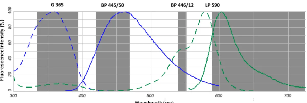
Detailed morphological analysis of neurons is crucial for studying the normal development of the dendritic and axonal arborization as well as the effect of drugs in the developing neurites. For that, images of neurons mounted in Mowiol were captured using a monochrome digital camera (AxioCamMR3, Zeiss) mounted on an inverted widefield fluorescence microscope (Zeiss Axiovert 200, Germany), with a 40x objective. As observed in Figure 3.1, the nuclear dye DAPI has a maximal absorption at 359nm and emission at 461nm, while Alexa Fluor 568 absorbs and emits at lower wavelengths with a maximum, respectively, at 578nm and 603nm. Thus, the filter sets (Figure 3.1) used were: a Zeiss filter set 15 (Excitation filter BP 546/12; Beam splitter FT 580; Emission filter LP 590) to detect Alexa Fluor 568 goat anti-mouse IgG antibody, in which allows to transmit light with a wavelength between 540-552nm and allows the transmission of wavelength greater than 590nM; and a Zeiss filter set 49 (Excitation filter G 365; Beam splitter FT 395; Emission filter BP 445/50) to detect DAPI bound to DNA.
Images were captured using the software AxioVision 4 (Carl Zeiss Imaging Systems). The pixel size in the object space was 0.25μm and the captured images (Figure 3.2) were 1400 x 1050 pixels size. Images were stored in uncompressed 8-bit Tiff format, each containing two channels, one with the representation of cellular nucleus, obtained with filter set 49 (Figure 3.2 DAPI), and other channel with the cellular structure, obtained with filter set 15 (Figure 3.2
MAP2). Images were analysed, in terms of neuronal morphometrics, using HCA-Vision software (CSIRO).
Figure 3.1 – Absorption (dashed line) and fluorescence emission (continuous line) spectra of the fluorophores
DAPI bound to DNA (blue) and Alexa Fluor 568 goat anti-mouse IgG antibody (green). The shading represents the excitation and emission intervals of the filter sets: Zeiss filter set 49, with excitation wavelength centred at 365 nm (G 365) and emission between 420-470 nm (BP 445/50); and Zeiss filter set 15, with excitation wavelength between 540-552 nm (BP 446/12) and emission greater than 590 nm (LP 590).
Figure 3.2 – Representative images of a control condition of cortical neurons with 7 days in culture. E18 rat
primary cultures of cortical neurons were plated at 4x10⁴cells/mL. At DIC 7, cultures were fixed and immunostained for microtubule-associated protein 2 (1/200 antibody dilution), MAP2, and with the DAPI nuclear stain (blue). Staining with anti-MAP2 antibody was revealed by Alexa Fluor 568 (red) labelled goat anti-mouse antibody (1/500 dilution). Images were taken with an inverted widefield fluorescence microscope (Zeiss Axiovert 200, Germany), with a 40x objective, and using the Zeiss filter sets 49 and 15 for detection of DAPI and Alexa Fluor 568 antibody conjugated, respectively.
HCA-Vision software has a module for Neurite Outgrowth which is a powerful tool for the analysis of neurite structure in fluorescence images. Through HCA-Vision it is possible to
BP 445/50 BP 446/12 LP 590
G 365
analyse images in a fully automated manner regarding the segmentation of neuron bodies and neurites. For that, Neurite analysis is divided into three steps (Figure 3.2), including cell body detection, neurite detection, and neurite analysis, and optimal parameters of each wizard have to be chosen by the user, so that the software can generate neurite traces, segment the cell soma and associate neurites with their respective soma, with a consequent report of a virtual image of each neuron. In the cell body detection step, the images of the neurons stained with MAP2 are treated to suppress the noise, background corrected, and cell bodies are identified, as well as cell debris are eliminated based on their small size. Then, the second step is neurite detection, in which linear features are identified by classifying pixels as belonging to a linear feature based on its intensity. Pixels with high intensity are connected and the connected components smaller than a user defined threshold are removed. Gaps in the skeleton are closed. Finally, neurite analysis step, apart from removing very short lateral branches that result from thinning artifacts, combines the result of the cell body detection and of the neurite detection to produce a virtual image of the neuronal cell. With this virtual image it will be possible for the software to make quantitative measurements and statistics biologically relevant which are reported on a per-cell basis (Vallotton et al., 2007). Some of the quantitative measurements with morphological importance upon neurite outgrowth are the total neurite length, maximum neurite length, the number of branch points, and the number of roots (Figure 3.4 and Figure 3.5).
To study neuronal morphometrics, the neuronal marker used was the antibody against MAP2, which labels dendrites (Bernhardt and Matus, 1984; Caceres et al., 1984). As no tau staining was used, it would be expected that axons were not taken into account (Binder et al., 1985). However, MAP2 staining is also able to label axons until DIC 7, although very weakly (Caceres et al., 1986). Nevertheless, despite axonal MAP2 staining being very weak at DIC 7 (Caceres et al., 1986), HCA-Vision software was able to detect it, and thus axons were taken into account. Thus, is possible that the maximal neurite length might be the neuronal axon, while the number of roots may vary depending on the number of dendrites formed.
In order to analyse neurons with HCA-Vision software, cells have to be plated at low density, with a range of 1-5x10⁴cells/mL, to avoid cell-cell contact (Vallotton et al., 2007). Therefore, in this work, cells were plated at 4x10⁴cells/mL. As a result, most of the cells
analysed were isolated neurons, with no contacts between them. When two cells overlap neurites, results are more subjective, being necessary to decide whether a neurite belongs to one or other of the two cells.
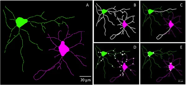
Figure 3.3 – Representative images of fluorescence image analysis of two DIC 7 cultured cortical neurons by
HCA-Vision software. Images with two channels (one for nucleus and other for cell structure) taken with an inverted widefield fluorescence microscope (Zeiss Axiovert 200, Germany), with a 40x objective, and using the Zeiss filter sets 49 and 15 for detection of DAPI and Alexa Fluor 568 antibody conjugated, respectively, were analyzed by HCA-Vision software. A three-step analysis was performed in order to obtain a virtual image of these two neurons: neuron body detection (A), neurite detection (B), and neurite analysis, where detected neurites in B are associated to neuron bodies already detected in A, forming thus two virtual neuronal images (C).
Figure 3.4 – Schematic representation of a neuron
analysed by HCA-Vision software. With HCA-Vision software analysis it is possible to obtain a virtual image of a neuron from which can be measured the morphological parameters: total neurite length, that are sum of the length of each neurite segment (S1 to S7); maximal neurite length, which corresponds to the the length of the longest path from a neuron body to an extreme segment (S6 + S7); number of branch points, the number of points where a neurite structure splits into two or more branches (B1 to B3); and number of roots, which are the the number of points where neurite structure touches a neuron body (R1 to R2). (image obtained from Bio Image Analysis Workshop (2008), CSIRO Biotech Imaging)
Figure 3.5 – Virtual images of two neurons obtained by HCA-Vision software. With a virtual image (A), it is
possible for the software to measure the morphological parameters: total neurite lengh (B), maximal neurite length (C), number of branch points (D) and number of roots (E). From these two virtual neuronal images, total neurite length is 374.86 and 339.55 μm for the green and pink neurons (B), respectively. In the case of maximal neurite length, green and pink neurons have respectively 62.49 and 69.68 μm (C). The number of branch points measured for green and pink neurons were 18 and 14 (D), and the number of roots 5 and 8 (E), respectively.
3.4 Electrophoresis and Immunoblotting
DIC 1, 3, 5 and 7 were lysated by adding to the 85-mm plate 250μL of 0.32M sucrose solution with 50mM Tris at pH 7.6, plus protease inhibitors (ROCHE) and scraping the plate with the help of a cell scraper. It was centrifuged at 1000g during 10 minutes at 4°C. The supernatant, corresponding to the dissociated cells, was collected. Protein was quantified according to Bradford assay (1976) using the Bio-Rad Protein assay kit. This sample was used for relative quantification of the proteins presented in Table 3.1.
Lysates were denaturated with 5x sample buffer (350mM Tris, 30% glycerol, 10% SDS, 600mM dithiothreitol, and 0.012% bromophenol blue, pH 6.8). 10% polyacrilamide gel samples were boiled at 100˚C and 12% polyacrilamide gel were heated at 80˚C, both with open cap, in order to concentrate samples and reach a 20μL and 150μL volume, respectively, before loading in the polyacrilamide gel. 10% polyacrilamide gel was performed with 10-wells,
A B C
while 12% with 5-well, to allow a higher amount of protein, namely A2A receptor, as it is very
difficult to detect in cortical neurons due to its low protein expression levels. Proteins were run in SDS-polyacrylamide gels, which were mounted in the electrophoresis apparatus filled with running buffer (25mM Tris, 192 glycine, 0.1% SDS). Equal volumes of samples were loaded into gels (Table 3.1). Samples were stacked by running at 80 V and resolved at 140 V.
Proteins in the polyacrylamide gel were transferred to polyvinylidene fluoride (PVDF) membranes, previously wet in methanol, during 1h 40min at room temperature in transfer buffer (24mM Tris, 182 mM glycine, 15% methanol). PVDF membranes are particularly effective for use with the chemiluminescent detection system that was employed, and because it has high affinity for proteins, it will allow some strippings of the membrane, so that it was possible to detect more than one protein in the same membrane. After the proteins had been transferred to the PVDF membrane, a blocking step was performed with 5% nonfat powdered milk dissolved in TBS (Tris Buffered Saline) (0.2mM Tris, 137 mM NaCl) with 0.1% Tween-20 (TBS-T) for 1h at room temperature, to saturate all the sites of the membrane not already bound with protein. After the proteins have been transferred to the PVDF membrane and the unoccupied sites have been blocked with milk protein, the incubations with the primary antibodies (presented in Table 3.1) against the protein of interest were performed overnight at 4°C, all of them diluted in 3% BSA in TBS-T and 0.02% sodium azide (NaN3), which
functions as a bacteriocide. During the incubation, the antibody binds to the protein of interest; however it can also bind nonspecifically to other proteins on the membrane. Nonspecific binding of the antibody is decreased or prevented by the disruption of these interactions by the presence of TBS containing a non-ionic (nondenaturating) detergent, polyoxyethylenesorbitan monolaurate (Tween 20) (4 x 10 min). The membrane was then incubated for 1h, at room temperature, with the secondary antibody directed to the species-specific IgG portion of the primary antibody, diluted in the blocking buffer. This secondary antibody was conjugated with the enzyme horseradish peroxidase (HRP), the critical component necessary for the luminescent chemical reaction. Any secondary antibody that had bound nonspecifically to proteins on the membrane other than the IgG of the primary antibody to be detected, was removed by washing again the PVDF membrane with TBS containg Tween 20 (4 x 10 min). After washing the PVDF membrane with a buffer containing