DOI: 10.1590/0004-282X20150110
ARTICLE
Clinical and ultrasonographic criteria for using
ventriculoperitoneal shunts in newborns with
myelomeningocele
Critérios clínicos e ultrassonográficos para a indicação de derivação ventrículo peritoneal
em neonatos portadores de mielomeningocele
Jose Roberto Tude Melo1, Pollyana Pacheco1, Emília Nunes de Melo1, Ângela Vasconcellos1, Rosane Klein Passos2
Myelomeningocele (MM) is the most common form of spinal dysraphism being the complex congenital malforma-tion of the central nervous system (CNS) compatible with life, of higher incidence1,2,3,4. Despite prophylactic measures, such
as use of folic acid before and during pregnancy5,6, incidence
of MM remains high, ranging between 0.1 and 10 per 1,000 live births1,7. Moreover, MM is more common in countries
showing low socioeconomic development8. According to the
World Health Organization, among 41 evaluated countries,
Brazil ranks fourth in the incidence of spina biida, at a rate
of 1.1:1,000 live births7,8. Hydrocephalus is present in most
children with MM7,9, thus making it essential to establish
criteria for indicating ventriculoperitoneal (VP) shunts in these children9,10,11,12.
Brazil is a large country with socioeconomic imbalance13
and poor accessibility to radiological methods of diagnosis, such as computed tomography (CT) or magnetic resonance imaging (MRI), for routine examination of neonates with neurological
dis-eases in public health hospitals, which makes it diicult to apply
standardized protocols followed in developed countries9,11,14. his
study aimed to describe the epidemiological proile of children
with MM by considering the records of a public pediatric refer-ence hospital and identify clinical and transcranial ultrasono-graphic criteria for the indication of VP shunts in these children.
1Hospital Martagão Gesteira, Unidade de Neurocirurgia Pediátrica, Salvador BA, Brazil; 2Hospital Martagão Gesteira, Unidade de Radiologia, Salvador BA, Brazil.
Correspondence: Jose Roberto Tude Melo; Rua Jose Duarte, 114; 40000-000 Salvador BA, Brasil; E-mail: [email protected]
Conflict of interest: There is no conlict of interest to declare.
Support: Fundação de Amparo à Pesquisa do Estado da Bahia (FAPESB) for inancial support in a part of this research (disclosure of results). Received 03 November 2014; Received in inal form 29 April 2015; Accepted 18 May 2015.
ABSTRACT
Objective: Hydrocephalus is one of the main complications associated with myelomeningocele (MM). This study aimed to identify clinical and ultrasonographic criteria for using ventriculoperitoneal (VP) shunts in this group of patients. Method: A retrospective cohort study, based on established protocol for VP shunt implant in hydrocephalic children with MM. Parameters used to guide the indication of VP shunts included measurement of head circumference (HC), evaluation of fontanels, and measurement of lateral ventricular atrium (LVA) width by transcranial ultrasonography. Results: 43 children were included in the analysis, of which 74% had hydrocephalus and required a VP shunt. These children had LVA width ≥ 15 mm, showed increased HC, or had bulging fontanels. Conclusion: VP shunt is required in children with increased HC (≥ 2 standard deviation regarding age group), bulging fontanels, or LVA width of ≥ 15 mm after the closure of MM.
Keywords: hydrocephalus, epidemiology, congenital abnormalities, myelomeningocele.
RESUMO
Objetivo: Identiicar os critérios clínicos e ultrassonográicos para a recomendação do implante de derivações ventrículo peritoneais (DVP) em neonatos portadores de mielomeningocele (MM). Método: Estudo de coorte retrospectivo, com base no protocolo estabelecido para o implante de DVP em crianças com hidrocefalia associada a MM. Parâmetros utilizados para orientar a indicação de DVP incluíram a medida da circunferência craniana (CC), a avaliação das fontanelas e a medida da largura lateral do átrio ventricular (LAV), avaliado por ultrassonograia transcraniana. Resultados: 43 crianças foram incluídas na análise, dos quais 74% tinham hidrocefalia com recomendações para uso de DVP. Conclusão: O aumento da CC e o abaulamento de fontanelas foram os principais critérios para a indicação de DVP. A DVP é necessária em crianças com aumento da CC (≥ 2 desvios padrões para a idade), fontanelas abauladas, ou LAV ≥ 15 mm após o fechamento cirúrgico da MM.
METHOD
his research was approved by the Brazilian Research
Ethics Committee (registration number 14990213.5.0000.5543).
his retrospective cohort study included the medical
records of all newborns who underwent surgical closure of MM at a reference public pediatric hospital in Salvador da Bahia, Brazil, between 2009 and 2013. All the children in-cluded in this study underwent the same protocol for hy-drocephalus in case of MM, as those described in previous reports9,11,12. Transcranial ultrasonography (TUS) was
cho-sen as the radiological method for diagnosing hydroceph-alus in all neonates, as that described in previous stud-ies15,16,17, because of the diiculty in performing CT scan or
MRI in these children. TUS was performed with a 1.9- to 6 MHz curvilinear transducer (Toshiba Aplio 100®with col-or Doppler), with a classical trans-fontanelar window
ap-proach by a trained operator (senior radiologist with > 10
years experience in TUS).
Based on previously established measurements, a neo-nate was considered as having mild hydrocephalus when the transversal lateral ventricular atrium (LVA) width, mea-sured slightly above the level of the thalami at the level of the
choroid plexus, was < 15 mm (10-14mm)18. When this value
was ≥ 15 mm (15-19mm), the neonate was considered as hav-ing moderate hydrocephalus18. hus, the study population
was divided into two groups:
• Group A: Children with LVA width of < 15 mm (n = 12) • Group B: Children with LVA width of ≥ 15 mm (n = 31) Because hydrocephalus mostly develops in the irst 43
days after the closure of MM9, we recommended a
biweek-ly follow-up during the irst two months after the closure of
MM to measure head circumference (HC) and LVA width (by TUS). We consider the measure of HC and evaluation of the fontanels as the most important clinical signs to be observed in these children, according to previously published stud-ies11,15,16,17,19. Infant girls with HC of 37.7 cm and infant boys
with HC of 38.5 cm who showed an increase in the ventric-ular system were considered as having hydrocephalus since birth17,18,20. In this group, the proposal for treatment was of
closure of MM and VP shunt at the same surgical time. VP shunt was chosen as the method of choice for treating
hy-drocephalus in these children after considering the beneits
and drawbacks of endoscopic third ventriculostomy (ETV) in neonates with MM12,17,21.
Statistical analyses
Based on previous study on the incidence of hydroceph-alus in children with MM9, our study included 43 children,
considering a sampling error of 5% and a conidence inter -val of 95%. Some results were exposed in descriptive manner, dispensing statistical analyses. Measures of central tenden-cy (mean, mode, and median) were calculated and present-ed where relevant. We compare groups (expospresent-ed and control
groups) and veriied the odds ration. he diferences were considered statistically signiicant when p < 0.05 (Z statis
-tic test). he sensibility, speciicity, predictive values and ef
-iciency (the proportion of correct predictions; sum of true
positives and true negatives) were calculated, considering
the LVA width, 15 mm cutof point.
RESULTS
Forty-seven newborns with MM were admitted for treat-ment during the proposed period. Six newborns (14%) were
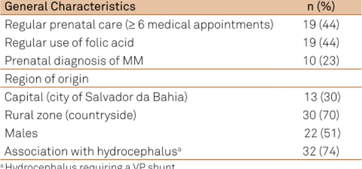
considered premature (< 37 weeks’ gestation). Follow-up was irregular in 4 babies; therefore, they were excluded from the inal analysis. Forty-three newborns were consecutively in -cluded in the study. Nineteen mothers (44%) received regular prenatal care (≥ 6 medical appointments) that involved regu-lar folic acid replacement during pregnancy and 70% came from the coutryside area. Eleven mothers (25%) underwent a transvaginal delivery. Our hospital did not have a maternity unit (obstetric department), and all the children were trans-ferred from other hospitals usually 24 h after birth. Among the newborns included in this study, 51% were boys and 95% had lumbar and lumbosacral lesions. MM was present as a ruptured lesion in 50% of our sample. Intrauterine correc-tions were not performed for the closure of MM in our hos-pital until the end of this research. Table 1 shows the main epidemiological aspects of children with MM that were eval-uated in this study.
he overall incidence of hydrocephalus and the need of
VP shunts were observed in 32 (74%) children. LVA width was measured by performing TUS at 48-72h after the clo-sure of MM. VP shunt were required in 50% (6/12) children
with an LVA width of < 15 mm (group A) and in 84% (26/31) children with an LVA width of ≥ 15 mm (group B; p = 0.02; OR = 5.2). Evaluating the sensitivity and speciicity of LVA
width as a measure to guide the indication of VP-shunt, we found sensitivity of 81%, a positive predictive value of
84%, and eiciency of 75% considering 15 mm cutof value
(Table 2).
Progressive increase in HC and bulging fontanels were the earliest signs of intracranial hypertension (ICH)
(ob-served in 94% children; 30/32); these signs were predominant
in children with LVA width of ≥ 15 mm. In the remaining two
children (6%; 2/32), VP shunts were indicated based on oth -er signs of ICH such as respiratory distress, hypotonia, and
drowsiness; these were consistent with the increases in the ventricular system identiied using TUS before hospital dis
-charge. he average duration for the development of hydro -cephalus was 35 days (range, 0-180 days) after the closure of MM. Figure shows the protocol for VP shunt in children with MM after considering TUS observations and clinical criteria.
hree children simultaneously underwent closure of MM and
an HC above the percentile 90% corrected for age (HC ≥ 2
standard deviations for age) and TUS showing an LVA width
of > 20 mm (considered as a severe hydrocephalus).
hree (7%) children who underwent surgical closure
of MM developed some local problems, as leakage or in-fection. After VP shunting, complications were observed
in 15% children. he complications were considered to be
mechanical in 7.5% and infectious in the remaining 7.5% children. Two children who developed mechanical
compli-cations associated with VP shunts, which were identiied during the follow-up period, underwent ETV. here was a
death in our sample, resulting from complications due to Chiari II malformation.
DISCUSSION
A previous study identiied the irregularity and deicien -cy of prenatal monitoring in pregnant women, assisted by a poor public health system, in the state of Bahia (Brazil)17
and indicated that these may be associated with the high in-cidence of spinal dysraphism5,22,23. We recommend, as
sug-gested by other authors, that at least 400 µg of a folic acid
supplement should be consumed daily; patients with an in -termediate to high risk of neural tube defects (such as pa-tients with a previous well-known history of neural tube de-fects) should consume 4-5 mg folic acid5,22,23. Investigation of
proposed type of delivery revealed high rates (25%) of
vagi-nal delivery. he study suggested that women undergoing
transvaginal delivery may receive irregular prenatal care that would consequently lead to the failure of diagnosing MM in the fetus, which in turn may lead to misguided management based on the type of delivery.Vaginal delivery may increase the risk of rupture and infection of MM during the passage
of the fetus from the birth canal; however, indication of ce
-sarean delivery in these pregnant women and its beneits in
newborn are still controversial12,24,25.
In our study, all the children were transferred from other hospitals in the city of Salvador da Bahia or from a
country-side, which is justiied by the fact that our hospital is a referral
center for treating children with MM in this region. Because of the lack of an obstetric department in our hospital, these
children usually arrive at our hospital 24 h after birth. his
delays the immediate closure of the lesions, which should in fact be corrected immediately12, and hinders the training of a
multidisciplinary team involved in intrauterine closure and correction of MM1. he same situation is observed in other
Brazilian states where many maternity hospitals do not have a unit for pediatric neurosurgery, thus making it necessary to transfer these children to other hospitals22,26.
he relationship of gender distribution of children with MM can be veriied in other studies as well as the prepon -derance of lumbar and lumbosacral region involvement9,12,26.
Probably, the fact that all children with MM who were eval-uated in this study have awaited transfer for treatment, be-sides the relatively high number of vaginal delivery, justify the large number of ruptured MM (50%). MM, particularly when ruptured, must be considered as a neurosurgical emergency because of the high risk of CNS infections12, which is a large
public health concern in our state. All children with MM un-dergo surgical closure of the MM as soon as they are trans-ferred to our hospital to prevent the risk of infection and the risk associated with keeping the placode exposed11,12,22,27.
Data indicate that the incidence of hydrocephalus and the requirement of VP shunts in these children vary from
52% to 90%; however, some of these data do not provide a clear deinition of the optimal timing and criteria for CSF
diversion in children with MM7,9,11,28. We veriied that 74%
children with MM who underwent an institutionalized pro-tocol based on the clinical and TUS criteria required VP shunts, which is the same as the percentage of children who underwent CT scan-based protocols11. he mean du
-ration of 35 days between the closure of MM and develop-ment of hydrocephalus observed in our study was similar to that observed in other studies9,11, which emphasizes the
im-portance of regular follow-up, especially in the irst 60 days
after the closure of MM. However, it should be noted that in some children, hydrocephalus may develop later (within 180 days after the closure of MM).
VP shunt should be considered after a careful clinical and radiological evaluation. According to the results reported here, LVA width of 15 mm, as per the TUS, could be consid-ered as a threshold to classify children with a lower or higher Table 1. Epidemiological aspects of 43 children with
myelomeningocele (MM) who were treated in a referral pediatric hospital in the state of Bahia, Brazil (2009-2013).
General Characteristics n (%)
Regular prenatal care (≥ 6 medical appointments) 19 (44)
Regular use of folic acid 19 (44)
Prenatal diagnosis of MM 10 (23)
Region of origin
Capital (city of Salvador da Bahia) 13 (30)
Rural zone (countryside) 30 (70)
Males 22 (51)
Association with hydrocephalusa 32 (74) a Hydrocephalus requiring a VP shunt.
Table 2. Comparison among the 43 newborns undergoing evaluation by transcranial ultrasonography (TUS) and lateral ventricular atrium (LVA) width, measured slightly above the level of the thalami at the level of the choroid plexus (15 mm cutoff), and the need of VP-shunt.
TUS VP shunt
Yes No
LVA ≥ 15 mm 26 5
LVA < 15 mm 6 6
risk of developing hydrocephalus and requiring VP shunts. Measurement of LVA width by using TUS was performed 48-72 h after the closure of MM in our study. It was observed that VP shunt were required in 84% children with LVA width
≥ 15 mm, which was statistically diferent (p = 0.02) from that
required by children with lesser LVA width, with a sensitivity of 81% and a positive predictive value of 84%. Because of the high incidence of hydrocephalus in these children, it is essen-tial to closely monitor them for signs of ICH after the closure of MM9,11,12,22,26. Besides the LVA width measured slightly above
the level of the thalami at the level of the choroid plexus, other criteria for hydrocephalus such as bifrontal diameter, bicau-date diameter, diameter of the body of the lateral ventricle, ventricular index, frontooccipital horn ratio, and thalamooc-cipital distance had already been studied in patients with MM and hydrocephalus11,29. Was not the aim of our study compari
-son between these methods and measures.
In children who still have opened fontanels and sutures, increased HC and bulging fontanels are the main clinical manifestations11,15,17,19 that require imaging methods to
con-irm the diagnosis and to implement an appropriate treat -ment11,12,16,17,30. In children with greater LVA width (≥ 15 mm),
signs such as progressive increase in HC or bulging fontanels occur more frequently, which lead to the indication of VP shunts 18. We emphasize that 6% of the children who required
a VP shunt showed progressive increase in the ventricular
system (identiied using TUS) without an increase in HC or
bulging fontanels, however showed signs suggestive of neu-rological impairment due to Chiari II malformation. Other
studies also highlight children with signs of ICH but without bulging fontanels11,15.
he best surgical time for VP shunt in these children
remains unknown (i.e., at the same time of the closure of MM or not). Results of other studies verify that closure of MM and VP shunt sometimes should not be performed in the same surgical time. It is emphasized that concomitant surgeries (closure of MM and VP shunt) should be avoided when MM is corrected 24 h after birth and that there is a possibility of infection at the site of MM and higher risk of complications associated with the shunt9,12. Complications
rates were similar to those reported by other authors, con-sidering CSF diversion7,15,17.
We recommend that these children should be moni-tored regularly by a multidisciplinary team in a systematic manner in the long-term mainly due to orthopedic and uri-nary malformations. Protocols should be followed correctly, according to the possibilities of each region (city, state, or country), to avoid overindication of VP shunt in these chil-dren and to avoid systematic realization of CT scan and un-necessary irradiation in these neonates. It should be not-ed that the results presentnot-ed here are from a single referral
center treating children with MM and may not relect the
reality in other regions. No single criteria can be considered completely safe, and TUS parameters used in the present study seeks to integrate a possible measure to be used, and under any circumstances overrides other previously pub-lished ventricular and ultrasound measurements. We plan to extend the protocol used in this study along with the Figure. Protocol for monitoring children with myelomeningocele (MM), considering the risk of developing hydrocephalus, from transcranial ultrasonography (TUS) and clinical criteria (head circumference [HC] and bulging fontanels [BF]).
Surgical closure of MM
Evaluation of HC and fontanels TUS 48-72h
VP-shunt
VP-shunt
CT scan or MRI in case of signs and symptoms associated with Chiari II malformation or between 6 and 12 months of life. Multidisciplinary follow-up (pediatrician, pediatric neurosurgery, pediatric neurology,
orthopedics, pediatric urology, physiotherapy and pediatric nutrology).
Group A (n = 12): 50% of incidence of hydrocephalus needs a VP-shunt Group B (n = 31): 84% of incidence of hydrocephalus needs a VP-shunt *Group A: TUS showing lateral ventricular atrium < 15mm
With no progressive increase of HC, nor BF nor signs of intracranial hypertension
Maintain outpatient follow-up biweekly, during the first 2 months, with measurement of HC and TUS
In case of progressive increase of HC, BF or other signs of intracranial hypertension
inclusion of transcranial Doppler to igure out the resistive
values of the cerebral vascular vessel and in postoperative follow-up study of these children to better identify the func-tioning of the VP shunts.
In conclusion, deinition of criteria that identify children
with MM who need a VP shunt is crucial to eliminate un-necessary CSF diversion. By following our institutionalized protocol, we observed that 74% children who had MM re-quired a VP shunt, which was closely the same observed with
other protocols based on CT scan. In our sample VP shunts
are usually indicated in children with an LVA width of ≥ 15
mm, increased HC (2 standard deviations for age), or bulging fontanels. We all know that no method or measure used in isolated form can be considered completely safe. We believe that the measures presented here considering the LVA width can be added to ventricular measurements and parameters previously described in the literature, assisting in the moni-toring of children with MM.
References
1. Adzick NS, Thom EA, Spong CY, Brock JW 3rd, Burrows PK, Johnson MP et al. A randomized trial of prenatal versus postnatal repair of myelomeningocele. N Engl J Med. 2011;364(11):993-1004. doi:10.1056/NEJMoa1014379
2. Bebbington MW, Danzer E, Johnson MP, Adzick NS. Open fetal surgery for myelomeningocele. Prenat Diagn. 2011;31(7):689-94. doi:10.1002/pd.2805
3. Dias MS, Partington M. Embryology of myelomeningocele and anencephaly. Neurosurg Focus. 2004;16(2):1-16. doi:10.3171/foc.2004.16.2.2
4. Talamonti G, D’Aliberti G, Collice M. Myelomeningocele: long-term neurosurgical treatment and follow-up in 202 patients. J Neurosurg. 2007;107(5):368-86. doi:10.3171/PED-07/11/368
5. MRC Vitamin Study Research Group. Prevention of neural tube defects: results of the Medical Research Council Vitamin Study. Lancet. 1991;338(8760):131-7. doi:10.1016/0140-6736(91)90133-A
6. Ryan-Harshman M, Aldoori W. Folic acid and prevention of neural tube defects. Can Fam Physician. 2008;54(1):36-8.
7. Bizzi JWJ, Machado A. [Meningomyelocele: basic concepts and recent advances]. J Bras Neurocirurg. 2012;23(2):138-51. Portuguese.
8. International Clearinghouse for Birth Defects Monitoring Systems, International Centrefor Birth Defects. World atlas of birth defects. 2nd ed. Geneve: World Health Organization; 2003.
9. Chakraborty A, Crimmins D, Hayward R, Thompson D. Toward reducing shunt placement rates in patients with myelomeningocele. J Neurosurg Pediatr. 2008;1(5):361-5. doi:10.3171/PED/2008/1/5/361
10. Drake JM. The surgical management of Pediatric hydrocephalus. Neurosurgery. 2008;62 Suppl 2:633-40. doi:10.1227/01.neu.0000316268.05338.5b
11. Phillips BC, Gelsomino M, Pownall AL, Ocal E, Spencer HJ, O’Brien MS et al. Predictors of the need for cerebrospinal luid diversion in patients with myelomeningocele. J Neurosurg Pediatr. 2014;14(2):167-72. doi:10.3171/2014.4.PEDS13470
12. Pinto FCG, Matushita H, Furlan ALB, Alho EJ, Goldenberg DC, Bunduki V et al. Surgical treatment of myelomeningocele carried out at ‘time zero’ immediately after birth. Pediatr Neurosurg. 2009;45(2):114-8. doi:10.1159/000209285
13. Associação Brasileira de Empresa de Pesquisa. Applying Criterion Brazil. São Paulo: Associação Brasileira de Empresa de Pesquisa; data [cited 2013 Jan 8]. Available from: http://www.abep.org/criterio-brasil
14. Righini A, Parazzini C, Doneda C, Arrigoni F, Rustico M, Re TJ et al. Fetal MRI features related to the Chiari malformations. Neurol Sci. 2011;32(3 suppl):279-81. doi:10.1007/s10072-011-0694-1
15. Juca CEB, Lins Neto A, Oliveira RS, Machado HR. [Treatment of hydrocephalus by ventriculoperitoneal shunt: analysis of 150 consecutive cases in the Hospital of the Faculty of Medicine of Ribeirão Preto]. Acta Cirurgica Brasileira. 2002;17 suppl 3:59-63. Portuguese. doi:10.1590/S0102-86502002000900013
16. Mandiwanza T, Saidlear C, Caird J, Crimmins D. The open fontanelle: a window to less radiation. Childs Nerv Syst. 2013;29(7):1177-81. doi:10.1007/s00381-013-2073-0
17. Melo JRT, Melo EN, Vasconcellos A, Pacheco P. Congenital hydrocephalus in the northeast of Brazil: epidemiological aspects, prenatal diagnosis, and treatment. Childs Nerv Syst. 2013;29(10):1989-93. doi:10.1007/s00381-013-2111-y
18. Lee CS, Hong SH, Wang KC, Kin SK, Park JS, Jun JK et al. Fetal ventriculomegaly: prognosis in cases in which prenatal neurosurgical consultation was sought. J Neurosurg Pediatr. 2006;105(4):265-70.
19. Zahl SM, Wester K. Routine measurement of head circumference as a tool for detecting intracranial expansion in infants: what is the gain? a nationwide survey. Pediatrics. 2008;121(3):e416-20. doi:10.1542/peds.2007-1598
20. Renier D, Sainte-Rose C, Pierre-Kahn A, Hirsch JF. Prenatal hydrocephalus: outcome and prognosis. Childs Nerv Syst. 1988;4(4):213-22. doi:10.1007/BF00270917
21. Ros B, Romero L, Ilbañez G, Iglesias S, Rius F, Pérez S et al. Success criteria in pediatric neuroendoscopic procedures. Proposal for classiication of results after 67 operations. Childs Nerv Syst. 2012;28(5):691-7. doi:10.1007/s00381-012-1689-9
22. Machado H, Oliveira RS. Simultaneous repair of myelomeningocele and shunt insertion. Childs Nerv Syst. 2004;20(2):107-9.
doi:10.1007/s00381-003-0853-7
23. Melo JRT, Pacheco P, Wanderley LE. Unusual spinal dysraphic lesions. Cases Rep Pediatr. 2013(2013):ID210301. doi:10.1155/2013/210301
24. Hamrick SEG. Cesarean delivery and its impact on the anomalous infant. Clin Perinatol. 2008;35(2):395-406. doi:10.1016/j.clp.2008.03.005
25. Lewis D, Tolosa JE, Kaufmann M, Goodman M, Farrell C, Berghella V. Elective cesarean delivery and long-term motor function or ambulation status in infants with meningomyelocele. Obstet Gynecol. 2004;103(3):469-73. doi:10.1097/01.AOG.0000113624.94710.ce
26. Salomao JF, Pinheiro JA, Carvalho JGS, Leibinger RD, Lucchesi G, Bomim V. [Myelomeningocele: Surgical treatment and results]. J Pediatr (Rio J.). 1995;71(6):317-21. doi:10.2223/JPED.799
27. Forgacs P, Geyer CA, Freidberg SR. Characterization of chemical meningitis after neurological surgery. Clin Infect Dis. 2001;32(2):179-85. doi:10.1086/318471
28. Hunt GM, Oakeshott P, Kerry S. Link between the CSF shunt and achievement in adults with spina biida. J Neurol Neurosurg Psychiatry. 1999;67(5):591-5. doi:10.1136/jnnp.67.5.591
29. Wakhlu A, Ansari NA. The prediction of postoperative hydrocephalus in patients with spina bíida. Childs Nerv Syst. 2004;20(2):104-6. doi:10.1007/s00381-003-0849-31