Instituto do Coração do Hospital das Clínicas - FMUSP
Mailing address: Fernando Bacal – InCor – Av. Dr. Enéas C. Aguiar 44 – 05403-000 – São Paulo, SP – Brazil
Objective - To determine the predictive values of nonin-vasive tests for the detection of allograft vascular disease.
Methods - We studied 39 patients with mean ages of 48±13 years and a follow-up period of 86±13 months. The diagnosis of allograft vascular disease was made by cine-coronary arteriography, and it was considered as positive if lesions existed that caused >50% obstruction of the lu-men. Patients underwent 24h Holter monitoring, thallium scintigraphy, a treadmill stress test, and dobutamine stress echocardiography. Sensitivity, specificity, and positive and negative predictive values were determined in per-centages for each method, as compared with the cine-co-ronary arteriography results.
Results - Allograft vascular disease was found in 15 (38%) patients. The Holter test showed 15.4% sensitivity, 95.5% specificity. For the treadmill stress test, sensitivity was 10%, specificity was 100%. When thallium scintigra-phy was used, sensitivity was 40%, specificity 95.8%. On echocardiography with dobutamine, we found a 63.6% sensitivity, 91.3% specificity. When the dobutamine echo-cardiogram was associated with scintigraphy, sensitivity was 71.4%, specificity was 87%.
Conclusion - In this group of patients, the combination of two noninvasive methods (dobutamine echocardiogra-phy and thallium scintigraechocardiogra-phy) may be a good alternative for the detection of allograft vascular disease in asymp-tomatic patients with normal ventricular function.
Key words: heart transplantation, allograft vascular di-sease, diagnostic methods
Arq Bras Cardiol, volume 76 (nº 1), 36-42, 2001
Fernando Bacal, N oedir Antonio Groppo Stolf, Viviane Cordeiro Veiga, W illiam A. Chalela, Cesar Grupi, Ana Clara Rodrigues, Eulógio E. Martinez, Alfredo Inácio Fiorelli, Luiz Felipe Pinho Moreira,
Edimar Alcides Bocchi, Giovanni Bellotti, José Antonio Franchini Ramires
São Paulo, SP - Brazil
Noninvasive Diagnosis of Allograft Vascular Disease after
Heart Transplantation
Allograft vascular disease is the main late complica-tion in the follow-up of patients who undergo heart trans-plantation.
Ever since the experimental period of heart transplanta-tion, Lower et al 1 described coronary atherosclerosis in a dog that underwent heart transplantation and proved that this complication could affect the late post-transplantation follow-up, yet the first description of coronary disease affecting human grafts was made by Thomson et al 2.
Allograft vascular disease is at this time responsible for most of the deaths that occur during the late posttrans-plantation follow-up, and its incidence amounts to an esti-mated 10% per postoperative year, reaching 40% to 50% in receptors by completion of their 5th postoperative year 3-7.
The first manifestation of the disease is often conges-tive heart failure or even sudden death, arising from an asymptomatic acute infarction of the myocardium, since hearts are denervated and have therefore no afferent sensitive fibers. In cases of coronary failure, the typical pre-cordial pain may not be reported, and only a few reports with a documented partial reinnervation have been publi-shed 8-10.
Another important aspect concerns the diagnosis of vascular disease of the graft. The method still commonly used is an annual angio-coronary angiography after the first postoperative year. Gao et al 11 proposed a classification based on the characteristics, location, and extension of the coronary lesions, defining them by their characteristics as diffuse, multiarterial, and predominantly distally affecting lesions. On angiography, coronary lesions are classically defined as type A, B, and C: type A - discrete proximal, me-dial, or distal lesions in major arteries or their branches; type B - diffuse, concentric lesions involving the medial and dis-tal thirds of major arteries or their branches; type C - asso-ciation between types A and B.
were underestimated by arteriographic analysis. Intravascu-lar ultrasound can also document endothelial alterations occurring early after transplantation, when the media and intima of the blood vessels and the endothelial response to the use of certain drugs with vasoactive properties are analyzed separately 12-14. Despite the potential benefit of this method for the detection of vascular disease of the graft, with high sensitivity and specificity, its applicability in Brazil is still limited, due to the small number of medical units that have it and its high cost. Noninvasive methods for the diagnosis of vascular disease of the graft have been studied in an attempt to replace the annual angiographic analysis, but the results are still controversial. Some groups have reported good diagnostic sensitivity with tests such as myocardial scintigraphy and stress echocardiography, but the great majority go on using annual arteriography due to the low sensitivity and specificity of those methods in their series of patients 15-18.
The objective of this study was to compare the nonin-vasive diagnosis of vascular disease in asymptomatic pat-ients with normal ventricular function, thus characterizing a population with a low suspicion of this disease.
Methods
We studied 39 patients who underwent orthotopic heart transplantation at the Instituto do Coração do Hospi-tal das Clínicas da Faculdade de Medicina da USP, with a mean age of 48±13 years, and a mean follow-up period of 86±31 months.
The inclusion criteria were: over two years of follow-up, normal ventricular function on echocardiogram at rest, absence of symptoms of angina or heart failure, absence of acute rejection, and compliance with the study protocol.
The following tests were performed: cine-coronary ar-teriography, 24-hour Holter monitor, treadmill stress test, myocardial thallium scintigraphy under physical stress (er-gometric test) or pharmacological stress (dipyridamole) in patients with exercise limitations or patients with permanent pacemakers, stress echocardiography with dobutamine. All the noninvasive tests were performed within the same week of the study.
The technique adopted for coronary angiography was that described by Sones and Shirey 19. Philips equipment model Optimus 1050 with a 6.5-to-9-inch image intensifier coupled with an Arritecno camera with a shooting rate of 30 pictures per second was used. The films were separately analyzed by two specialists of the InCor Hemodynamics service, who had no previous knowledge of the patients’ clinical and laboratory data. Each artery was analyzed, in an attempt to establish the degree of lumen reduction of the vessel on at least two incidences: oblique anterior right and oblique anterior left. This method was considered the gold standard for the definition of the presence or absence of vascular disease of the graft. The test was considered posi-tive for vascular disease of the graft whenever the lumen obstruction was ≥50% 20,21, after which the designations
disease “Yes” or “No”criteria were adopted. Lesions <50% were considered wall irregularities and disregarded. Based on the results of this method, sensitivity, specificity, and the positive and negative predictive values of the noninvasive diagnostic methods were calculated.
With regard to 24-hour electrocardiography (Holter), patients were monitored by means of a Marquette 8000 two-channel portable monitor with modulated amplitude wave recording from Marquette (Milwaukee, WI, USA), conta-ining a cassette tape sufficient for a continuous 24-hour study. The monitor was connected to the patient the mo-ment he or she entered the study, and patients were instruc-ted to report the occurrence of any possible symptoms du-ring these 24 hours, in order to have them recorded in an at-tached diary.
Once the monitor was removed, the tapes were analy-zed by a Marquette Laser SXP processor (1988) from Mar-quette (Milwaukee, WI, USA) and evaluated by the staff of the Holter service. Heart beats were classified and evaluated manually. For the diagnosis of ischemic events, an isoelectric point – the PQ interval – and a point 60 m distant from the J point were manually adjusted upon the automatic reading, in determine the baseline of the ST segment, starting from which the variations upwards, up-unlevelling, and down-wards, down-unlevelling, were considered.
To be diagnosed as an ischemic event, an elevation >2mm or a horizontal or descending depression >1mm of the ST segment from the baseline had to occur, lasting at least one minute, and returning to the baseline for at least one minute. The possible ischemic events identified in this man-ner were defined as ischemic episodes (positive test), and each one of them was recorded on paper, at a 25mm/s speed, with a 1mV standard, using a Marquette laser printer, from which the variations of the ST segment were manually reevaluated.
The exercise stress tests were performed according to criteria established by the Consenso Nacional de
Ergome-tria (National Stress Test Consensus) 22.
Exercise was interrupted when the patient attained the heart rate considered the maximum, obtained by the formula: maximum HR= 210-(agex0.65), corresponding to 100% of the patient’s heart rate during stress. Whenever this parameter was not reached, the test interruption criteria were the ones established by the American Heart Association 23.
A MAT 2100 treadmill with a movable ramp was used, coupled with an ML 8000 Stress System from Fukuda Den-shi Co. Ltd. The protocol used was Ellestad´s with a re-cording of 12 simultaneous classical derivations.
Blood pressure measurements were automatically ma-de every 60 s by a Colin ma-device, moma-del STBP 780, coupled with the ML 8000 software.
The radioactive drug used in myocardial perfusion scintigraphy was thallium-201 chloride (201TI). The perfu-sion study protocol encompassed two phases: 1) images were taken immediately after radionuclide injection on the peak of exercise. The injected dose was 111MBq, and tomo-graphy images were obtained with the patient in dorsal de-cubitus; 2) Four hours later and in the same way, redistribu-tion images were obtained.
To obtain the tomography images, a Siemens scintilla-tion camera, model Orbiter ZLC-Digitrac 750 was used, cou-pled with a Maxdelta computer (Microvax-3300). After the reconstruction of the images, transversal sections were ob-tained at every 6.09mm, reoriented to the heart axis, thus al-lowing the determination of the sections corresponding to the three planes mutually perpendicular to the system of coordinates set for the heart.
For the interpretation of the images, the heart was divi-ded into five regions: septal, anterior, inferior, apical, and la-teral. A value was attributed to each one of them by means of a random grade system, according to the uptake of the radioactive drug (0= normal uptake; 1= discrete underup-take; 3= pronounced underuptake). Uptake (perfusion) defects were considered if observed on at least two diffe-rent axes and two sequential sections. A decrease of at least one point of this grading in the analyzed region, when the images of physical stress are compared with the other pha-ses, was interpreted as a transient underuptake. It was con-sidered as partial if the value zero was not observed on the redistribution the reinjection, the rest images, or all of these. The maintenance or increase of this gradation was called persistent underuptake (fibrosis). Once transient underup-take was documented, the test was defined as positive for the detection of vascular disease of the graft.
The protocol for dobutamine stress echocardiogra-phy consisted of continuous dobutamine administration through an infusion pump, starting with a 10mcg/kg/min dose for 3 minutes, progressively increasing to 20, 30, and 40mcg/kg/min, until the patient attained the submaximum heart rate for his or her age (85% of the maximum heart rate). If this did not occur, atropin was given in 0.25mg boli up to a total dose of 1 mg. The reason for anticipating the end of the test was the occurrence of side effects, such as an increase in systolic blood pressure to more than 220mmHg, complex arrhythmia or important symptoms of intolerance to medication (headache, angina). Neither hypotension (a drop of systolic blood pressure >20mmHg with respect to the base level) nor the occurrence of segmental contractility alterations were considered as reasons to discontinue the protocol. Bidimensional echocardiograms were performed by means of an ATL echocardiograph model 3000 or 5000 and a 3.5 or 2.5 MHz transductor. Images were obtained at rest, at the end of stage 10 and 40 of dobutamine administra-tion, and upon recovery. The whole test was recorded on videocassette, and the data were stored on a computer for processing and ulterior analysis. Four basic images were obtained: parasternal long axis and short axis, apical two cameras and four cameras, and they were analyzed by two
observers with no knowledge about the cine-coronary arteriography and, whenever there was a disagreement, an evaluation by a third observer was requested. The result was considered as positive if any alteration of in regional wall motion appeared. The wall motion index was given by the following values: 1) normal: an increase in wall motion and systolic thickening after dobutamine; 2) hypokinesia: a decrease of systolic thickening and wall motion during dobutamine; 3) akinesia: the absence of thickening during dobutamine, and 4) dyskinesia: an outward movement of the analyzed segment after dobutamine. The 12-derivation electrocardiogram was made at rest and after the maximum dobutamine dose, and blood pressure was constantly monitored, being recorded at the end of each dobutamine stage 24.
Statistical analysis – In order to comparatively assess the noninvasive tests with the gold standard, catheteri-zation, for the diagnosis of coronary disease, sensitivity and specificity indexes, as well as the positive and negative predictive values, were calculated. To evaluate whether the disagreement proportion between each test and catheteri-zation was negligible, i. e., close enough to zero, a test for generalized linear models, based on Wald´s statistics 25, was used.
The level of significance established for the analysis was 5%, and all calculations were made according to the SAS (Statistical Analysis System) 26.
Results
In 15 (38.4%) of the 39 studied patients, cine-coronary arteriography showed a vascular disease of the graft, cha-racterized by a >50% obstructive coronary lesion, in at least one vessel. Fifteen patients were studied during the period from the 2nd to the 5th postoperative year, 17 from the 6th to the 9th year and seven after more than 10 years from the operation. As for the characteristics of the coronary lesions found, five were triarterial, seven were biarterial and three were uniarterial. From the viewpoint of Gao et al’s 11 arteriographic classification, 11 patients presented with type C lesions, two patients with type A, and two patients with type B.
Thirty-five patients had the 24-hour electrocardiogram (Holter) carried out. Excluded from the analysis were those who had a block of the D branch and those who wore a pa-cemaker, due to the difficulties in analyzing the alterations of ventricular repolarization. Ischemia was the result found in three patients, two of whom had vascular disease of the graft. Of the 32 patients with normal results, 21 did not have the disease and 11 had it. The sensitivity of the test was 15.4%, specificity was 95.5%, positive predictive value was 66.7%, and negative predictive value was 65.6% (Table I).
the 30 patients with a normal result, 21 did not have the disease and nine had it. The sensitivity of this test was 10%, its specificity was 100%, its positive predictive value was 100%, and its negative predictive value was 70% (Table II). All patients underwent scintigraphy with thallium-201 (Thallium). Seven patients had a positive test result, and six of them had a vascular disease of the graft. Of the 32 patients with normal results, nine had a vascular disease of the graft, as documented by cine-coronary arteriography. The sensitivity of the test was 40%, its specificity was 95.8%, its positive predictive value was 85.7%, and its negative predictive value was 71.9% (Table III).
Thirty-four patients underwent stress electrocardio-graphy with dobutamine (Echo-Dobu). Those who were unable to have the test done due to technical difficulties were excluded, mainly because of the lack of an electrocar-diographic window favorable for analysis. Nine patients had a positive test result, and seven of them had the disease. Of the 25 patients who tested normal, only four had a vascular disease of the graft. The sensitivity of this test was 63.6%, its specificity was 91.3%, its positive predictive value was 77.8%, and its negative predictive value was 84% (Table IV). A joint analysis was performed of the two diagnostic methods with the best sensitivity for the detection of vascular disease of the graft (dobutamine
echocardiogra-phy and thallium scintigraechocardiogra-phy). The result was considered as positive whenever at least one of them showed ischemia. The results found are shown in Table V and figure 1.
Discussion
Coronary angiography is still the gold standard for the diagnosis of presence or absence of coronary artery di-sease in the general population. In patients who undergo heart transplantation, the role of this test in the diagnosis of vascular disease of the graft also seems well established, and therefore an annual angiography is recommended after the first postoperative year.
The incidence of allograft vascular disease is estima-ted at 10% per year, amounting to a total of 40% to 50% by the end of the 5th year after operation 27,28. Cases, however, do exist where evolution is rapid, with diffuse and severe lesions even before the first year after transplantation has elapsed, and the causes of this occurrence are still obscure, although histological aspects similar to arteritis have been shown in such cases. In our analysis, we found a 38% inci-dence of vascular disease of the graft documented by an-giography in asymptomatic patients with normal ventricular function, thus characterizing a population with a low suspi-cion rate for this disease. The distribution pattern of the Table I - Results of 24h Holter in the detection of vascular disease of
the graft
Disease Sensitivity 15.4% Test Result No Yes Total Especificidade 95.5%
Holter No 21 11 32 % Concordance 65.7% Yes 1 2 3 % Discordance 34.3% Total 22 13 35 Predictive value + 66.7% Predictive value - 65.6%
Table II - Results of treadmill stress test in the detection of vascular disease of the graft
Disease Sensitivity 10.0% Test Result No Yes Total Specificity 100.0%
Exercise ECG No 21 9 30 % Concordance 71.0% Yes 0 1 1 % Discordance 29.0% Total 21 10 31 Predictive value+ 100.0%
Predictive value - 70.0%
Table III - Results of thallium scintigraphy in the detection of vascular disease of the graft
Disease Sensitivity 40.0% Test Result No Yes Total Specificity 95.8%
Thallium No 23 9 32 % Concordance 74.4% Yes 1 6 7 % Discordance 25.6% Total 24 15 39 Predictive value+ 85.7% Predictive value - 71.9%
Table IV - Results of stress echocardiography in the detection of vascular disease of the graft
Disease Sensitivity 63,6% Test Result No Yes Total Specificity 91,3%
Stress Echo No 21 4 25 % Concordance 82,4% Yes 2 7 9 % Discordance 17,6% Total 23 11 34 Predictive value+ 77,8% Predictive value - 84,0%
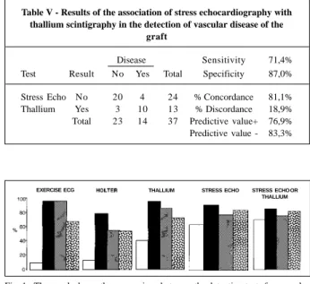
Table V - Results of the association of stress echocardiography with thallium scintigraphy in the detection of vascular disease of the
graft
Disease Sensitivity 71,4% Test Result No Yes Total Specificity 87,0%
Stress Echo No 20 4 24 % Concordance 81,1% Thallium Yes 3 10 13 % Discordance 18,9% Total 23 14 37 Predictive value+ 76,9% Predictive value - 83,3%
Fig. 1 - The graph shows the comparison between the detection tests for vascular disease of the graft. Sensitivity, specificity, positive predictive value, negative predictive value.
obstructive lesions of the coronaries was similar to that fo-und by other investigators, i. e., multiarterial, diffuse le-sions, frequently affecting the distal bed. On the other hand, Uretsky et al. 29 showed that angiographic analysis might underestimate the existing lesions; in their study, they documented a 28% occurrence of cardiac events in patients with a normal angiography. Some aspects have been pro-posed for the standardization of this method, mainly the need to use the same angle in the analysis of each study, the taking of measurements at the same point in each vessel and under the same vasomotor tonus and, preferably, the use of the computer-assisted method for a better definition of the degree of obstruction of the vessel. Analyses should be made by at least two observers and possibly by a third one, if any disagreement occurs between the first ones. Cur-rently, with the additional information given by the in-travascular ultrasound, lesions can be better defined, es-pecially when they are discrete. Early studies with this me-thod, made as soon as within the first weeks after trans-plantation, allowed the identification of endothelial dys-functions, probably due to immunological alterations, which may predispose in the future to the occurrence of vascular disease of the graft 29-31. As already mentioned, the characteristic coronary lesions are diffuse, affecting main and secondary branches, with a frequent distal involve-ment, making therapeutic propositions such as angioplasty and surgical myocardial revascularization very difficult. In practice, doctors are faced with a big dilemma, i. e., to know that the patient has a severe coronary affliction and to be, so far, unable to intervene effectively both in prevention and in treatment. In cases where the diffuse coronary affliction is accompanied by ventricular dysfunction, the therapeutic indication is a new transplant, which has given poorer re-sults than the first transplant in a number of series in the literature 6,7. This fact, associated with low organ availabili-ty, leads us to ponder about this indication, even from an ethical point of view, because the chance of the first trans-plant can be given to patients who have been on the waiting list longer. The use of noninvasive diagnostic methods will become more important if, in the future, their effectiveness in detecting patients at greater risk of developing cardiac events further on will be proven, thus allowing detection of those with severer conditions, who will need an invasive diagnostic study.
The use of stress electrocardiography for the detec-tion of ischemia in convendetec-tional coronary patients is well established; yet, its usefulness in patients who undergo cardiac transplantation is still controversial. Reports of other authors have shown low sensitivity and specificity of this method, which corresponds to the findings of this study 32,33. In this series, the sensitivity of the method was only 10%, which makes it difficult to use in this specific group of patients on a daily medical practice basis. Some factors seem to have an influence on these results. First of all, given the fact that the transplanted heart is denerved, the
lack of innervation has an influence on the physiological response of the subject to exercise, so mainly a low chrono-tropic response is observed, associated with the presence of high ventricular filling pressures and increased levels of circulating catecholamines, attempting to compensate for that chronotropic deficiency. The alterations described here, together with blood hypertension that often accompa-nies the patients, may lead to diastolic dysfunction, impai-ring the tolerance of transplanted individuals to stress. In practice, patients show difficulties with their heart rate res-ponse, and this represents a limitation of the method for the detection of ischemia, because the triggering of ischemic events upon physical stress depends directly on the increase of the heart rate.
The use of a 24-hour electrocardiogram (Holter) as a diagnostic method for ischemic events seems to be well es-tablished, especially for the detection of the so-called silent ischemia, defined as the occurrence of ischemic events with no medical expression that might identify them. In studies with patients with coronary disease, most of the ischemic events identified by Holter are said to be asymptomatic 34,35. Patients who undergo cardiac transplantation fit the model of silent ischemia perfectly, because their denerved hearts have no afferent sensitive fibers, and the first manifestation of coronary failure is frequently sudden death or even a picture of heart failure. Yet, in practice, Holter use in patients who undergo heart transplantation has shown a low diagnostic sensitivity. In this series, we found a15% sensi-tivity, with most patients being able to have the test done, except for those who had permanent pacemakers or a block of the right branch who had to be excluded because of the difficulties in analyzing the ventricular repolarization. Some important aspects are worth considering in the attempt to explain our results. Asymptomatic ischemic events occur mostly due to alterations of the myocardial oxygen con-sumption, therefore, being preceded by elevations of the heart rate36. Because the denerved patient has no physiolo-gic chronotropic response and has a low R-R variability, the triggering of ischemic events, documented by Holter through an underunlevelling of the ST region, after the ele-vation of the heart rate, becomes highly evident, giving the method a low diagnostic sensitivity, as shown in several published series and in agreement with our results 24, 37,38.
studying the chronotropic response of these patients to exercise. Pharmacological stress was indicated for those patients who had exercise limitations or had a permanent pacemaker. In our study, the sensitivity of this test was 40% and its specificity was 96%, values that are lower than those found by other groups who used pharmacological dipyri-damole stress and found a 60 to 70% diagnostic sensitivity, according to the series of patients 19,20,39.40. The fact that the patients had a major decrease in the chronotropic response due to the posttransplant denervation, which can be shown by the inability to reach maximum heart rate, could explain in part the low sensitivity of the method. Another important aspect to consider is the fact that the pattern of the vascular disease of the graft is frequently diffuse and affects the distal part of the vessels, leading to the so-called balanced ischemia, which may be difficult to detect by imaging diag-nostic methods that compare the differences in radioiso-tope uptake by the different regions of the heart. The diag-nostic method becomes more sensitive as the maximum stress for the triggering of ischemia is attained, and it is pos-sible that, in this specific population, the use of myocardial dipyridamole scintigraphy could show better diagnostic sensitivity values.
The use of dobutamine stress echocardiography for the diagnosis of coronary disease has increased over the last few years and is based on the concept that high and progressive doses of dobutamine have an influence on the increase in heart labor, due to the positive inotropic and chronotropic effects. So the ischemic events are triggered as the heart rate increases. The sensitivity of this method for conventional coronary disease varied from 80% to 95%, ac-cording to the series, being more sensitive for the detection of ischemias related to the anterior descending and the right coronary arteries and less sensitive in the region of the circumflex artery 41,42. As for the detection of vascular disease of the graft after cardiac transplantation, the results are still controversial. Spes et al. 43 obtained a 79% sensiti-vity and an 83% specificity with this method in the detection of vascular disease of the graft, and Derumeaux et al. 44 found an 86% sensitivity and a 95% specificity in their series. In our study, the diagnostic sensitivity was 63% and the specificity was 91%, with all patients who had the test done reaching maximum heart rate, thus characterizing an effective test. These patients, however, had normal ventri-cle function and no segmental hypocontractility on echo-cardiography at rest during the inclusion period of the stu-dy and, when this subgroup of patients was analyzed in other studies, a lower sensitivity was observed as well,
simi-lar to the one we found. Another aspect that may influence the analysis of the diagnostic methods based on qualitative rather than quantitative criteria is the observer’s experience, which makes it desirable that the analysis be performed by two observers and, on request, by a third one, if any diag-nostic disagreement occurs between the first two ob-servers. Lately, studies are under way in coronary patients to assess the impact of the findings of dobutamine stress echocardiography on the prevention of cardiac events su-ch as heart failure, unstable angina, acute myocardial infarc-tion, retransplantainfarc-tion, and death of cardiac origin. Negative test findings were related to a good prognosis in several studies, suggesting that this diagnostic method may help identify patients with a functionally important coronary disease and at risk of suffering some event in the future 45-47. In patients who underwent cardiac transplantation this concept is not yet well established. In this study, when two diagnostic methods (stress echocardiography and myocar-dial scintigraphy) are analyzed together, considering the test as positive whenever at least one of the tests was alte-red, diagnostic sensitivity increases to 71%. With the possi-bility of differentiating patients at higher risk for coronary events, especially in selected groups of patients, the use of these noninvasive diagnostic methods may become an effective option in the attempt to replace periodic angio-graphic evaluation in patients who undergo cardiac trans-plantation, but further studies with long-term follow-up are needed to confirm this proposition. In spite of its lower cost, the association of the two methods still carries some doubts regarding the therapeutic procedures to be pro-posed, yet signs are present indicating that normal tests and normal ventricle function may be predictive of a good prognosis.
Limitations of the study – The small number of pa-tients (15) with angiographic documentation of their di-sease is a limiting factor for the analysis of the results of the noninvasive diagnostic methods, therefore requiring fur-ther studies to obtain more accurate predictive values for the employed diagnostic methods and for the confirmation of our results.
1. Lower RR, Kontos HA, Kosek JC, Sewell DH, Graham WH. Experiences in heart transplantation. Am J Cardiol 1968; 22: 766-71.
2. Thomson JG. Production of severe atheroma in a transplanted heart. Lancet 1969; 2: 1088-92.
3. Fiorelli A, Stolf N, Graziosi P, et al. Incidência de coronariopatia após o trans-plante cardíaco ortotópico. Rev Bras Cir Cardiovasc 1994; 9: 69-80. 4. Gao SZ, Alderman EL, Schroeder JS, et al. Clinical and laboratory correlates of
ac-celerated coronary artery disease in the cardiac transplant patient. Circulation 1987; 76: 56-61.
5. Grattan MT, Moreno Cabral CE, Starnes VA, Oyer PE, Stinson EB, Shumway NE. Eight year results of cyclosporine treated patients with cardiac transplants. J Thorac Cardiovasc Surg 1990; 99: 500-9.
6. Greenberg ML, Uretsky BF, Reddy PS, et al. Long term hemodynamic follow-up of cardiac transplant patients treated with cyclosporine and prednisone. Circu-lation 1985; 71: 487-94.
7. Bocchi E, Vilas-Boas F, Pedrosa AA, et al. Doença coronariana após transplante cardíaco ortotópico. Arq Bras Cardiol 1994; 62: 195-200.
8. Gao SZ, Schroeder JS, Hunt AS, Billingham ME, Valentine HA, Stinson EB. Acute myocardial infarction in cardiac transplant recipients. Am J Cardiol 1989; 64: 1093-7.
9. Starke RP, Mcginn AL, Wilson RF. Chest pain in cardiac transplant recipients: evidence of sensory re-innervation after cardiac transplantation. N Engl J Med 1991; 324: 1791-4.
10. Halpert I, Goldberg AD, Levine AB, Kornberg R, Kelly C, Lesch M. Reinnerva-tion of the transplanted human heart as evidenced from heart rate variability stu-dies. Am J Cardiol 1996; 77: 180-3.
11. Gao SZ, Alderman EL, Schroeder JS, Silverman JF, Hunt SA. Accelerated coro-nary vascular disease in the heart transplant patient: corocoro-nary arteriographic findings. J Am Coll Cardiol 1988; 12: 334-40.
12. Valantine H, Pinto FJ, Goar FG, Alderman EL, Popp RL. Intracoronary ultra-sound imaging in heart transplant recipients: the Stanford experience. J Heart Lung Transpl 1992; 11: 60-4.
13. Rickenbacher PR, Kemna MS, Pinto FJ, et al. Coronary artery intimal thickening in the transplanted heart. An in vivo intracoronary ultrasound study of immuno-logic and metabolic risk factors. Transplantation 1996; 61(suppl 1): 46-53. 14. Kapadia SR, Nissen SE, Tuzcu EM. Impact of intravascular ultrasound in
unders-tanding transplant coronary artery disease. Curr Opin Cardiol 1999; 14: 140-50. 15. Rodney RA, Johnson LL. Myocardial perfusion scintigraphy to acess heart
transplant vasculopathy. J Heart Lung Transpl 1992; 11: 74-8.
16. Verhoeven PPAM, Lee FA, Ramahi TM, et al. Prognostic value of noninvasive testing one year after orthotopic cardiac transplantation. J Am Coll Cardiol 1996; 28: 183-9.
17. Spes CH, Klauss V, Mudra H, et al. Diagnostic and prognostic value of serial do-butamine stress echocardiography for noninvasive assessment of cardiac allo-graft vasculopathy: a comparison with coronary angiography and intravascular ultrasound. Circulation 1999; 100: 509-15.
18. Akosah KO, Mcdaniel S, Hanrahan JS, Mohanty PK. Dobutamine stress echo-cardiography early after heart transplantation predicts development of allograft coronary artery disease and outcome. J Am Coll Cardiol 1998; 31: 1607-14. 19. Sones FM, Shirey EK, Prondfit WL, Westcott RN. Cine-coronary arteriography.
Circulation 1959; 20: 773.
20. Uretsky BF, Murali S, Reddy S, et al. Development of coronary artery disease in cardiac transplant patients receiving immunosupressive therapy with cyclos-porine and prednisone. Circulation 1987; 76: 827-34.
21. Smart FW, Ballantyne CM, Cocanougher B, Farmer JA, Sekela ME, Noon GP, Young JB. Insensitivity of noninvasive tests to detect coronary artery vasculo-pathy after heart transplant. Am J Cardiol 1991; 67: 243-7.
22. Consenso Nacional de Ergometria. Arq Bras Cardiol 1995; 65: 191-211. 23. ACC/AHA Guidelines for exercise testing. A report of the American College of
Cardiology/American Heart Association. Taske Force on Practise Guidelines. J Am Coll Cardiol 1997; 30(suppl 1): 260-315.
24. Bourdillon PDV, Broderick TM, Sawada SG, et al. Regional wall motion index for infarct and non-infarct regions after reperfusion in acute myocardial infartic comparison with global wall motion index. J Am Soc Echocardiogr 1989; 2: 398-407.
25. Agresti A. Categorical Data Analysis. New York: John Wiley & Sons, 1990: 71-97.
26. SAS Institute Inc., SAS/STATâ User’s Guide. Version 6, fourth edition, volume 1, Cary, NC: SAS Institute Inc., 1989.
27. Pennock JL, Oyer PE, Reitz BA, et al. Cardiac transplantation in perspective for the future. J Thorac Cardiovasc Surg 1982; 83: 168-77.
28. Barnhart GR, Pascoe EA, Mills AS. Accelerated coronary atherosclerosis in car-diac transplant recipients. Transplant Ver 1988; 1: 31-46.
29. Fitzgerald PJ, Goar FG, Connolly AJ, et al. Intravascular ultrasound imaging of coronary arteries. Is three layers the norm? Circulation 1992; 86: 154-8. 30. Goar FG, Pinto FJ, Aldermanl EL, Valantine HA, et al. Intracoronary ultrasound
in cardiac transplant recipients. Circulation 1992; 85: 979-87.
31. Rickenbacher PR, Kemna MS, Pinto FJ, et al. Coronary artery intimal thickening in the transplanted heart. Transplantation 1996; 61: 46-53.
32. Martin TW, Gaucher J, Pupa LE, Seaworth JF. Response to upright exercise after cardiac transplantation. Clin Cardiol 1994; 17: 292-300.
33. Kao AC, Van Trigt P, Shaeffer-MCCAL GS, et al. Central and peripheral limita-tions to upright exercise in untrained cardiac transplant recipients. Circulation 1994; 89: 2605-15.
34. Stern S, Tzivoni D. Early detection of silent ischaemia heart disease by 24 hours eletrocardiographic monitoring of active subjects. Br Heart J 1974; 36: 481-5. 35. Epstein SE, Quyyumi AA, Bonow RO. Myocardial ischaemia: silent or
sympto-matic. N Engl J Med 1988; 318: 1038-43.
36. Deanfield JE, Selwyn AP, Chierchia S, et al. Myocardial ischaemia during daily life in patients with stable angina: Its relation to symptoms and heart rate chan-ges. Lancet 1983; 1: 753-8.
37. Halpert I, Goldberg AD, Levine AB, et al. Reinnervation of the transplanted hu-man heart as evidenced from heart rate variability studies. Am J Cardiol 1996; 77: 180-3.
38. Bigger JT, Steinman RC, Rolnitzky LM, Fleiss JL, Albrecht P, Cohen RJ. Power low behavior of RR-interval variability in healthy middle aged persons, patients with recent acute myocardial infarction, and patients with heart transplants. Cir-culation 1996; 93: 2142-51.
39. Richter J, Herreros J, Serena A, Domper M, Ramirez JC, Arias R. Thallium scinti-graphy in human transplants: a way to detect myocardial damage. J Heart Lung Transplant 1991; 10: 33-7.
40. Smart FW, Grinstead WC, Cocanougher B, et al. Detection of transplant arterio-pathy: does exercise thallium scintigraphy improve noninvasive diagnostic capabilities? Transplant Proc 1991; 23: 1189-92.
41. Segar DS, Brown SE, Sawada SG, Ryan T, Feigenbaum H. Dobutamine stress echocardiography: correlation with coronary lesion severity as determined by quantitative angiography. J Am Coll Cardiol 1992; 19: 1197-202. 42. Marcovitz PA, Armstrong WF. Accuracy of dobutamine stress
echocardiogra-phy in detecting coronary artery disease. Am J Cardiol 1992; 69: 1269-73. 43. Spes CH, Mudra H, Schnaack SD, et al. Dobutamine stress echocardiography for
noninvasive diagnosis of cardiac allograft vasculopathy: a comparison with an-giography and intravascular ultrasound. Am J Cardiol 1996; 78: 168-74. 44. Derumeaux G, Redonnet M, Mouton S, et al. Dobutamine stress
echocardiogra-phy in orthotopic heart transplant recipients. J Am Coll Cardiol; 25: 1665-72. 45. Akosah KO, Olsovsky M, Kirchberg D, Salter D, Mohanty PK. Dobutamine
stress echocardiography predicts cardiac events in heart tranplant patients. Cir-culation 1996; 94: 283-8.
46. Lewis JF, Selman SB, Murphy JD, Mills RM, Geisen EA, Conti CR. Dobutamine echocardiography for prediction of ischemic events in heart transplant recipients. J Heart Lung Transplant 1997; 16: 390-3.
47. Spes CH, Mudra H, Schnaack SD, et al. Prognostic value of dobutamine stress echocardiography after heart transplantation. J Am Coll Cardiol 1996; 29: 290.