Leandro Silva Marques(a) Mônica Costa Armond(a) Maria Letícia Ramos-Jorge(b) Raquel Gonçalves Vieira de Andrade(b)
Ana Maria Bolognese(c)
(a) Department of Orthodontics, School of Dentistry, Vale do Rio Verde University (UNINCOR), Três Corações, MG, Brazil.
(b) Department of Pediatric Dentistry, School of Dentistry, Federal University of Vales do Jequitinhonha e Mucuri, Diamantina, MG, Brazil.
(c) Department of Orthodontics, School of Dentistry, Federal University of Rio de Janeiro, Rio de Janeiro, RJ, Brazil.
Corresponding author:
Leandro Silva Marques R. Arraial dos Forros, 215 Diamantina - MG - Brazil CEP: 39100-000
E-mail: [email protected]
Received for publication on Jun 23, 2010 Accepted for publication on Oct 12, 2010
Correlations between dentoskeletal
variables and deep bite in Class II
Division 1 individuals
Abstract: This study aimed to evaluate the cephalometric pattern of Class II Division 1 individuals with deep bite, and to determine possible correlations between dentoskeletal variables and deep bite. Comparisons were also made between genders and cases that were to be treated both with and without premolar extraction. A total of 70 lateral cephalograms were used, from both male (n = 35) and female (n = 35) individuals with an average age of 11.6 years, who simultaneously presented with ANB
≥ 5º and overbite ≥ 4 mm. Statistical analysis involved parametric (t-test) and non-parametric (Mann-Whitney) tests for independent samples, as well as the Spearman correlation test (p ≤ 0.05). The values of Go-Me, Ar-Pog, PM-1 and PM-CMI were higher in males (p < 0.05). However, no signiicant differences were found among the averages of the cepha-lometric measurements when the sample was divided by treatment with and without extraction. Deep bite was positively correlated to the PM-1 and SNA measurements, and negatively correlated to the Go-Me, Ar-Pog, SNB and SNGoMe measurements. The main factors associated with the determination of deep bite in Angle’s Class II Division 1 cases were: greater lower anterior dentoalveolar growth and/or lower incisor extru-sion, horizontal growth pattern, maxillary protrusion and mandibular retrusion.
Descriptors: Orthodontics; Malocclusion; Tooth Extraction; Radiography.
Introduction
The main factors involved with the establishment of Class II Divi-sion 1 maloccluDivi-sion are: maxillary protruDivi-sion with normal mandibular position, mandibular retrusion with normal maxillary position, a combi-nation of maxillary protrusion and mandibular retrusion, and posterior rotation of the mandible.1 In turn, deep bite has been related to a lack
of vertical growth in the molar and premolar regions, and/or the supra-eruption of incisors and canines.2-5
The treatment of Class II Division 1 malocclusion can be accom-plished by several methods.6-10 Treatment considerations include the
pa-tient’s facial proile, skeletal pattern, growth potential, and severity of the malocclusion.6 A deep overbite can be corrected by intrusion of
an-terior teeth or extrusion of posan-terior teeth, or a combination of both.10
bite; however, it might lead to a more severe Class II molar relationship.6
In this context, the Class II Division 1 malocclu-sion with deep overbite may be linked to a combina-tion of a large number of factors. Determining the individual inluence of such factors provides a valu-able resource for orthodontic diagnosis, and can make the difference between the success or failure of treatment.
Therefore, the present study proposes to evalu-ate the pre-treatment cephalometric pattern of Class II Division 1 individuals with deep bite, with par-ticular attention to the identiication of factors cor-related to the determination of deep bite.
Material and Methods
The teleradiographs used in the present study were gathered from the orthodontic documentation collection of the Department of Orthodontics at the Federal University of Rio de Janeiro. The sample comprised 70 patients, 35 male and 35 female. Indi-viduals had an average age of 11.6 years (minimum of 9.8 years and maximum of 14.9 years). The inclu-sion criteria were as follows:
• Simultaneous presence of Class II Division 1 mal-occlusion with ANB ≥ 5o and deep bite ≥ 4 mm.
• Absence of any other type of prior treatment, as this could inluence the vertical development of the alveolar process or the dimensions of the mid-face structures.
• All were Caucasian Brazilians, to avoid ethnic differences in the craniofacial morphology.
• All presented teleradiographs of satisfactory quality.
The sample was also divided according to the type of treatment to be carried out – with or with-out the extraction of four premolars.
Lateral teleradiographs were traced and mea-sured by hand, by a single investigator (LSM), using a 17.5 x 17.5 cm sheet of acetate (Ultraphan, Berlin, Germany), 0.5 mm Pentel mechanical pencil, pro-tractor, square, eraser, and millimetric ruler (Faber Castell, São Carlos, Brazil). All radiographs were obtained using the same mobile X-Ray system (10 Orthoceph - Siemens, set to 62 KV and 16 mA, with exposure time of 1.3 seconds).
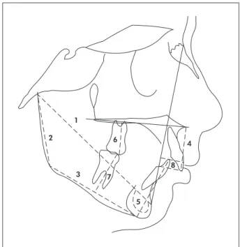
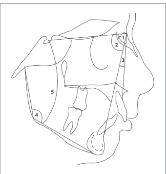
Thirteen cephalometric variables (8 linear and 5 angular) were catalogued as follows (Figures 1 and 2):
1. Overbite (mm): distance between the edges of the upper and lower incisors, perpendicular to the Downs facial plane.
2. Height of the mandibular ridge (Ar-Go): lin-ear distance between the Articular and Gonion points.
3. Length of the mandibular body (Go-Me): lin-ear distance between the Gonion and Menton points.
4. Total mandibular length (Ar-Pog): linear dis-tance between the Articular and Pogonion points.
5. PP-1: Perpendicular distance in millimeters from the edge of the upper permanent incisor to the palatal plane.
6. PP-CMS: Perpendicular distance in millimeters from the mesiobuccal cusp of the irst upper per-manent molar to the palatal plane.
7. PM-1: Perpendicular distance in millimeters from the incisal edge of the lower incisor to the Steiner mandibular plane.
Figure 1 - Cephalometric landmarks, reference lines, and linear measurements used in the study. 1. Ar-Pog. 2. Ar-Go. 3. Go-Me. 4. PP-1. 5. PM-1. 6. PP-CMS. 7. PM-CMI. 8. Overbite.
1
2
3
4
5 6
7
8. PM-CMI: Perpendicular distance in millimeters from the mesiobuccal cusp of the irst lower per-manent molar to the Steiner mandibular plane.
9. Degree of protrusion or retrusion of the maxilla in relation to the base of the cranium - SNA: an-gle formed by the Sella-Nasion and Nasion lines - Point A.
10. Degree of protrusion or retrusion of the mandi-ble in relation to the base of the cranium - SNB: angle formed by the Sella-Nasion and Nasion lines - Point B.
11. Anterior-posterior relation between the maxilla and mandible - ANB: angle formed by the Sella-Nasion and Sella-Nasion lines - Point B.
12. Gonial angle (ArGo-Me): angle formed by the Ar-Go and Go-Me lines.
13. Mandibular plane in relation to the base of the cranium (SNGoMe): angle formed by the man-dibular plane (Go-Me) and the Sella-Nasion line. Statistical analysis was done using the Software Package for Social Sciences (SPSS for Windows, version 12.0, SPSS Inc., Chicago, IL, USA). The arithmetic average and standard deviation were cal-culated for each variable. The data obtained were submitted to a variance homogeneity test (Levene’s
test) and a normality test (Kolmogorov-Smirnov). Next, the variables that would be analyzed, using the parametric (T test) and non-parametric (Mann-Whitney) tests, were determined. The correlation of initial deep bite with the other variables was estab-lished using the non-parametric Spearman correla-tion test.
Three weeks after the initial records were taken, 20 teleradiographs were randomly selected, retraced and new measures were taken. Next, the T test for paired samples was applied. Differences between the irst and second records of the 20 teleradiographs proved to be insigniicant.
Results
Initially, we sought to determine if there were differences between genders regarding the cepha-lometric characteristics of Class II Division 1 mal-occlusion with deep bite. As Table 1 displays, sig-niicant differences appeared between the Go-Me (p = 0.007), Ar-Pog (p = 0.009), PM-1 (p = 0.008) and PM-CMI (p = 0.047) measures. Table 1 also provides the average and standard deviation, as well as the minimum and maximum, of each cephalo-metric measurement proposed in the present study.
Table 2 displays the descriptive analysis and comparison of the cephalometric measurements ac-cording to treatment with or without the extraction of four premolars. This analysis conirms that there were no signiicant differences between the groups.
Table 3 displays the correlations between deep bite and the other cephalometric variables. A posi-tive correlation was observed between deep bite and the PM-1 (p = 0.027) and SNA (p = 0.023) variables. Deep bite presented a negative correlation with the Go-Me (p < 0.001), Ar-Pog (p < 0.001), SNB (p = 0.005) and SNGoMe (p = 0.034) variables.
Discussion
Overbite
The dificulty of correcting deep bite has been recognized for decades. Over the years, opinions have differed regarding the etiology of this altera-tion and, consequently, regarding how it should be treated.11 This controversy can be explained by the
lack of standardization among studies, thereby
con-Figure 2 - Cephalometric landmarks, reference lines, and angular measurements used in the study. 1. SNA. 2. SNB. 3. ANB. 4. ArGo-Me. 5. SNGoMe.
1 2
3
Table 2 - Descriptive analysis and comparison of pre-treatment cephalometric measurements according to treatment with and without extraction of four premolars.
Descriptive statistics
p-value
Mean SD Minimum Maximum
E NE E NE E NE E NE
Line
ar
(mm
)
Overbite 5.67 5.88 1.06 1.24 4.00 4.00 8.00 9.00 0.463T
Ar-Go 39.63 41.20 3.79 4.52 32.00 33.00 47.00 51.00 0.130T
Go-Me 65.73 66.60 4.41 4.59 58.00 60.00 79.00 77.00 0.429T
Ar-Pog 100.70 101.83 5.44 5.77 91.00 93.00 113.00 117.00 0.411T
PP-1 28.87 29.05 3.22 2.69 21.00 22.00 35.00 34.00 0.797T
PP-CMS 21.87 21.38 2.19 1.94 17.00 17.00 29.00 25.00 0.325T
PM-1 41.97 40.98 2.48 3.60 37.00 33.00 46.00 50.00 0.200T
PM-CMI 29.30 29.68 2.69 3.65 23.00 24.00 37.00 45.00 0.637T
A
ngu
lar
(de
gre
e
s) SNA 82.90 83.33 4.02 4.19 74.00 72.00 91.00 95.00 0.671T
SNB 75.70 76.43 3.87 4.01 67.00 70.00 82.00 87.00 0.451T
ANB 7.20 6.90 1.99 2.35 4.5 4.5 12.00 12.00 0.575T
ARGo-Me 131.53 130.32 6.03 4.96 120.00 121.00 144.00 143.00 0.361T
SNGoMe 37.17 35.38 5.72 5.71 26.00 23.00 50.00 47.00 0.199T
Note: 30 patients treated with extraction and 40 without extraction. T p-value obtained through the T test for independent samples. E - Extraction. NE - Non
extraction.
Table 1 - Descriptive analysis and comparison of pre-treatment cephalometric measurements according to gender.
Descriptive statistics
p-value
Mean SD Minimum Maximum
M F M F M F M F
Line
ar
(m
m)
Overbite 6.06 5.51 0.99 1.27 4.0 4.0 8.0 9.0 0.051T
Ar-Go 40.34 40.71 4.70 3.85 32.0 33.0 51.0 49.0 0.719T
Go-Me 67.66 64.80 4.27 4.32 60.0 58.0 79.0 75.0 0.007T
Ar-Pog 103.09 99.60 5.66 5.08 94.0 91.0 117.0 111.0 0.009T
PP-1 29.46 28.49 2.68 3.09 22.0 21.0 35.0 34.0 0.165T
PP-CMS 21.74 21.43 1.57 2.45 18.0 17.0 25.0 29.0 0.595U
PM-1 42.40 40.40 3.34 2.73 33.0 34.0 50.0 45.0 0.008T
PM-CMI 30.29 28.74 3.54 2.78 23.0 24.0 45.0 37.0 0.047T
A
ngu
lar
(de
gre
e
s) SNA 83.77 82.51 4.38 3.74 74.0 72.0 95.0 91.0 0.202T
SNB 76.46 75.77 4.18 3.72 67.0 69.0 87.0 82.0 0.471T
ANB 7.31 6.7 2.05 2.32 4.50 4.50 12.0 12.0 0.279T
ArGo-Me 131.00 130.69 5.47 5.47 120.0 121.0 143.0 144.0 0.811T
SNGoMe 36.40 35.89 6.35 5.15 23.0 27.0 50.0 45.0 0.711T
Note: 35 male patients and 35 female patients. T p-value obtained through the T test for independent samples. U p-value obtained through the Mann-Whitney
fusing the interpretation of results and their clinical applications. Thus, the present study offers a sin-gular advantage over others found in the literature due to the size and homogeneous nature of the se-lected sample. Only Class II Division 1 cases with deep bite were included. Furthermore, the sample is larger than that of any previous study found in the literature, enabling it to be divided into subgroups (gender, and treatment with or without extraction).
The manifestation of deep bite presented no sig-niicant difference between genders. This result con-fers homogeneity to the sample, and is in agreement with results found by Simons and Joondeph.12
Furthermore, no signiicant differences were ob-served among the averages of deep bite regarding the cases that were to be treated, either with or without the extraction of four premolars. This result demon-strates that the magnitude of pre-treatment overbite exercised no inluence over the decision for treat-ment, either with or without extraction. A number of authors12-15 also corroborate the principle that
pre-treatment deep bite per se does not necessarily counter-indicate an extraction approach. However, all authors state that such a principle is valid only as long as treatment is performed with planning and adequate mechanics.
When the 70 cases were evaluated together, it was observed that deep bite was positively correlat-ed to the PM-1 and SNA measurements, and nega-tively correlated to the Go-Me, Ar-Pog, SNB, and SNGoMe measurements. The interpretation of these results suggests that deep bite in Class II Division 1 individuals is principally related to greater den-toalveolar growth in the lower incisor region and/ or the extrusion of these teeth.1,11 Furthermore, it is
also related to the pattern and amount of maxillary-mandibular growth; overbite is more pronounced when there is more horizontal growth and less man-dibular growth.1 In summary, the results from the
present study support the conclusion that the more severe the Class II Division 1 malocclusion is, the greater the chances are of developing deep bite. These indings corroborate indings by Bjork16 that
the development of deep bite depends on the relation between the upper and lower incisors: if the lower incisor has adequate contact with the lingual surface of the permanent upper incisor, there is less chance of developing deep bite. Other authors also consider the supra-eruption of lower incisors to be a deter-minant factor for deep bite.17-20 Lewis,21 Steadman22
and Popovich23 attribute the occurrence of deep bite
to a lack of vertical growth in the molar and premo-lar regions.
Class II
An evaluation of the results from the pre-treat-ment phase of the present study suggests there is a common dentoskeletal pattern among males and fe-males regarding Class II Division 1 characterization and deep bite (Table 1). Although there were statisti-cally signiicant differences in the total length of the mandible (Ar-Pog) and the length of the mandibular body (Go-Me), these were relative differences: there were in fact no signiicant differences among the principal determinant Class II factors (SNA, SNB and ANB). Another relevant aspect is that there were also no signiicant differences among the av-erages of the mandibular plane angle (SNGoMe), suggesting that the spatial coniguration of the man-dible is similar between male and female subjects in the determination of a Class II Division 1 mal-occlusion with deep bite. These results differ from
Table 3 - Coefficient of Spearman correlation between deep bite and other cephalometric measurements, for all 70 cases of the sample.
Cephalometric Measurements Spearman Correlation Coefficient p-value
Line
ar
(m
m)
Ar-Go 0.214 0.076
Go-Me −0.458 < 0.001
Ar-Pog −0.496 < 0.001
PP-1 0.020 0.867
PP-CMS 0.099 0.415
PM-1 0.264 0.027
PM-CMI 0.035 0.776
A
ngu
lar
(de
gre
e
s) SNA 0.272 0.023
SNB −0.335 0.005 ANB −0.149 0.220
ARGo-Me −0.021 0.864
those found by Lima-Filho et al.,24 who identiied
signiicant differences between the genders regard-ing the SNA and ANB angle; however, the indregard-ings are in agreement with these same authors regard-ing the angle of the mandibular plane (SNGoMe). It is worth noting that the sample Lima-Filho et al.24
used comprised 40 Class II Division 1 patients, but these patients did not present with deep bite. In the literature researched, there were no studies found that allow comparisons with ours.
Another relevant observation, made from the re-sults of the present study in regard to Class II Di-vision 1, was the absence of signiicant differences between the group to be treated without extraction and the group to be treated with extraction (Table 2). These results clearly indicate that the parameters used in the deinition of the diagnosis and conse-quent treatment plan were associated mainly with dental aspects, most likely the amount of crowd-ing, as the dentoskeletal characteristics were similar for both groups. These indings were not expected, and once again reveal an exclusive pattern regard-ing the characteristics and approach to the Class II Division 1 malocclusion with deep bite. In this context, it is important to compare the results from this study to those found by Basciftci and Usumez,25
who observed that the Class II Division 1 group was different with regard to various parameters before treatment. The pre-treatment values showed a more divergent growth pattern and more proclined inci-sors among the group treated with extraction, and a smaller mandibular body with greater overjet and overbite among the group treated without extrac-tion. The differences between the results of these two studies can be explained by the fact that the Basciftci and Usumez25 study used just Class II
Divi-sion 1 cases without deep bite (initial overbite in the group treated with extraction had an average of just
1.44 mm). Furthermore, the initial records of the patients treated with extraction showed an average age of 17.5 years.
In this study, eight patients were in the mixed dentition stage and the second molars had not yet erupted. Since the average overbite was 6.06 (± 0.99) in males and 5:51 (± 1.27) in females (Table 1), the authors do not believe that including these patients would inluence the results of this study. Additional-ly, longitudinal evaluation developed by Bergensen26
shows that overbite decreases with the eruption of second molars and subsequent eruption of third mo-lars by an average of 0.58 (± 1.06 mm), decreasing the amount of the gap from 4:34 (± 1.18 mm) to 3.76 mm (± 1.39 mm).
A critical analysis of the literature reveals that most studies do not address pre-treatment dento-skeletal characteristics in a speciic manner. They state that that this is not the main focus of the study, and omit data that would certainly contribute sig-niicantly to a more precise diagnosis of the factors involved in the determination of a Class II Division 1 malocclusion with deep bite.
Conclusions
• The Class II Division 1 malocclusion with deep bite developed in a different manner among males and females.
• The severity of Class II Division 1 and deep bite had no inluence on the treatment option, with or without the extraction of premolars, among the sample studied.
• The main factors associated with the determi-nation of deep bite were: greater lower anterior dentoalveolar growth and/or lower incisor extru-sion, horizontal growth pattern, maxillary pro-trusion and mandibular repro-trusion.
References
1. Proffit WR, Fields HW, Sarver DM. Contemporary Ortho-dontics. 4th ed. St Louis: Mosby Elsevier; 2007. 751 p.
2. Hering K, Ruf S, Pancherz H. Orthodontic treatment of open-bite and deepopen-bite high-angle malocclusions. Angle Orthod. 1999 Oct;69(5):470-7.
4. Hammond AB 3rd. Treatment of a Class II malocclusion
with deep overbite. Am J Orthod Dentofacial Orthop. 2002 May;121(5):531-7.
5. Van Steenbergen E, Burstone CJ, Prahl-Andersen B, Aartman IH. The role of a high pull headgear in counteracting side ef-fects from intrusion of the maxillary anterior segment. Angle Orthod. 2004 Aug;74(4):480-6.
6. Marques LS, Ramos-Jorge ML, Araujo MT, Bolognese AM. Class II Division 1 malocclusion with severe overbite: cepha-lometric evaluation of the effects of orthodontic treatment. World J Orthod. 2008 Winter;9(4):319-28.
7. Siara-Olds NJ, Pangrazio-Kulbersh V, Berger J, Bayirli B. Long-term dentoskeletal changes with the Bionator, Herbst, Twin Block, and MARA functional appliances. Angle Orthod. 2010 Jan;80(1):18-29.
8. Janson G, Camardella LT, Araki JD, de Freitas MR, Pinzan A. Treatment stability in patients with Class II malocclusion treat-ed with 2 maxillary premolar extractions or without extrac-tions. Am J Orthod Dentofacial Orthop. 2010 Jul;138(1):16-22.
9. Garbui IU, Nouer PR, Nouer DF, Magnani MB, Pereira Neto JS. Cephalometric assessment of vertical control in the treat-ment of class II malocclusion with a combined maxillary splint. Braz Oral Res. 2010 Jan-Mar;24(1):34-9.
10. Woods MG. Sagittal mandibular changes with overbite cor-rection in subjects with different mandibular growth direc-tions: late mixed-dentition treatment effects. Am J Orthod Dentofacial Orthop. 2008 Mar;133(3):388-94.
11. Hans MG, Kishiyama C, Parker SH, Wolf GR, No-achtar R. Cephalometric evaluation of two treat-m e nt s t r at e g i e s fo r d e e p ove rbit e c o r r e c t io n . Angle Orthod. 1994 Aug;64(4):265-74.
12. Simons ME, Joondeph DR. Change in overbite: a ten-year postretention study. Am J Orthod. 1973 Oct;64(4):349-67. 13. Magill JM. Changes in the anterior overbite relationship
fol-lowing orthodontic treatment in extraction cases. Am J Or-thod. 1960 Oct;46(10):755-88.
14. El-Mangoury NH. Orthodontic relapse in subjects with vary-ing degrees of anteroposterior and vertical dysplasia. Am J Orthod. 1979 May;75(5):548-61.
15. Burzin J, Nanda R. The stability of deep overbite correction. In: Nanda R, Burstone CJ, editors. Retention and stability in orthodontics. 1th ed. Philadelphia: Saunders; 1993. p. 61-80.
16. Bjork A. Prediction of mandibular growth rotation. Am J Orthod. 1969 Jun;55(6):585-99.
17. Strang RHW. An analyses of the overbite-problem malocclu-sion. Angle Orthod. 1934 Jan;4(1):65-84.
18. Kim YH. Overbite depth indicator with particular reference to anterior open-bite. Am J Orthod. 1974 Jun;65(6):586-611. 19. Fleming HB. An investigation of the vertical overbite dur-ing the eruption of the permanent dentition. Angle Orthod. 1961 Jan;31(1):53-62.
20. Samuelson G, Garner LD, Potter R. Tooth movements as-sociated with deep overbite correction in class II division 1 malocclusions. Int J Orthod. 1989 Fall-Winter;27(3-4):3-8. 21. Lewis P. Correction of deep anterior overbite. A report of three
cases. Am J Orthod Dentofacial Orthop. 1987 Apr;91(4):342-5.
22. Steadman SR. Overbites and overjets. Angle Orthod. 1974 Apr;44(2):156-61.
23. Popovich F. Cephalometric evaluation of vertical overbite in young adults. J Can Dent Assoc. 1955 Apr;21(4):209-22. 24. Lima-Filho RM, Lima AL, Oliveira Ruellas AC. Longitudinal
study of anteroposterior and vertical maxillary changes in skeletal class II patients treated with Kloehn cervical headgear. Angle Orthod. 2003 Apr;73(2):187-93.
25. Basciftci FA, Usumez S. Effects of extraction and nonextrac-tion treatment on class I and class II subjects. Angle Orthod. 2003 Feb;73(1):36-42.