From the Department of Cardiopneumology of the Heart Institute, Hospital das Clínicas, Faculty of Medicine, University of São Paulo.
Received for publication on October 07, 2002.
DURA MATER MITRAL AND TRICUSPID
BIOPROSTHESES: 30 YEARS OF FOLLOW-UP
Luiz Boro Puig, Carlos Manuel de Almeida Brandão, Lauro Kawabe, Geraldo Verginelli, José Antonio Francchini Ramires and Sérgio Almeida de Oliveira
PUIG LB et al. - Dura mater mitral and tricuspid bioprostheses: 30 years of follow-up. Rev. Hosp. Clín. Fac. Med. S. Paulo 58(3):163-168, 2003.
PURPOSE: The dura mater bioprosthesis was developed in the Department of Cardiopneumology of the Hospital das Clínicas of the University of São Paulo Medical School in 1971. Here, we present the clinical results of the dura mater bioprosthesis over 30 years of follow-up.
METHODS: We studied 70 consecutive patients who underwent mitral or tricuspid valve replacement with a dura mater bioprosthesis between January 1971 and August 1972.
RESULTS: The early mortality was 10% (7 patients). The up was 87% complete (9 patients were lost to follow-up). Two patients were alive and asymptomatic 30 years after valve replacement; 33 patients underwent reoperations due to valve dysfunction, and 19 died during the follow-up period. At 30 years, the actuarial survival was 49.2 ± 8.6%; freedom from rupture, 27.0 ± 10.2%; freedom from calcification, 78.8 ± 8.6%; and freedom from reoperation, 18.8 ± 7.5%.
CONCLUSIONS: The dura mater bioprosthesis played an important role in the treatment of patients with mitral and tricuspid valve disease. The low rate of thromboembolism and the long period of follow-up without evidence of valve dysfunction, which occurred for several of our patients, are important characteristics of these bioprosthesis.
DESCRIPTORS: Dura mater. Bioprosthesis. Glycerol. Valve disease. Valve surgery.
The dura mater bioprosthesis was an original contribution developed in 1971 in the Department of Cardio-pneumology of the Hospital das Clínicas, University of São Paulo Medical School1,2. In the 1960s, the
Starr-Edwards ball-valve prosthesis was the only valve substitute avail-able. This prosthesis was associated with a high incidence of thromboem-bolic events, mainly involving mitral valve replacement, with a prevalence of cerebrovascular accidents over 4 years of evolution of 27.4%3. The
ho-mologous dura mater bioprosthesis was developed for the purpose of pre-venting this serious complication. The initial and midterm results with this bioprosthesis were satisfactory. After 6 years of follow-up, some patients
pre-sented with fatigue injuries, mani-fested as tears in the dura mater cusps. Despite this complication, the low rate of thromboembolism indicated the use of this prosthesis instead of the Starr-Edwards prosthesis.
In the early 1980s, the use of the dura mater bioprosthesis was discon-tinued. The heterologous porcine and pericardial bioprosthesis, prepared with glutaraldehyde, became the first choice for use in heart valve replace-ment due to the higher availability,
su-perior quality control, and lower rates of associated structural valve deterio-ration observed during short-term fol-low-up.
In this paper, we present the clini-cal evolution of the first series of 70 patients with dura mater bioprosthesis in the mitral and tricuspid positions, 30 years after the operation.
PATIENTS AND METHODS
Table 2 - Reoperation: causes, time of event, and follow-up period of events.
Cause No. of cases Time of event Follow- up period of event
(years) 0-10 y 11–20 y 21-30 y
Rupture 23 (70%) 3 – 28 9 (40%) 7 (30%) 7 (30%)
(mean= 15)
Calcification 5 (15%) 8 – 17 1 (20%) 4 (80%)
(mean= 12.4)
Technical failure 3 (9%) 1 – 3 3 (100%)
(mean= 2)
Paravalvar leak 1 (3%) Immediate 1 (100%)
Hemolysis 1 (3%) 8 1 (100%)
TOTAL 33 (100%)
Table 1 - Causes of Late Mortality.
Cause of mortality Patients %
Heart failure 7 36.8
Cerebrovascular accident 2 10.5
Arrhythmia 2 10.5
Bronchopneumonia 2 10.5
Endocarditis 2 10.5
Acute abdominal disease 1 5.3 Mesenteric thrombosis 1 5.3
Unknown 2 10.5
TOTAL 1 9 100
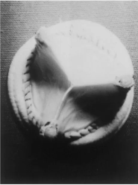
Figure 2 - Actuarial survival. Figure 1- Model of implanted dura mater
bioprosthesis.
Forty-two patients (60%) were fe-male and 28 (40%) were fe-male. The age of the patients ranged from 17 to 54 years. The valve lesions identified preoperatively were as follows: mitral insufficiency in 26 patients (37.1%), double mitral lesions in 20 (28.6%), mitral stenosis in 3 (4.3%), tricuspid insufficiency in 1 (1.4%), thromboem-bolism due to a Starr-Edwards mitral prostheses in 18 (25.7%), infective en-docarditis in 1 (1.4%), and Ebstein’s disease in 1 (1.4%).
Clinical information was obtained from the patients through clinical ex-amination, letter, or telephone contact, or from clinical cardiologists’ reports. The follow-up was completed on 87% of patients (9 patients were lost to fol-low-up) completed over a closing pe-riod of 6 months, with a cumulative follow-up period of 555 years and an average follow-up interval of 10.3 years.
Definitions of complications, as well as data analysis and reporting of results, are in accordance with recom-mended guidelines4.
RESULTS
Hospital mortality was 10% (7 pa-tients), which was attributable to heart failure in 5 patients (71.4%), a stroke in 1 patient (14.3%), and bronchop-neumonia in 1 patient (14.3%). Five of the 7 patients presented preoperatively
with neurologic deficits caused by Starr-Edwards valve thrombosis and underwent emergency operations in a state of hemodynamic instability.
Nineteen died during the late fol-low-up period from a variety of causes (Table 1). Late mortality was considered as valve-related in 5 patients; 2 patients had prosthetic valve endocarditis, 2 pa-tients had a stroke, and 1 patient had mesenteric thrombosis. Thirty-three pa-tients underwent reoperations during the follow-up period due to valve dys-function (Table 2).
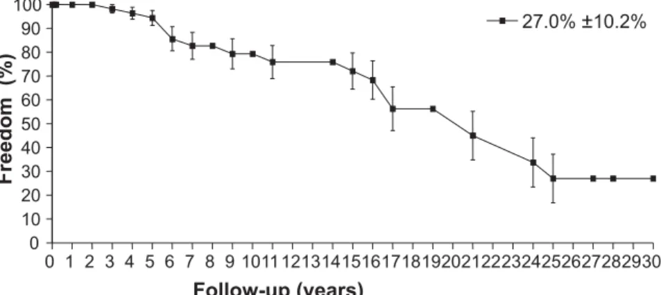
Figure 3 - Actuarial freedom from rupture.
Figure 5 - Actuarial freedom from structural valve dysfunction. Figure 4 - Actuarial freedom from calcification.
78.8% ±8.6% (Fig. 4); freedom from structural valve deterioration, 20.9% ±8.3% (Fig. 5); freedom from reoperation, 18.8% ±7.5% (Fig. 6); and freedom from thromboembolism, 91.4% ±4.3% (Fig. 7).
DISCUSSION
The thromboembolic events asso-ciated with Starr-Edwards prostheses represent one of the most serious com-plications related to mechanical
pros-theses. In mitral valve replacement, the rate of thromboembolism is high, and several patients have experienced per-manent neurologic sequelae. The new mechanical prostheses offer low rates of thromboembolism, although pa-tients are kept under anticoagulant therapy and are subjected to its risks. New materials and structural modifica-tions of mechanical and biological prostheses have been evaluated in sev-eral clinical and experimental studies to identify means of reducing the risk of thromboembolic events.
Following experimental studies on homologous dura mater preserved in glycerol by Pigossi et al.5, this
biologi-cal tissue was used as an implant by several surgical groups in the early 1970s. After experimental study, we observed that homologous dura mater was an appropriate tissue for making bioprosthetic cusps for heart valve re-placement during cardiac surgery. The structure of dura mater, consisting of 2 layers of collagen fibers disposed in many directions, confers great resist-ance and pliability, which are both es-sential to valve function.
The initial clinical results with dura mater bioprostheses were very favorable, and this prosthesis soon be-came our first choice for use in heart valve replacement. The presentation of our results stimulated a great number of surgeons and institutions to use or study this bioprosthesis6-12. Harasaki et
al.13, comparing aortic bovine valves
treated with glutaraldehyde and hu-man dura mater bioprostheses treated with glycerol, showed that dura mater bioprostheses were more durable.
dur-Figure 6 - Actuarial freedom from reoperation.
Figure 7 - Actuarial freedom from thromboembolism.
Figure 8 - Dura mater bioprosthesis recovered 25 years after implantation. Note the preservation of the tissue in a great portion of the valve.
ing the late postoperative period (5 years) and therefore were not related to the sterilization method.
The dura mater bioprosthesis is as-sociated with low thrombogenicity, but structural deterioration constitutes the main reason for reoperation14,15. The
main complication was tissue tears, and these were observed after 6 years of follow-up. A higher incidence of this complication occurred after 10 to 25 years of follow-up in patients who un-derwent mitral valve replacement. Rupture did not occur in patients who underwent tricuspid valve replace-ment, perhaps because of the lower hemodynamic stress associated with right ventricular systole16. The
occur-rence of rupture between 10 and 25
years postoperatively is an observation favorable to the dura mater bioprosthesis and indicative of its du-rability. The actuarial freedom from rupture at 30 years was 27.0% ±10.2%. Dura mater bioprostheses are asso-ciated with low rates of calcification. This is probably because they are com-posed of a homologous tissue, and are dehydrated with 98% glycerol without chemical interaction with the amino radicals that compose the collagen fi-bril protein structures. At 30 years, the actuarial freedom from calcification was 78.8% ±8.6%.
With the Carpentier-Edwards stand-ard porcine bioprosthesis, Jamieson et al. demonstrated that freedom from structural valve deterioration after mi-tral valve replacement was 21.% ±3.9% at 15 years17 and 8.5% ±3.4% at 18
years18. In contrast, freedom from
struc-tural dysfunction was 20.9% ±8.3% at 30 years in our study, although we must consider the limited number of patients compared with the other se-ries. Whereas our data do not demon-strate overall superiority of the dura mater prosthesis, they do suggest that calcification rates are lower and that rupture occurs later with this bioprosthesis than with others.
prepared to implant Starr-Edwards prostheses, which do not require atten-tion to the posiatten-tion of the fixaatten-tion stitches. In the case of bioprostheses, misplaced stitches can interfere with the frame, holding the cusps and caus-ing valvular insufficiency.
Using electron microscopy, Allen et al.19 studied 12 dura mater
bioprostheses removed in reoperations after 23 to 108 months of implanta-tion. These authors observed evidence of cellular remodeling and concluded that the long durability of these bioprostheses may relate to factors
such as the preservation of collagen fibers by glycerol and the viability of fibroblasts and macrophages inside the dura mater tissue. These findings indi-cate that dura mater bioprostheses are structures actively involved in the process of surface degeneration and remodeling.
At our institution, a protocol study for the selective fixation of dura mater with glutaraldehyde is being devel-oped in order to improve resistance to rupture without compromising cusp pliability. Three patients received these new dura mater bioprostheses 5
years ago and they have experienced a normal evolution up until now.
In conclusion, the dura mater bioprosthesis may offer some advan-tages over other prostheses including low thrombogenicity, superior durabil-ity, and good pliability. However, the dura mater bioprosthesis has 2 opera-tional problems that limit its use—the reliability of the sterilization process and the low availability. Only by solv-ing these problems will we be able to resume the use of dura mater bioprostheses.
RESUMO
PUIG LB e col. - Bioprótese de dura mater mitral e tricúspide: 30 anos de acompanhamento. Rev. Hosp.
Clín. Fac. Med. S. Paulo 58(3):
163-168, 2003.
A bioprótese de dura-mater foi de-senvolvida no Hospital das Clínicas da Faculdade de Medicina da Universida-de Universida-de São Paulo em 1971. Este traba-lho apresenta os resultados clínicos com 30 anos de seguimento.
MÉTODOS: Foram estudados 70
pacientes consecutivos com biopró-tese mitral ou tricúspide, operados de janeiro de 1971 a agosto de 1972.
RESULTADOS: A mortalidade
imediata foi 10% (7 pacientes). Dois pacientes evoluem bem com a biopró-tese de dura-máter, 9 não tem seguimen-to atualizado, 33 apresentaram disfun-ção da bioprotese e foram reoperados e 19 faleceram durante a evolução tardia. A curva atuarial de sobrevida foi de 49,2 ± 8,6%, livre de rotura, 27,0 ± 10,2%, livre de calcificação, 78,8 ± 8,6%
e livre de reoperação, 18,8% ± 7,5%.
CONCLUSÕES: A bioprótese de
dura-mater teve mais importante papel no tratamento de pacientes com lesão das valvas mitral e tricúspide. A baixa taxa de tromboembolismo e o longo período de seguimento sem disfunção valvar em vários pacientes são impor-tantes características desta bioprótese.
REFERENCES
1. PUIG LB, VERGINELLI G, BELLOTTI G, KAWABE L et al. -Homologous dura-mater cardiac valve. Preliminary study of 30 cases. J Thorac Cardiovasc Surg 1972;64:154-160. 2. PUIG LB, VERGINELLI G, KAWABE L et al. - Valva cardíaca de
dura-mater homóloga. Rev Hosp Clín Fac Med São Paulo 1974;29:85-89.
3. STOLF NAG, DALLAN LA, PUIG LB et al. - Clinical results of valve replacement by the Starr-Edwards prosthesis. Artif Organs 1980;4:24-26.
4. EDMUNDS LH Jr, CHAIRMAN, CLARK RE et al. - Guidelines for reporting morbidity and mortality after cardiac valvular operations. Ann Thorac Surg 1996; 62:932-935.
5. PIGOSSI N, RAIA A, LEX A et al. - Estudo experimental e clínico sobre o emprego como implante, da dura-mater homogenea conservada em glicerina a temperatura ambiente. Rev Assoc Med Bras 1971; 17:263.
6. HIGHISON GL, ALLEN DJ, DiDIO LJA et al. - Ultrastructural morphology of dura-mater aortic allografts after 44-73 months of implantation. J Submicros Cytol 1980; 12:165-187. 7. ZERBINI EJ, PUIG LB - The dura-mater allograft valve. In:
IONESCU MI- Tissue Heart Valves. London, Butterworth, 1979. v.7, p.253-301.
8. ZERBINI EJ, PUIG LB - Experience with dura-mater allograft. In: SEBENING F- Bioprosthetic Cardiac Valves. Munchen, Deutsches Herzzentrun, 1979. v.3, p.179-90.
9. OYER PE, STINSON EB - Biologic valves. In: GLEN WWL et al. - Thoracic and Cardiovascular Surgery. Connecticut, Appleton-entury Crafts, 1983. v.95, p.1362-1369.
10. ZERBINI EJ, PUIG LB, VERGINELLI G - Dura-mater valve. In: YANGKAI WE, PETERS RM - International Practice in Cardiac Surgery. Beijing, Science Press Book, 1985. v.83, p.966-977.
11. KIRKLIN JW & BARRATT-BOYES BG - Aortic Valve Disease. Cardiac Surgery. 2nd ed. Londo, Churchill, 1983. v.12,
p.491-571.
12. BURAKORVSKY BI, BOKER YA - Cirurgia Cardiovascular. Moscou, Medicina, 1989. p. 424.
13. HARASAKI H, KIRALY RJ, JACOBS GB et al. - Bovine aortic and human dura mater valves: a comparative study in artificial hearts in calves. J Thorac Cardiovasc Surg 1980;79 :125-137.
14. PUIG LB, VERGINELLI G, IRYIA K et al. - Homologous dura-mater cardiac valves. Study of 533 surgical cases. J Thorac Cardiovasc Surg 1975; 69:722-728.
15. PUIG LB - Bioprótese Cardíaca de Dura-Mater. Arq Bras Cardiol 1997; 69(3): 153-154.
16. PUIG LB, BRANDÃO CMA, POMERANTZEFF PMA et al. -Tricuspid dura mater bioprostheses: more than 20-year follow-up of 3 patients. Ann Thorac Surg 2001; 72:615-617. 17. JAMIESON WR, MUNRO AI, MIYAGISHIMA RT et al.
-Carpentier-Edwards standard porcine bioprosthesis: clinical perfomance to seventeen years. Ann Thorac Surg 1995; 60:999-1007.
18. JAMIESON WR, BURR LH, MUNRO AI et al. - Carpentier-Edwards standard porcine bioprosthesis: a 21-year experience. Ann Thorac Surg 1998; 66:S40-3.