169
REV. HOSP. CLÍN. FAC. MED. S. PAULO 58(3):169-172, 2003
CASE REPORT
From the Departments of Pathology and Clinical Medicine, Infectious and Parasitic Disease Service, Antonio Pedro University Hospital, Faculty of Medicine, Fluminense Federal University, Rio de Janeiro/RJ, Brazil.
Received for publication on May 16, 2002.
MENINGEAL CARCINOMATOSIS AS THE INITIAL
MANIFESTATION OF A GALLBLADDER
ADENOCARCINOMA ASSOCIATED WITH A
KRUKENBERG TUMOR
Tizuko Miyagui, Luciana Luchemback, Graça Helena Maia do Canto Teixeira and Kátia Martins Lopes de Azevedo
MIYAGUI T et al. - Meningeal carcinomatosis as the initial manifestation of a gallbladder adenocarcinoma associated with a Krukenberg tumor. Rev. Hosp. Clín. Fac. Med. S. Paulo 58 (3):169-172, 2003.
A case of malignant neoplasm is described in which the initial manifestations were mental dysfunction and meningeal irritation, mimicking chronic or subacute meningitis. Physical examination showed cranial nerve involvement and a pelvic tumor. There was progressive deterioration, and death occurred in 2 weeks. The autopsy revealed a gallbladder adenocarcinoma, meningeal carcinomatosis, and ovarian metastasis presenting as a Krukenberg tumor. The authors emphasize the importance of including meningeal carcinomatosis as a possibility in the differential diagnosis of non-characteristic clinical pictures, as well as the importance of the cerebrospinal fluid cytologic examination, repeated as needed, in order to confirm this diagnosis.
DESCRITORS: Ovarian neoplasms. Gallbladder cancer. Carcinomatosis. Metastasis. Meningitis.
Meningeal carcinomatosis (MC) is an infrequent manifestation of malig-nant neoplasia1,2. Pure MC implies in
the involvement of the leptomeninges with no other metastasis elsewhere in the central nervous system (CNS)2,3.
Meningeal involvement may be focal, multifocal, or diffuse2. The incidence
of MC appears to be increasing, per-haps due to longer survival of patients with malignant neoplasms. Neverthe-less, MC is still considered infrequent, and its variable clinical manifestations make antemortem diagnosis difficult to achieve1,4,5. Headache, signs of
menin-geal irritation, abnormalities of the mental status, cranial nerve deficits, and seizures may indicate the lep-tomeningeal dissemination of a neo-plasm primarily located outside the CNS3-5. Clinical objective signs related
to the CNS can be more prominent than patient complaints, with findings cor-responding to lesions involving the CNS at more than 1 anatomic site5.
The presence of neoplastic cells in the cerebrospinal fluid (CSF) is diagnos-tic. Repeated CSF cytological exami-nations may be necessary4,5. We
de-scribe a case of gallbladder adenocar-cinoma with 2 uncommon features: initial presentation as MC and ovarian dissemination as a Krukenberg tumor.
CASE REPORT
170
REV. HOSP. CLÍN. FAC. MED. S. PAULO 58(3):169-172, 2003 Meningeal carcinomatosis as the initial manifestation of a gallbladder
Miyagui T et al.
film was normal, and a cranial CT scan showed cerebral edema with mild ven-tricular dilatation. Cerebrospinal fluid (CSF) was mildly xanthochromic with 75 cells/mL (60% neutrophils). Glucose and protein levels were 36 mg/dL and 123 mg/dL, respectively. Viral or tuber-culous meningitidis was suspected, and she was started on tuberculostatic drugs. A further CSF specimen with the same biochemical and cytologic char-acteristics gave a positive latex reac-tion to Cryptococcus neoformans.
Am-photericin B was then substituted for tuberculostatic drugs, and mannitol was added. The patient’s condition wors-ened, with torpor, bilateral amaurosis, and right facial nerve palsy. She pro-gressed to coma and died 2 weeks later from cardiorespiratory arrest.
At autopsy both ovaries presented with solid tumors, the right one meas-uring 17 x 11 x 7 cm and weighing 650 g and the left one measuring 19 x 17 x 7 cm and weighing 1,400 g. The cut surface showed compact areas with a fascicular pattern intermingled with cystic areas containing a yellow ge-latinous fluid. This was initially sup-posed to be a malignant ovarian pri-mary neoplasm. There were para-aor-tic and peripancreapara-aor-tic metastapara-aor-tic lymph node enlargements. The gall-bladder was filled with small calculi and had thickened walls with an ir-regular mucosal surface. The brain was edematous and weighed 1,400 g. There was a hernia of the right cerebel-lar tonsil and opacity of the leptome-ninges, mainly on the basal areas.
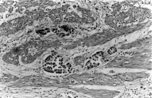
Microscopic examination revealed a moderately differentiated adenocar-cinoma of gallbladder (Fig. 1), with a scanty mucinous component. Normal ovarian architecture was entirely re-placed by neoplastic cells disposed in alveolar and trabecular patterns; there were also mucinous and signet ring cells without any organization amidst a proliferated stroma. These findings
completely supported the diagnosis of Figure 2 - Ovary: Compact neoplastic infiltration and stromal proliferation with areas of mucinsecreting cells in acinar arrangement (Hematoxilyn and eosin x 100). Figure 1 - Gallbladder: Neoplastic glandular structures infiltrating the muscular layer (Hematoxilyn and eosin x 100).
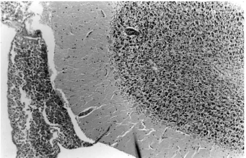
a Krukenberg tumor (Fig. 2). There was diffuse infiltration of the subarachnoid space (Fig. 3) that extended into the cortical parenchyma along the pen-etrating vessels. Other neoplastic changes were noted in lymph nodes, large intestine, and vertebral bone. A few emboli were found in small pul-monary arteries. The neoplastic cells showed characteristics of glandular cells, with positivity to PAS (Periodic Acid Schiff) and Alcean-blue stains.
DISCUSSION
Meningeal carcinomatosis, an un-common complication of disseminated neoplasia, can nevertheless present as its initial manifestation1,6,7. It occurs
more frequently with adenocarcinomas but can also be associated with squamocellular carcinomas and sarco-mas3,5. Meningeal carcinomatosis is
171
REV. HOSP. CLÍN. FAC. MED. S. PAULO 58(3):169-172, 2003 Meningeal carcinomatosis as the initial manifestation of a gallbladder Miyagui T et al.
is only one in which MC was the ini-tial clinical manifestation6. Meningeal
carcinomatosis is more common in lung and breast carcinomas and in melanomas2,3,5,7. The paucity of
neuro-logic manifestations can make diagno-sis difficult, but the diversity of sig-nals and symptoms as well as multifocal involvement, sometimes in-cluding cranial nerves or spinal roots, are both suggestive of MC5,7. Since
CSF laboratory findings are similar, often the diagnostic problem is to dis-tinguish MC from tuberculous or fun-gal meningitis5,8. Lumbar puncture
yields clear or mildly turbid fluid with elevated pressure, low glucose levels, and high protein concentrations1,7.
The presence of neoplastic cells in the CSF confirms the diagnosis. Repeated cytologic examinations may be neces-sary5,7,8. Positive diagnosis can be
en-hanced by tests for immunocyto-chemical reactions to carcinoem-bryonic (CEA) and anti-epithelial membrane (EMA) antigens and to cytokeratins, if they are available1,8,9.
At gross examination, the brain may appear normal, or the leptomeninges can present milky opacity on the ba-sal portions and along the vessels3,5.
Other findings are the presence of small nodules or focal granular changes in the meninges, mild hydro-cephalus, and cerebral edema, as well as focal subarachnoid hemorrhage and laminar infarctions3-5. Microscopically,
there can be nonspecific changes such as meningothelial reaction, fibrosis, and inflammatory infiltration of the meninges. In the subarachnoid space, the neoplastic cells may be disposed in layers, clustered in small nodules, grouped in glandular structures, or dif-fusely spread, tending to concentrate in the basal areas near the brain stem or in non-plane areas such as the hip-pocampus sulci and cauda equina3,5.
Neoplastic perivascular infiltration can extend to the cortical paren-chyma2,4,5.
The pathways by which neoplastic cells reach the meninges are still sub-ject to controversy. Proposed routes are vascular (arterial or trough choroid and Batson venous plexuses), perivas-cular and perineural lymphatics, or spread by contiguity with adjacent bone or from CNS parenchymatous metastasis2,3,6. Each of these routes
could alone explain MC, but this com-plication is more probably due to a
combination of them. Cytokines se-creted by tumor cells are also consid-ered to play an important role in this kind of dissemination. There is evi-dence of synergic activity between TNF-α and cytokines in the develop-ment of MC10. In our clinical case, it
was difficult to establish the route of dissemination. Since the ovaries were involved, it could be that neoplastic cells reached the leptomeninges through perineural lymphatics, due to the proximity of the ovaries to the lumbosacral nerves. Other possibilities are spread by contiguity with vertebral bone metastasis or dissemination by the neoplastic emboli that were de-tected in the pulmonary arteries (the hematogenous route).
Another infrequent complication present in this case of gallbladder ade-nocarcinoma was ovarian metastasis manifested as a Krukenberg tumor. This kind of metastasis is rare in gall-bladder adenocarcinoma, being more common in adenocarcinoma of stom-ach or intestine, due to the higher fre-quency of mucinous or signet ring cells in these neoplasias. These types of neoplastic cells induce the infiltrat-ing pattern of dissemination that is present in Krukenberg tumor and are not prone to develop nodular metastases11.
It is important to emphasize the exceptionality of a case of MC present-ing as the initial manifestation of a gallbladder adenocarcinoma. The iden-tification of a pelvic mass pointed to the possibility of a neoplasia. In spite of this, the clinical work-up was di-rected to the neurologic disease be-cause of its severity. Some factors hin-dered the diagnosis of neoplastic dis-ease, including the presence of lithi-asis; the absence, excepting for the pelvic mass, of findings related to the malignant disease; and the false-posi-tive CSF reaction to Cryptococcus neoformans. The diagnosis of
neopla-sia, of its primary localization, and of Figure 3 - Cerebellum: Subarachnoid space filled by neoplastic cells (Hematoxilyn and eosin
172
REV. HOSP. CLÍN. FAC. MED. S. PAULO 58(3):169-172, 2003 Meningeal carcinomatosis as the initial manifestation of a gallbladder
Miyagui T et al.
its dissemination to the leptomeninges was performed only at the postmortem examination.
Meningeal carcinomatosis is a neoplastic dissemination with poor
prognosis, requiring intrathecal chemotherapy3,7. It is important that
MC be included in the differential di-agnosis of mycotic, tuberculous, and other forms of chronic meningitides5.
Cytologic examination for detection of neoplastic cells in the CSF must be performed, since these cells readily confirm the diagnosis, enabling spe-cific treatment4,5,7,12.
RESUMO
MIYAGUI T e col. – Carcinomatose meníngea como manifestação ini-cial de um adenocarcinoma de vesí-cula biliar com tumor de Krukenberg. Rev. Hosp. Clín. Fac. Med. S. Paulo 58 (3):169-172, 2003.
Descreve-se um caso de neoplasia maligna cuja manifestação inicial foi distúrbio de comportamento e quadro de irritação meníngea, simulando uma
meningite subaguda ou crônica. Na in-vestigação clínica foram detectados o comprometimento de pares cranianos e a presença de massa tumoral pélvica. Houve piora progressiva, com evolução para o óbito em duas semanas. No exa-me post-mortem foram diagnosticados
adenocarcinoma de vesícula biliar com componente mucinoso, carcinomatose meníngea e metástase ovariana sob a forma de um tumor de Krukenberg. Os
autores mostram a importância da inclu-são da carcinomatose meníngea no diagnóstico diferencial de quadros neu-rológicos incaracterísticos, e a necessi-dade de exames citológicos do liquor, às vezes repetidos, para a confirmação desta hipótese diagnóstica.
DESCRITORES: Neoplasia ovaria-na. Câncer de vesícula biliar. Car-cinomatose. Metástase. Meningite.
REFERENCES
1. DEEB LS, YAMOUT BI, SHAMSEDDINE AI, et al. - Meningeal carcinomatosis as the presenting manifestation of gastric adenocarcinoma. Am J Gastroenterol 1997; 92(2):329-31. 2. KOKKORIS C P - Leptomeningeal carcinomatosis. How does
cancer reach the pia-arachnoid? Cancer 1983; 51(1):154-60. 3. GONZALES-VITALE JC, GARCIA-BUNUEL R - Meningeal
carcinomatosis. Cancer 1976; 37(6):2906-11.
4. WASSWERSTROM WR,GLASS JP, POSNER JB - Diagnosis and treatment of leptomeningeal metastases from solid tumors: experience with 90 patients. Cancer 1982; 49(4):759-72. 5. OLSON ME, CHERNIK N L, POSNER JB - Infiltration of the
leptomeninges by systemic cancer- a clinical and pathologic study. Arch Neurol 1974; 30(2):122-37.
6. TANS RJJ, KOUDSTAAL J, KOEHLER PJ - Meningeal carcinomatosis as presenting symptom of a gallbladder carcinoma. Clin Neurol Neurosurg 1993; 95(3):253-6 7. HUFFMAN JL, YEATMAN TJ, SMITH JB - Leptomeningeal
carcinomatosis: a sequela of cholangiocarcinoma. Am Surg 1997; 63(4):310-3.
8. THOMAS JE, FALLS E, VELASCO ME et al. - Diagnostic value of immunocytochemistry in leptomeningeal tumor dissemination- a report of two cases. Arch Pathol Lab Med 2.000; 124(5):759-61.
9. JORDA M, GANJEI-AZAR P, NADJI M - Cytologic characteristics of meningeal carcinomatosis. Arch Neurol 1998; 55(2):181-94.
10. NAKAMURA S, NAGANO I, YOSHIOKA M, et al. -Immunocytochemical detection of tumor necrosis factor- a in infiltrating tumor cells in the cerebrospinal fluid from five patients with leptomeningeal carcinomatosis. Acta Neurol Scand 1995; 91(2):137-40.
11. FOX H - Metastatic Tumours of the Ovary. In: HAYNES M, TAYLOR C, FOX H ed. - Obstetrical and Gynecological Pathology. 3th ed. Edinburg, Livingstone, 1985. p. 714-23.