Copyright © 2012 The Korean Society of Plastic and Reconstructive Surgeons
This is an Open Access article distributed under the terms of the Creative Commons Attribution Non-Commercial License (http://creativecommons.org/
licenses/by-nc/3.0/) which permits unrestricted non-commercial use, distribution, and reproduction in any medium, provided the original work is properly cited. www.e-aps.org
INTRODUCTION
An alveolar clet occurs in 75% of patients with clet lip, but it cannot be corrected using a standard operation for clet lip or clet palate [1]. Generally, the alveolar bone grat is considered
to be an essential serial treatment for restoration of maxillary continuity and canine eruption.
he result of an alveolar bone grat is inluenced by the timing of surgery, type of grated bone, donor site, clet type, size of the defect, status of tooth eruption on the clet site, and skill of the
Inluence of the Alveolar Clet Type on Preoperative
Estimation Using 3D CT Assessment for Alveolar
Clet
Hang Suk Choi, Hyun Gon Choi, Soon Heum Kim, Hyung Jun Park, Dong Hyeok Shin,
Dong In Jo, Cheol Keun Kim, Ki Il Uhm
Department of Plastic and Reconstructive Surgery, Konkuk University School of Medicine, Seoul, Korea
Correspondence: Ki Il Uhm Department of Plastic and Reconstructive Surgery, Konkuk University School of Medicine, 120 Neungdong-ro, Gwangjin-gu, Seoul 143-729, Korea
Tel: +82-2-2030-5235 Fax: +82-2-2030-5249 E-mail: [email protected]
Background The bone graft for the alveolar cleft has been accepted as one of the essential treatments for cleft lip patients. Precise preoperative measurement of the architecture and size of the bone defect in alveolar cleft has been considered helpful for increasing the success rate of bone grafting because those features may vary with the cleft type. Recently, some studies have reported on the usefulness of three-dimensional (3D) computed tomography (CT) assessment of alveolar bone defect; however, no study on the possible implication of the cleft type on the difference between the presumed and actual value has been conducted yet. We aimed to evaluate the clinical predictability of such measurement using 3D CT assessment according to the cleft type.
Methods The study consisted of 47 pediatric patients. The subjects were divided according to the cleft type. CT was performed before the graft operation and assessed using image analysis software. The statistical signiicance of the difference between the preoperative estimation and intraoperative measurement was analyzed.
Results The difference between the preoperative and intraoperative values were -0.1 ± 0.3 cm3 (P = 0.084). There was no signiicant intergroup difference, but the groups with a cleft
palate showed a signiicant difference of -0.2 ± 0.3 cm3 (P < 0.05).
Conclusions Assessment of the alveolar cleft volume using 3D CT scan data and image analysis software can help in selecting the optimal graft procedure and extracting the correct volume of cancellous bone for grafting. Considering the cleft type, it would be helpful to extract an additional volume of 0.2 cm3 in the presence of a cleft palate.
Keywords Alveoloplasty / Bone transplantation / Cone-beam computed tomography
Received: 15 Jun 2012 • Revised: 11 Jul 2012 • Accepted: 25 Jul 2012
pISSN: 2234-6163 • eISSN: 2234-6171 • http://dx.doi.org/10.5999/aps.2012.39.5.477 • Arch Plast Surg 2012;39:477-482
The authors gratefully acknowledge the Korean Association of Plastic Surgeons Foundation for inancial support of this article.
No potential conlict of interest relevant to this article was reported.
surgeon [2]. he mixed dentition period, when a 1/4 to 1/2 of the canine root has formed, is commonly seen as a satisfactory period in which to perform a bone grat [3,4]. Autogenous can-cellous bone is widely used for the grat because it contains the functionality, including osteogenesis, osteoinduction, and os-teoconduction, needed for a successful outcome [5]. Although autogenous bone may be harvested from various areas, the iliac crest is most frequently used because it provides abundant can-cellous bone with high success rates of over 95% [6].
As mentioned above, the success rate of a bone grat may be af-fected by the alveolar clet type and size because the bone defect can vary with the shape and size of the clet. Several studies have noted that the absorption rate of a grated bone can be altered according to the cleft type [2]. Therefore, the preoperative measurement of the shape and size of the bone defect will be quite useful for a successful bone grat. Some recent studies have reported on the relationship between preoperative measure-ment of the clet bone defect size using three-dimensional (3D) computed tomography (CT) assessment and the actual amount of grafted bone. However, no studies have considered the im-pact of the clet type, which could cause erroneous diferences between the defect size and the amount of grated bone [7-9].
herefore, we classiied the alveolar clet by whether clet pal-ate accompanied and compared the preoperative estimpal-ated vol-ume (PEV) with the intraoperative measured volvol-ume (IMV) to determine whether the presence of the clet palate could give rise to disparity in each value and how to apply the measure-ments to patients clinically.
METHODS
he study included 47 pediatric patients in their mixed dentition
period who underwent alveolar bone grats from March 2011 to May 2012. he patients were grouped according to bilaterality and concomitant clet palate (Table 1).
A CT scan of the alveolar clet was performed one month pri-or to the grat operation. he spacing of the CT images was set at 0.67 mm. Two programs, Radipia 3D 2.8 (Ininit Healthcare, Seoul, Korea) and Ondemand 3D 1.0 (Cybermed Inc., Seoul, Korea), were used for calculating the bone defect volume of the alveolar clet in order to minimize errors. hese programs have been used for calculating the volume in various clinical medical research fields [10]. These have worked by summing the area computed on each axial cut ater determining the height of the defect on both the coronal and sagital views. he formula was as follows [11]: Volume = [A1× S]+[A2× S]+. . . +[An× S] (A,
area; S, space of the image; 0.67 mm and n, number of images) he height of the alveolar clet was decided from the loor of the adjacent alveolar ridge to the highest point of the alveolar cleft or floor of the pyriform aperture when the cleft was ex-tended to the pyriform aperture. he anterior-posterior dimen-sion was set to the same thickness as the circumjacent normal
Characteristics Values
Age (yr)
Average (range) 9.8 (8-11)
Sex
Male 29
Female 18
Cleft type
Unilateral cleft lip and alveolus group 13 Unilateral cleft lip and palate group 18 Bilateral cleft lip and alveolus group 2 Bilateral cleft lip and palate group 14 Table 1. Patient characteristics (n = 47)
A D
B
C Fig. 1. Preoperative computed tomography data
Fig. 2. Preoperative 3D reconstruction of the alveolar cleft
(A) Reconstruction of the cleft lip and alveolar in a patient. (B) Recon- struction of the cleft lip and palate in a patient. The defect size was larger and the bone defect extended to the hard palate.
A B
Fig. 3. Intraoperative photograph
(A) The harvested cancellous bone chips approximately 2 × 2 × 2 mm in size. (B, C) The harvested bone was illed in syringe with 2 mL normal saline to measure the volume. (D) The alveolar bone defect prior to the cancellous bone graft. (E) The defect illed with cancellous bone chips. The cancellous bone was grafted in accordance with the maxillary contuor to avoid overcorrection.
A
D
B C
E alveolar ridge. he bone defect in the hard palate was included
in the calculations when a clet palate accompanied the alveolar clet (Fig. 1). All the measurements were performed by single plastic surgeon, and the mirroring system of the programs was applied to the unilateral cases. he staged operations were per-formed for the bilateral cases; however, only the data of the irst operation was included in this study. he irst operation site was selected randomly. he 3D architecture of the bone defect was
reconstructed with the same programs (Fig. 2).
Patient Sex/Age Cleft type program 1PEV of program 2PEV of Mean of PEV IMV
1 F/10 UCLP 1.6 2.0 1.8 1.5
2 M/9 BCLA 1.4 1.2 1.3 1.0
3 M/11 BCLP 1.8 1.8 1.8 2.1
4 F/11 UCLA 1.0 1.0 1.0 1.1
5 M/11 UCLP 1.3 1.3 1.3 1.7
6 F/11 UCLP 0.8 1.2 1.0 1.2
7 M/8 UCLP 1.0 1.1 1.1 0.8
8 M/10 UCLP 0.8 1.2 1.0 1.5
9 F/10 UCLA 0.6 0.6 0.8 0.6
10 F/9 UCLA 1.3 1.1 1.2 0.8
11 F/9 UCLP 1.3 2.0 1.8 2.0
12 M/9 BCLP 1.4 0.7 1.0 1.1
13 F/9 UCLP 1.5 1.1 1.3 1.6
14 M/10 BCLP 1.7 1.0 1.3 1.5
15 M/10 BCLP 1.8 1.2 1.5 1.8
16 F/10 UCLP 1.5 1.5 1.5 1.2
17 F/11 UCLP 1.0 1.3 1.2 1.8
18 M/10 UCLP 1.3 1.3 1.3 1.6
19 F/9 UCLP 1.4 1.5 1.5 1.6
20 F/10 BCLP 0.9 1.1 1.0 1.3
21 F/9 BCLA 1.1 1.0 1.0 1.2
22 F/11 UCLP 1.8 1.8 1.8 2.3
23 M/11 UCLA 1.8 1.3 1.6 1.3
24 M/10 BCLP 0.7 0.9 0.8 0.7
25 M/9 UCLA 0.9 0.8 0.9 0.9
26 M/11 UCLP 1.1 1.2 1.2 1.4
27 M/9 UCLA 1.0 0.7 0.8 0.8
28 M/10 BCLP 1.9 1.5 1.6 1.3
29 F/8 BCLP 1.8 1.8 1.8 2.2
30 F/8 BCLP 0.5 0.7 0.6 0.6
31 F/9 UCLP 1.0 1.0 1.4 1.1
32 M/9 UCLA 1.5 1.2 1.3 1.0
33 M/10 UCLA 0.7 0.7 0.7 0.9
34 M/11 UCLP 0.8 1.3 1.1 1.5
35 M/10 UCLA 1.0 0.8 0.9 0.6
36 F/9 UCLA 1.0 0.6 0.7 0.7
37 M/10 UCLA 1.0 0.8 0.9 1.3
38 M/10 BCLP 1.9 1.9 1.9 1.5
39 M/11 BCLP 2.0 2.1 2.0 2.3
40 M/9 UCLP 1.5 1.9 1.7 2.1
41 F/11 UCLP 0.6 1.0 0.8 1.3
42 M/9 UCLP 1.3 1.3 1.3 1.6
43 M/10 UCLA 1.3 1.3 1.3 1.8
44 M/9 UCLA 1.0 0.7 0.8 0.5
45 M/11 BCLP 1.4 1.4 1.4 1.5
46 M/10 BCLP 1.7 1.9 1.8 1.6
47 M/11 BCLP 1.6 1.7 1.7 1.6
Mean 9.8 1.3 1.2 1.2 1.3
P-value 0.686a) 0.084a)
PEV, preoperative estimated volume; IMV, intraoperative measured volume; UCLP, unilateral cleft lip and palate group; BCLA, bilateral cleft lip and alveolus group; BCLP, bilateral cleft lip and palate group; UCLA, unilateral cleft lip and alveolus group.
a)Paired-sample t-test.
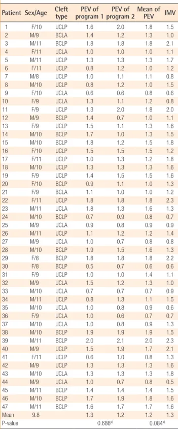
Table 2. Individual data including PEV and IMV overilling never ocurred (Fig. 3).
The statistical analysis of PEV and IMV was first performed to verify the two variables of bilaterality and presence of cleft palate, respectively. For statistical analysis, the paired sample t-test was performed with 95% confidence, and the Wilcoxon signed-rank test was performed when the number of patients was smaller than 30. If statistical significance was shown, the efect of the other variable on the mean diference between the PEV and IMV was veriied using the Mann-Whitney U test. All statistical analysis was performed with SPSS ver. 17 (SPSS Inc., Chicago, IL, USA).
RESULTS
he mean age of the patients was 9.8 years (range, 8 to 11 years), and 29 males and 18 females were included in this study. All 47 patients were categorized into 4 groups: unilateral clet lip and alveolus (UCLA, n = 13), unilateral clet lip and palate (UCLP, n = 18), bilateral clet lip and alveolus (BCLA, n = 2), and bilat-eral clet lip and palate (BCLP, n = 14) (Table 1).
Individual data including PEV and IMV are shown in Table 2. here was no signiicant diference between PEVs calculated by the two programs: Radipia 3D 2.8 and Ondemand 3D 1.0 (Ta-ble 2). Overall, the mean values of PEV and IMV were 1.2±0.4 cm3 and 1.3 ±0.5 cm3, respectively. he diference between the
two values was -0.1 ±0.3 cm3, which had no statistical
signifi-cance with a P-value of 0.084 on a paired sample t-test. Com-pared with bilaterality, the diference between PEV and IMV was -0.1 ±0.3 cm3 (P =0.094) in the unilateral group and -0.1 ±0.2
cm3 (P =0.474) in the bilateral group. Considering the type of
accompanying clet palate, the diference between PEV and IMV was 0.1±0.3 cm3 (P=0.439) in the clet lip and alveolus (CLA)
group and -0.2 ±0.3 cm3 (P =0.007) in the cleft lip and palate
(CLP) group. Only the CLP group showed a statistically signii-cant difference on both the paired sample t-test and Wilcoxon signed-rank test (Table 3). The difference between PEV and IMV in the CLP group had no statistical signiicance (P=0.197) on the Mann-Whitney U test with bilaterality (Table 4).
DISCUSSION
Secondary alveolar bone grating has many advantages such as excellent periodontal atachment to the adjacent teeth, restoring maxillary continuity, inducing dental eruption at the clet site, giving aesthetic improvement through gingival recovery, and minimizing the interruption of facial growth [12,13]. hese re-sults can be achieved using an adequate volume of bone grating material. An inadequate volume of grated bone can cause grat
Value (cmPEV 3) (cmIMV 3) Difference (cm3) P-value
Unilateral (n=31) 1.2±0.3 1.3±0.5 -0.1±0.3 0.094a) Bilateral (n=16) 1.4±0.4 1.5±0.5 -0.1±0.2 0.474b) CLA (n=15) 1.0±0.3 0.9±0.3 0.1±0.3 0.439b) CLP (n=32) 1.3±0.4 1.5±0.4 -0.2±0.3 0.007a) Total (n=47) 1.2±0.4 1.3±0.5 -0.1±0.3 0.084a)
PEV, preoperative estimated volume; IMV, intraoperative measured volume; CLA, cleft lip and alveolus; CLP, cleft lip and palate.
a)Paired-sample t-test; b)Wilcoxon signed-rank test.
Table 3. Bone volume comparison by cleft type
Bilaterality N Average of PEV-IMV SD P-value
UCLP 18 -0.22 0.31 0.197
BCLP 14 -0.13 0.26
CLP 32
CLP, cleft lip and palate; PEV, preoperative estimated volume; IMV, intraoperative measured volume; SD, standard deviation; UCLP, unilateral cleft lip and palate; BCLP, bilateral cleft lip and palate.
Table 4. Mann-Whitney U test with group factor of bilat-erality in CLP group
bone or repeating the procedure with a small amount can also increase donor site morbidity. For these reasons, several studies have reported on the usefulness of the preoperative estimation of the bone defect volume on the alveolar clet using 3D CT as-sessment. As mentioned above, the shape and size of the bone defect of an alveolar clet can be adjusted according to the alveo-lar cleft type, but the alveoalveo-lar cleft type that can influence the prediction procedure has never been considered.
herefore, this study proceeded with the hypothesis that the alveolar cleft type has a certain effect on 3D CT assessment. The results showed that the overall PEV was not statistically different from the IMV (P = 0.084) in all of the groups taken together, but there was a significant difference between them in the CLP group (P < 0.05). The Mann-Whitney U test with bilaterality within the CLP group showed that there was no significant difference between the UCLP and BCLP groups (P = 0.197) (Table 4). he underestimated volumes by 3D CT were -0.2 cm3 in both the UCLP and BCLP groups.
Consider-ing that the overall mean value of the IMV was 1.3 cm3, -0.2 cm3
is not negligible. In several cases, ater donor site repair, the iliac donor site was opened again to harvest 0.2 cm3 more bone
dur-ing the operation; this can lead to donor site complications such as scarring and infection.
he size and structural disparity between the CLA and CLP group seemed to be an important factor for explaining the dif-ference between the two groups. The mean value of the IMV (1.5 ± 0.4 cm3) in the CLP group was larger, by as much as
about 0.6 cm3, than that of the CLA group. his is regarded as
one of the causes of the larger diference between PEV and IMV in the CLP group. he architecture of an alveolar bone defect is a pyramidal shape posteriorly bounded by an aveolus and the palate. On a reconstructed 3D image, when accompanied by a clet palate, the bone defect was larger and extended to the hard palate (Fig. 2). In the past, some have advocated that strong compression and ine bony particles were necessary for a suc-cessful bone graft, but recently, several studies have reported that excessive crushing of cancellous bone can be harmful to the blood supply to the graft core and bony particles that are too tiny easily fail to revascularize and can be resorbed [14-16]. Thus, we tried not to apply excessive compression in order to avoid crushing the grat, and prepared the grat with bone chips of 2 × 2 × 2 mm to induce good revascularization (Fig. 3A) [16]. However, in the CLP group, packing the grat posteriorly tended to overpack into the soft tissue of the lingual side that had a weak support structure without strong compression (Fig. 4). It was thought that this phenomenon explained the signii-cant diference between the PEV and IMV in the CLP group. Fig. 4. Sagittal view of the postoperative computed tomog-raphy scan
The white arrow indicates an overpacked bone graft toward the lingual side.
Fig. 5. Preoperative computed tomography scan
Another structural disparity was the lingual process that was is a bony ridge on the lingual side of the alveolar clet (Fig. 5). he process provides strong posterior support, which reduced the gap between the PEV and IMV in the CLA group. We observed the process in 13 of the 15 patients in the CLA group (87%), but only in 1 of the 32 patients in the CLP group (3%).
There is only one study that has reported on the usefulness of 3D CT assessment for alveolar clet in vivo. Shirota et al. [7] performed late secondary bone grating in 13 patients and sug-gested the usefulness of preoperative CT images by using image analysis software. Their patients’ mean age was 22 years and the patients were in the period of late secondary bone grating, not the mixed dentition period. Moreover, their study did not consider the clet type. he results of the study showed a statisti-cally insigniicant diference of -0.3 cm3 between PEV and IMV
because the study only included adult patients; thus the larger margin of error could be permited. Recently, the alveolar bone graft has been commonly performed in the mixed dentition period because the bone grat in this period never disturbs the maxillary growth, and the erupting canine gives functional stress to the grat, which increases the success rate [17]. Our study can support future guidelines for alveolar clet treatment because we included only the patients in the mixed dentition period with a mean age of 9.8 years and prior to canine eruption.
he 3D CT assessment is a very reliable investigation tool, but it cannot interpret the variables of soft tissue and its elasticity because it focuses on the bony structure [8]. The purpose of this study is to demonstrate the application of 3D CT assess-ment clinically. As a result, the clet type should be considered for determining the grated volume using preoperative 3D CT assessment. Harvesting about 0.2 cm3 more bone for patients
with a concomitant clet palate is desirable based on our results.
REFERENCES
1. Bell WH, Proit WR, White RP. Residual alveolar and pala-tal clets. In: Bell WH, Proit WR, White RP, editors. Surgi-cal correction of dentofacial deformities. 1st ed. Philadelphia: Saunders; 1980. p. 1330.
2. van der Meij AW, Baart JA, Prahl-Andersen B, et al. Out-come of bone grating in relation to clet width in unilateral clet lip and palate patients. Oral Surg Oral Med Oral Pathol Oral Radiol Endod 2003;96:19-25.
3. Ochs MW. Alveolar clet bone grating (Part II): secondary bone grating. J Oral Maxillofac Surg 1996;54:83-8.
4. Kim SK. Secondary alveoloplasty using iliac cancellous bone grat in the clet lip and palate patients. J Korean Clet
Palate-Craniofac Assoc 2004;5:85-93.
5. Jun SH, Padwa BL, Jung YS. Alveolar cleft graft. J Korean Assoc Maxillofac Plast Reconstr Surg 2009;31:267-72. 6. Hofman WY, Mount D. Clet palate repair. In: Mathes SJ,
editor. Plastic surgery. 2nd ed. California: Saunders; 2006. p. 264-6.
7. Shirota T, Kurabayashi H, Ogura H, et al. Analysis of bone volume using computer simulation system for secondary bone grat in alveolar clet. Int J Oral Maxillofac Surg 2010; 39:904-8.
8. Albuquerque MA, Gaia BF, Cavalcanti MG. Oral clet volu-metric assessment by 3D multislice computed tomographic images. Int J Oral Maxillofac Surg 2011;40:1280-8.
9. Feichtinger M, Mossbock R, Karcher H. Evaluation of bone volume following bone grafting in patients with unilateral clefts of lip, alveolus and palate using a CT-guided three-dimensional navigation system. J Craniomaxillofac Surg 2006;34:144-9.
10. Oh SH, Lee SS, Lee KJ, et al. Volume measurement of ves-tibular schwannoma using new sotware. Korean J Otolar-yngol-Head Neck Surg 2002;45:451-5.
11. Feichtinger M, Mossbock R, Karcher H. Assessment of bone resorption ater secondary alveolar bone grating using three-dimensional computed tomography: a three-year study. Clet Palate Craniofac J 2007;44:142-8.
12. Kim JR, Jin SJ, Cho YC, et al. Clinical study of autogenous secondary bone grating in clet maxilla. J Korean Assoc Max-illofac Plast Reconstr Surg 2001;23:162-8.
13. Lee C, Crepeau RJ, Williams HB, et al. Alveolar cleft bone grats: results and imprecisions of the dental radiograph. Plast Reconstr Surg 1995;96:1534-8.
14. Stassen LF. Alveolar bone grafting-how I do it. In: Booth PW, Hausamen JE, Schendel SA, editors. Maxillofacial sur-gery. Edinburgh: Churchill Livingstone; 1999. p. 1047-55. 15. Kim KR, Kim S, Baek SH. Change in grafted secondary
alveolar bone in patients with UCLP and UCLA. A three-dimensional computed tomography study. Angle Orthod 2008;78:631-40.
16. Fonseca RJ, Nelson JF, Clark PJ, et al. Revascularization and healing of onlay particulate allogeneic bone grats in primates. J Oral Maxillofac Surg 1983;41:153-62.