1. clinical Biomechanical and rehabilitation engineering research center, aja university of medical sciences
2. clinical research development center at modarres Hospital, shahid Beheshti university of medical sciences
3. school of medicine, isfahan university of medical sciences 4. dr. mahmoudi’s acquired Brain injury rehabilitation center 5. Birjand university of medical sciences
IntroductIon
Fibromyalgia (FM) is the ringleader of the big family known as central sensitivity syndrome, manifesting with widespread pain, hyperalgia and unpleasant sen-sation after physical contact or thermal exposure. It is considered a multifactorial disorder for which central nervous system sensitization, infection, trauma, stress and genetics are suggested as the contributing factors1.
Cognitive symptoms, irritable bowel syndrome, heada -che, unrefreshing sleep, fatigue and a number of other somatic symptoms, altered the definition from a “pe-ripheral pain” defined disease to a “systemic symptoms” based disease2. It imposes a great burden to patients
comparable to osteoarthritis3and rheumatoid
arthri-tis4, and affects personal relationships, physical daily
activities and ability to work as well as mental health. Economic burdens are also comparable to diabetes and hypertension, and mostly influence the pre-diagnosed stage of illness5.
FM affects 2% of the population of all ages (3.4% in women and 0.5% in men). The prevalence rate in-creases with age, most prominently in women com-pared to men (7.1% vs. 1.2%), aged 60-696. The most
recent estimates from United States suggest that FM in-volves up to 5% of all women and is the third most common rheumatic disorder after low back pain and osteoarthritis6.
The classic criteria proposed by American College of Rheumatology (ACR)7emphasizes the evaluation of
tender points but it has proved nonsufficient8. The new
ACR criteria in 2010 have 83% specificity for correct dia gnosis9. The main limitation is that they cannot be
applied to patients with FM secondary to other disor-ders such as rheumatoid arthritis or systemic lupus ery-thematosus. Despite the efforts in recent years, there is no approved and optimal paraclinical tool in order to facilitate the diagnosis and followup of FM. Some stu -dies have been performed in order to investigate
elec-Thenar muscles H reflex in patients with fibromyalgia:
a case control study
Azma K1, Raeissadat SA2, Hosseini A3, Mahmoudi H4, Sepehrian Mh5, Salehi Z4
acta reumatol port. 2016;41:145-150
AbstrAct
Objective: To delineate Hoffman reflex (H-reflex) pa-rameters and spe cify the diagnostic accuracy measures of thenar muscle H-reflex in fibromyalgia (FM).
Methods: The study was a cross sectional study per-formed on 30 subjects with FM and 30 healthy volun-teers in two major referral hospitals. We recorded the number of obtainable thenar Hreflexes and their mini -mum latency, threshold and amplitude in each group.
Results: There was a significantly more chance to eli -cit the H-reflex in patients with FM. H-reflex threshold and minimum latency were lower in FM group but no significant difference was shown for H wave amplitude. According to our study, thenar H-reflex has 46.7% sen-sitivity, 86.7% specificity and 66.7% diagnostic accuracy to detect FM. It also has moderate predictive va -lues and positive likelihood ratio but low negative likelihood ratio.
Conclusion: Higher rate of thenar muscle H-reflex in FM can be interpreted as a confirmatory finding to cen-tral sensitization theory for this disorder. Obtaining H-reflex from thenar muscles could be a helpful diagnos tic tool for FM that increases the confidence in diagn osis. Although it is a weak tool for screening be-cause of low sensitivity, it has a relatively high speci-ficity.
tromyography, Magnetic Resonance Imaging (MRI) and electroencephalography responses in patients with FM10-17. However, a consensus about optimal
diagnos-tic method is still lacking.
Hoffman reflex (Hreflex) is the result of submaxi -mal stimulation of type Ia sensory fibers. The poten-tial enters the posterior horn of the spinal cord and passes through the synapses with alpha-motor neu-rons. Finally, a compound muscle action potential is generated and is recorded as H wave. It seems that the reflex is dependent upon the balance between aug-menting and inhibitory factors. H-reflex is mostly recorded from gastrocnemius-soleus complex and, sometimes, flexor carpi radialis muscles18. The
intrin-sic muscles of the foot or hand have also been report-ed as sites for eliciting the H-reflex19, 20, but to our
knowledge the thenar muscles have not been assessed as the site of H-reflex origin.
Despite the lack of literature on eliciting H-reflex from thenar area, due to our random findings and con-sidering the theoretical suprasegmental facilitating ef-fects of FM on H-reflex, the current study was per-formed to evaluate possible differences in H-reflex recording between patients with FM and normal po -pulation. Also we wanted to define diagnostic accura-cy measures of obtainable thenar muscle H-reflex and clarify if this non-invasive tool can be used as an ad-junctive measure to help diagnosis of FM.
methods
We carried out a matched comparative cross-sectio nal study with two groups of volunteers, one of FM pa-tients and other of healthy controls. The study was conducted from September 2010 to February 2011 in outpatient clinics of physical and rehabilitation medicine at two large referral academic hospitals.
PArtIcIPAnts And sAmPlIng
We recruited patients and healthy subjects between 20 to 60 years of age. First we recruited 5 FM patients. Hreflex could be obtained in 2 of them. Assuming po -wer of 80%, alpha of 0.05 and confidence interval of 95%, we calculated the sample size of 30 for each arm. So, 30 subjects with FM and 30 age and sex frequen-cy matched healthy subjects entered the study. We re-cruited the subjects using the convenient sampling method. Overall, 13 pairs of patients with their matched controls were examined in one hospital, and
17 were examined in the other hospital. All researchers who performed physical examination and the investi-gators who carried out electrodiagnostic studies were expert and qualified.
At the beginning of the session, the subjects were briefed. A trained general practitioner asked for the medical history of the patient, performed a general exa -mination and evaluated laboratory findings including Complete Blood Count (CBC), Thyroid Function Tests (TFT), Erythrocye Sedimentation Rate (ESR), C-Reactive Protein (CRP), Creatine Phosphokinase (CPK) and Lactate Dehydrogenase (LDH). Then a phys-ical and rehabilitation medicine specialist completed the medical history and physical examination looking for symptoms and signs of FM based on the ACR crite-ria. Because of the admissibility and popularity of clas-sic ACR criteria that numerous previous studies were done based on, we applied it in our study. After the di-agnosis of FM based on the first general and second specific history taking and physical examination accor -ding to ACR criteria and also ensuring of normal blood tests, the participants were led to Electromyography (EMG) la boratory to complete the investigation.
InclusIon And exclusIon crIterIA
All the subjects signed the provided informed consent before the first physician visit. Exclusion criteria were pain due to disorders other than FM, other rheumatologic, immunorheumatologic, inflammatory and hormonal di -sorders interfering with diagnosis of FM, any condition that made relaxing of the upper limb impossible such as spasticity, conditions potentially affecting the as-sessment of H-reflex such as radiculopathies, upper motor neuron diseases, compressive neuropathies, especially Carpal Tunnel Syndrome (CTS), and peri -pheral neuropathies. Besides, patients with limited range of motion in upper limb and muscular weak-ness did not enter the study. Recruited participants were screened to ensure that they have not used medi -cations, which increase serum serotonin level within the last one-month before the study. We excluded in-dividuals if they could not tolerate the procedure of H-reflex measurement.
rAndomIzAtIon And blIndIng
was no need to blind the subjects because the H-reflex is an objective measure and the subjects could not in-terfere with the results.
Protocols And Procedures
In the beginning of the session a general practitioner asked for medical history including baseline demo-graphic characteristics, features and duration of pain, sleep disturbances, fatigue, absence from work, symp-toms of co-morbidities, and alternative diagnoses. The physical and rehabilitation medicine specialist per-formed physical examination according to the classic ACR criteria. The tender points were examined over the occipital, low cervical, trapezius, supraspinatus, second rib, lateral epicondyle, gluteus, greater trochanter and at the medial fat pad of the knee; bilate -rally. The Physician considered each point as positive if patient reported the point as painful by applying less than 4kg pressure with a pressure algometer device.
We elicited H-reflex in a quiet environment with convenient temperature and after 5 minutes of rest. We instructed the participants to lie in a semi-reclined po-sition with the head and arms supported on a firm sur-face to reduce variability of the H-reflex and to provide comfort throughout the testing. A Medelec EMG de-vice with the following settings was used to stimulate the median nerve: stimulation duration (1 millise -conds), frequency (0.1 Hz), sweep speed (5 ms/divi-sion), sensitivity (500 microvolts/divims/divi-sion), and filter setting (2 Hz to 10 KHz). Stimuli were applied 10 seconds apart to reduce the effects of post-activation depression. E1 recording electrode was put over the belly of thenar muscle area and the E2 on 1stmet a
-carpo-phalangeal joint area.
We stimulated the median nerve above the wrist, 10 cm proximal to the site of recording. We also put a ground electrode just proximal to E1 recording electro -de. In order to measure the H-wave, first we set the stimulation intensity to zero and then gradually in-creased the intensity until we found the lowest stimu-lation intensity with highest H-wave amplitude. We re-peated H-wave measurement with this intensity several times to be sure of reproducibility and steady onset la-tency. H-wave was defined as triphasic wave with on-set roughly between 20 and 30 milliseconds which comes after an initial compound muscle action poten-tial and its amplitude becomes maximum at a sub-maximal stimulation intensity. The amplitude shortens with increasing the intensity.
We decided that H-reflex is not obtainable in a
par-ticular subject if after 3 trials and gradually increasing the intensity to supramaximal intensity performed on both hands, no H-reflex could be observed. In order to avoid tester bias all measurements achieved and record-ed by digital device.
ethIcAl consIderAtIons
Ethics approval was obtained from common institu-tional review boards of the two centres and the study protocol was carried out in accordance with the De -claration of Helsinki. The rationale of the study was ex-plained to all participants. Patients were informed that they were free to withdraw from the study at any time. A trained study nurse accompanied patients and pro-vided verbal information, if needed, and a leaflet on the diagnostic procedures to eligible participants. Pa-tients gave written informed consent at the screening visit and did not pay for the diagnostic procedures. They were referred to other departments for appropri-ate treatment if needed.
stAtIstIcAl AnAlysIs
The collected data were analyzed using SPSS-20 soft-ware. We used Kolmogorov-Smirnov test to evaluate the normality of variable distribution. Then we used Chi Square test to compare the frequency of obtainable H-reflex in FM and control group. Distribution of am-plitude, threshold and minimum latency were not nor-mal so we used Mann-Whitney test to compare these variables between groups. Finally we drew a 2 by 2 table showing the frequency of obtainable and unobtai -nable H-reflexes in FM and control groups to calculate the diagnostic accuracy measures.
results
descrIbIng the sAmPle
The sample was composed of 30 sex and age matched pairs. Nineteen (63%) of these pairs were female. Mean age was 40 ± 11.2 and 40 ± 11.3 years for the FM and control groups, respectively, with no statistically sig-nificant difference (P = 0.95). The upper limb length was 62 ± 6.5 cm in FM group and 62 ± 6.3 cm in con-trol group, which did not differ significantly (P = 0.86).
h-reflex meAsures
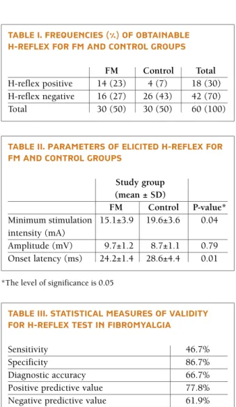
control groups. Minimum stimulation intensity for elici ting H-reflex was 15.1 ± 3.9 millivolts in FM group, which was lower than 19.6 ± 3.6 millivolts in control group (P=0.04). Minimum onset latency was also low-er in FM group (24.2 ± 1.4 ms) compared with 28.6 ± 4.4 ms in control group (P=0.01). There was no statis-tically significant difference between the H-reflex am-plitudes of the two groups (P=0.79). Table II summa-rizes the H-reflex parameters in the study population.
dIAgnostIc AccurAcy meAsures
Using the values of Table I, we calculated important statistical measures of validity for thenar muscle H-re-flex in patients with FM. The proportion of individu-als with elicited H-reflex out of all individuindividu-als with FM (sensitivity) was 46.7% while the proportion of
indi-viduals without elicited H-reflex out of all normal sub-jects (specificity) was 86.7%. The likelihood that a person with positive thenar Hreflex actually has FM (po -sitive predictive value) is 77.8% and the likelihood that a person who has negative thenar H-reflex actually has not FM (negative predictive value) is 61.9%. To inves-tigate how much more likely it is that a person has FM after the H-reflex test is done (post-test probability), we calculated positive and negative likelihood ratios which were 3.5 and 0.62, respectively. For easier un-derstanding and comparison, we have summarized the measures of validity in Table III.
dIscussIon
h-reflex PArAmeters
The results of our study showed that the chance of ob-taining H-reflex from thenar muscles is significantly higher in patients with FM compared to normal sub-jects. This confirms the theory of increased central sen-sitivity in FM. H-reflex displays the late response of A fibers (large myelinated nerve fibers) and since in pa-tients with FM central hypersensitivity may occurs in all kinds of nerve fibers including A fibers, higher rate of H-reflex in FM could be justifiable and interpreted. Minimum stimulation intensity and onset latency of Hreflex were lower in patients with FM. Lower stimu -lation intensity also is consistent with central sensiti vity theory suggesting lower threshold of intraspinal synapses due to increased excitatory or decreased in-hibitory factors. Smaller onset latency in patients with FM suggests that this condition possibly affects fast fiber terminals more than the slow fiber terminals, so the overall latency will be reduced. This finding could help us for better understanding the nature of this mul-tifactorial condition.
The unexpected finding of our study was similar am-plitude of H waves in FM and control subjects. Changes in H-reflex amplitude after applying a conditioning stimulus have been used to evaluate post-synaptic events or to assess changes in the amount of the presy-naptic inhibition acting on Ia afferent terminals. It has been suggested that the amplitude of the test reflex de-pends on the motor neuron excitability and the exis ting pre-synaptic inhibition of Ia fibers18. Our finding
suggests that factors other than motor neuron pool exci -tability may contribute to form the H wave amplitude. Another explanation lies in the H wave amplitude itself as it is the most variable parameter of H-reflex. It is pos-tAble I. frequencIes (%) of obtAInAble
h-reflex for fm And control grouPs
FM Control Total
H-reflex positive 14 (23) 4 (7) 18 (30) H-reflex negative 16 (27) 26 (43) 42 (70) Total 30 (50) 30 (50) 60 (100)
tAble II. PArAmeters of elIcIted h-reflex for fm And control grouPs
Study group (mean ± SD)
FM Control P-value*
Minimum stimulation 15.1±3.9 19.6±3.6 0.04 intensity (mA)
Amplitude (mV) 9.7±1.2 8.7±1.1 0.79 Onset latency (ms) 24.2±1.4 28.6±4.4 0.01
*The level of significance is 0.05
tAble III. stAtIstIcAl meAsures of vAlIdIty for h-reflex test In fIbromyAlgIA
Sensitivity 46.7%
Specificity 86.7%
Diagnostic accuracy 66.7%
sible that equal H wave amplitude in FM and normal subjects is due to technical issues. For example, if we increased the number of attempts to detect a typical H wave, we could record the waves with higher ampli-tudes in FM group.
dIAgnostIc AccurAcy meAsures
Several studies have been performed in order to eva -luate electromyography and nerve conduction study (EMG & NCS) parameters in FM. Some of them failed to show any differences, while others found dissimi-larities in EMG & NCS parameters10-14. Among the va
-rious measures, surface and needle EMG are the most widely investigated parameters. These studies are very heterogeneous regarding the target investigated mus-cles and the target EMG & NCS parameters. This can partially explain why the results are not consistent.
Our results show that thenar muscle H-reflex has a low sensitivity (46.7%) and a relatively high specifici-ty (86.7%). This means that the probabilispecifici-ty of obtain-ing an H-reflex in patients with FM is not very high, so it is not a useful test for screening. On the other hand, the probability that H-reflex cannot be obtained in per-sons without FM is high, so the false positive results are low for this test.
The results also show that obtaining H-reflex from thenar muscles has 77.8% positive predictive value and 61.9% negative predictive value. Both positive and nega tive predictive values for thenar H-reflex are wi thin intermediate range. This shows that this test cannot be used as a single tool for detecting FM, but it might be helpful as an adjunct to increase the accuracy of other tools.
Eventually, to see if obtaining H-reflex can be used by clinicians to revise their pre-test estimates of the disea se (post-test probabilities), we calculated the likeli hood ratios. Regarding to the estimated 3.5 for positive likelihood ratio and 0.62 for negative likeli-hood ratio, we can say that ability to obtain thenar H--reflex may help to improve our confidence that a pa-tient actually has FM, but negative thenar H-reflex does not have any important information and it will not in-crease our confidence that the patient has not FM. The likelihood ratios have advantage over sensitivity, speci-ficity and predictive values because they are indepen-dent of disease prevalence and, therefore, can be ap-plied across settings and patients.
study lImItAtIons
As a preliminary investigation, our results suggested
that H-reflex could be useful to help diagnosis of FM. Meanwhile, the procedure has an inherent limitation. The procedure for obtaining H-reflex has not been fully standardized and clinicians may use slightfully diffe -rent techniques for this purpose. As a result inter-rater bias could be high if no standard procedure is used. Some studies have described the appropriate methods to elicit the H-reflex and examined the reliability of this measurement in different muscles. However, when us-ing the H-reflex as a measurement tool, it is necessary to describe certain details regarding how the reflex has been elicited and the responses recorded, so that con-clusions can be drawn with more confidence
conclusIon
Our results are partly confirming the central sensitivi-ty theory for FM. Higher rate of thenar muscle H-reflex in FM can be interpreted as a confirmatory finding to central sensitization theory for this disorder. Obtaining H-reflex from thenar muscles area could be a helpful diag nostic tool for FM that increases the confidence in diagnosis. Although it is a weak tool for screening because of low sensitivity, it has a relatively high specificity. It also may be that Hreflex be used for monito -ring of treatments too. These hypotheses require tes ting in future research.
corresPondence to
Alireza Hosseini
School of Medicine, Isfahan University of Medical Sciences, Isfahan, Iran
Email: [email protected]
references
1. Smith H. Fibromyalgia syndrome: review of the epidemiology and mechanism involved. Adv Stud Med. 2009;9(4):108-114. 2. Mease P. Fibromyalgia syndrome: review of clinical presenta-tion, pathogenesis, outcome measures, and treatment. J Rheumatol Suppl. 2005;75:6-21.
3. Kleinman N, Harnett J, Melkonian A, Lynch W, Kaplan-Mach-lis B, Silverman SL. Burden of fibromyalgia and comparisons with osteoarthritis in the workforce. J Occup Environ Med. 2009;51:1384-1393.
4. Silverman S, Dukes EM, Johnston SS, Brandenburg NA, Sa-dosky A, Huse DM. The economic burden of fibromyalgia: com-parative analysis with rheumatoid arthritis. Curr Med Res Opin. 2009;25(4):829-840.
5. Doron Y, Peleg R, Peleg A, Neumann L, Buskila D. The clinical and economic burden of fibromyalgia compared with diabetes mellitus and hypertension among Bedouin women in the Negev. Fam Pract. 2004;21(4):415-419.
14. Casale R, Sarzi-Puttini P, Atzeni F, Gazzoni M, Buskila D, Rain-oldi A. Central motor control failure in fibromyalgia: a surface electromyography study. BMC Musculoskelet Disord. 2009;10:78.
15. Bell IR, Baldwin CM, Stoltz E, Walsh BT, Schwartz GE. EEG beta 1 oscillation and sucrose sensitization in fibromyalgia with chemical intolerance. Int J Neurosci. 2001;108(1-2):31-42. 16. Cook DB, Lange G, Ciccone DS, Liu WC, Steffener J, Natelson
BH. Functional imaging of pain in patients with primary fi-bromyalgia. J Rheumatol. 2004;31(2):364-378.
17. Gracely RH, Petzke F, Wolf JM, Clauw DJ. Functional magne tic resonance imaging evidence of augmented pain processing in fi-bromyalgia. Arthritis Rheum. 2002;46(5):1333-1343. 18. Knikou M. The H-reflex as a probe: pathways and pitfalls. J
Neurosci Methods. 2008;171(1):1-12.
19. Palmieri RM, Ingersoll CD, Hoffman MA. The Hoffmann reflex: methodologic considerations and applications for use in sports medicine and athletic training research. J Athl Train. 2004; 39(3):268-277.
20. Wolfe F, Ross K, Anderson J, Russell IJ, Hebert L. The prevalence and characteristics of fibromyalgia in the general population. Arthritis Rheum. 1995;38(1):19-28.
United States. Part II. Arthritis Rheum. 2008;58(1):26-35. 7. Wolfe F, Smythe HA, Yunus MB, et al. The American College of
Rheumatology 1990 criteria for the classification of fibromyal-gia. Report of the Multicenter Criteria Committee. Arthritis Rheum. 1990;33:160-172.
8. Fitzcharles MA, Boulos P. Inaccuracy in the diagnosis of fi-bromyalgia syndrome: analysis of referrals. Rheumatology (Ox-ford). 2003;42(2):263-267.
9. Wolfe F, Clauw DJ, Fitzcharles MA, et al. The American College of Rheumatology preliminary diagnostic criteria for fibromyal-gia and measurement of symptom severity. Arthritis Care Res (Hoboken). 2010;62(5):600-610.
10. Anders C, Sprott H, Scholle HC. Surface EMG of the lumbar part of the erector trunci muscle in patients with fibromyalgia. Clin Exp Rheumatol. 2001;19(4):453-455.
11. Bazzichi L, Dini M, Rossi A, et al. Muscle modifications in fi-bromyalgic patients revealed by surface electromyography (SEMG) analysis. BMC Musculoskelet Disord. 2009;10:36. 12. Maquet D, Croisier JL, Dupont C, et al. Fibromyalgia and
relat-ed conditions: electromyogram profile during isometric muscle contraction. Joint Bone Spine. 2010;77(3):264-267.