Disclosure
The authors report no conflicts of interest.
References
1 Neves FS, Malloy-Diniz LF, Romano-Silva MA, Aguiar GC, de Matos LO, Correa H. Is the serotonin transporter polymorphism (5-HTTLPR) a potential marker for suicidal behavior in bipolar disorder patients? J Affect Disord. 2010;125:98-102.
2 Neves FS, Malloy-Diniz LF, Barbosa IG, Brasil PM, Correˆa H. Bipolar dis-order first episode and suicidal behavior: are there differences according to type of suicide attempt? Rev Bras Psiquiatr. 2009;31:114-8. 3 Leverich GS, Post RM. Course of bipolar illness after history of
childhood trauma. Lancet. 2006;367:1040-2.
4 Grassi-Oliveira R, Stein LM, Pezzi JC. [Translation and content validation of the Childhood Trauma Questionnaire into Portuguese language]. Rev Saude Publica. 2006;40:249-55.
5 Labonte´ B, Turecki G. Epigenetic effects of childhood adversity in the brain and suicide risk. In: Dwivedi Y, editor.The neurobiological basis of suicide. Boca Raton: CRC Press; 2012. p.256-84.
New-onset panic attacks
after deep brain stimulation
of the nucleus accumbens
in a patient with refractory
obsessive-compulsive and
bipolar disorders: a case
report
Rev Bras Psiquiatr. 2015;37:182–183 doi:10.1590/1516-4446-2014-1581
New-onset panic attacks (PA) have been described in patients with obsessive-compulsive disorder (OCD) receiving deep brain stimulation (DBS), mostly during the intraoperative period or a few weeks after device implantation.1,2We report the case of a 39-year-old, right-handed man with severe treatment-refractory OCD and bipolar disorder type I (BD-I), beginning at age 17 (without any other psychiatric disorder), who developed late-onset PA after DBS implant placement.
The patient presented with obsessions of doubt, cleaning, and disgusting thoughts accompanied by checking and cleaning compulsions, with an intense need for reassurance and avoidance. Due to poor response to multiple drugs and to cognitive-behavioral therapy (Table 1), the patient underwent surgical evaluation for DBS. Implantation was performed after the patient and relatives had signed an informed consent form and following authorization from the Federal Council of Medicine. At baseline, the Yale-Brown Obsessive Compulsive Scale (Y-BOCS) score was 363and the Beck Depression Inventory (BDI) score was 35.4
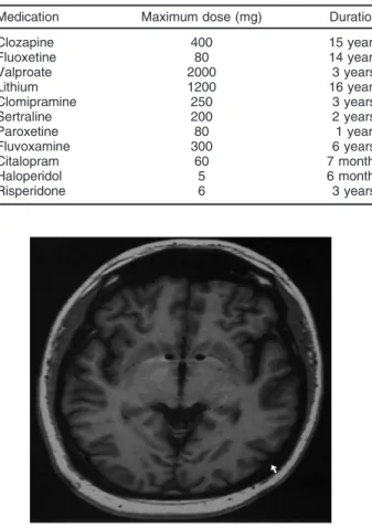
Bilateral DBS electrodes were inserted through the anterior limb of the internal capsule into the nucleus accumbens (NAcc) near the anterior commissure (Figure 1).
Intraoperative evaluation of the DBS electrodes was carried out using bipolar stimulation at each contact. Pulse width and stimulation frequency ranged from 90 to 210 ms and
100 to 180 Hz, respectively. Voltage varied between 0 and 4 V, while bilateral stimulation was 3+/0-, 3+/1-, 3+/2-, and 0+/3-. The patient did not notice any change in mood or anxiety during stimulation. Testing occurred for approxi-mately 2 to 4 minutes at each setting and the voltage was turned off before testing each contact. The patient was discharged from the hospital with the DBS regulated at 4.2 V, 150ms, 150 Hz both sides, LL 3+, zero and 1 Neg,
RR 7+, 4 and 5 Neg. Final adjustment was performed after several trials with on-off checking. Five months after surgery, the patient had experienced significant improve-ment of both OCD (Y-BOCS = 17) and depression (BDI = 9). Suddenly, within 12 hours of a follow-up visit involving a parameter adjustment for better control of OCD symptoms (4 V, 180ms, 120 Hz both sides, LL C+, zero
and 1 [-], RR C+, 4 and 5 [-]), the patient began to have Figure 1 Magnetic resonance imaging scan showing the deep brain stimulation electrodes (Medtronic model 3387) inserted bilaterally through the anterior limb of the internal capsule into the nucleus accumbens near the anterior commissure. Cartesian coordinates of the distal end of the deepest contact relative to the mid-commissural point were: left and right: 6 mm lateral to midline, 3 mm anterior to mid-commissural point, and in the anterior commissure-posterior commissure plane.
Table 1 Medications previously taken by the patient
Medication Maximum dose (mg) Duration
Clozapine 400 15 years
Fluoxetine 80 14 years
Valproate 2000 3 years
Lithium 1200 16 years
Clomipramine 250 3 years
Sertraline 200 2 years
Paroxetine 80 1 year
Fluvoxamine 300 6 years
Citalopram 60 7 months
Haloperidol 5 6 months
Risperidone 6 3 years
Rev Bras Psiquiatr. 2015;37(2)
severe panic attacks, which were controlled after new adjustments in association with clonazepam 1 mg/day. The adjustments involved more ventral connectivity with bipolar stimulation, instead of a dorsal stimulation, and were performed because they elicited better OCD control, but possibly triggered PA. The device was turned off; however, due to patient request, it was immediately reset to the previous settings, thus limiting conclusions of causality.
Shapira et al. and Okun et al. only observed the occurrence of panic attacks by activating the most ventral contact that is located next to the NAcc.1,2When this region was stimulated at contact zero, it probably caused amygdala activation, thus evoking the experience of panic.1,2This may have occurred because of the role of the NAcc as an interface for limbic projections from the amygdala, hippocampus, and cingulate cortex, which receives input from dopaminergic-containing nuclei, while mediating the behavioral and affective changes induced by DBS.2,5 Additionally, the patient’s comorbid BD-I could have facilitated affective side effects with NAcc stimulation.
Marcelo B. Sousa,1Telmo Reis,2Alexandre Reis,2 Paulo Belmonte-de-Abreu1,2,3
1Psychiatry Service, Hospital de Clı´nicas de Porto Alegre (HCPA),
Porto Alegre, RS, Brazil.2Neurosurgery Service, Hospital Moinhos de Vento (HMV), Porto Alegre, RS, Brazil.
3Department of Psychiatry, School of Medicine, Universidade
Federal do Rio Grande do Sul (UFRGS), Porto Alegre, RS, Brazil
Submitted Oct 10 2014, accepted Jan 29 2015.
Disclosure
The authors report no conflicts of interest.
References
1 Shapira NA, Okun MS, Wint D, Foote KD, Byars JA, Bowers D, et al. Panic and fear induced by deep brain stimulation. J Neurol Neurosurg Psychiatry. 2006;77:410-2.
2 Okun MS, Mann G, Foote KD, Shapira NA, Bowers D, Springer U, et al. Deep brain stimulation in the internal capsule and nucleus accumbens region: responses observed during active and sham programming. J Neurol Neurosurg Psychiatry. 2007;78: 310-4.
3 Goodman WK, Price LH, Rasmussen SA, Mazure C, Fleischmann RL, Hill CL, et al. The Yale-Brown Obsessive Compulsive Scale. I. Development, use, and reliability. Arch Gen Psychiatry. 1989;46: 1006-11.
4 Beck AT, Ward CH, Mendelson M, Mock J, Erbaugh J. An inventory for measuring depression. Arch Gen Psychiatry. 1961;4:561-71. 5 Gorman JM, Kent JM, Sullivan GM, Coplan JD. Neuroanatomical
hypothesis of panic disorder, revised. Am J Psychiatry. 2000;157: 493-505.