Relative Biological Efectiveness (RBE) of
131I
Radiation Relative to
60Co Gamma Rays
Ali Neshasteh-Riz, Ph.D.1*, Ali Mahmoud Pashazadeh, M.Sc.2, Seyed Rabie Mahdavi, Ph.D.1
1. Faculty of Allied Medicine, Department of Radiation Sciences, Tehran University of Medical Sciences ,Tehran, Iran 2. The Persian Gulf Nuclear Medicine Research Center, Bushehr University of Medical Sciences, Bushehr, Iran
* Corresponding Address: P.O. Box: 14155-6183, Faculty of Allied Medicine, Department of Radiation Sciences, Tehran University of Medical Sciences, Tehran, Iran
E mail: [email protected]
Received: 31/Jan/2012, Accepted: 07/Jan/2013
Abstract
Objective: To assess relative biological effectiveness (RBE) of 131I radiation relative to 60Co gamma rays in glioblastoma spheroid cells.
Materials and Methods: In this experimental study, glioblastoma spheroid cells were exposed to 131I radiation and 60Co gamma rays. Radiation induced DNA damage was evaluated by alkaline comet assay. Samples of spheroid cells were treated by radiation from 131I for four different periods of time to ind the dose-response equation. Spheroid cells were also exposed by 200 cGy of 60Co gamma rays as reference radiation to induce DNA damage as endpoint.
Results: Resulted RBE of 131I radiation relative to 60Co gamma rays in 100 µm giloblas-toma spheroid cells was equal to 1.16.
Conclusion: The inding of this study suggests that 131I photons and electrons can be more effective than 60Co gamma rays to produce DNA damage in glioblastoma spheroid cells.
Keywords: RBE, Glioblastoma, Spheroid, Photons, Electrons
Cell Journal(Yakht eh), Vol 15, No 3, Aut um n 2013, Pages: 224- 229
Citation: Neshasteh-Riz A, Pashazadeh AM, Mahdavi SR. Relative biological effectiveness (RBE) of 131 I radiation
relative to 60co gamma rays. Cell J. 2013; 15(3): 224-229.
Introduction
In nuclear medicine, 131I is one of the most
com-monly used radioisotopes in treatment of thy-roid. Application of this radioisotope in treatment of thyroid-related diseases has been one of the most successful treatments in radiation therapy. Recently some studies have been performed in order to assess the treatment capability of 131I
ra-diation in central nervous system (CNS) tumors.
Application of 131I labeled with
metaiodobenzyl-guanidine (MIBG) has shown promising therapeu-tic effects in treatment of neuroblastoma. In one study, performed on 42 patients with advanced neuroblastoma, the overall survival for stage three and stage four were 75 and 69% respectively (1). Post-surgery radioimmunotherapy of patients with glioblastoma has also shown promising results in controlling the progression of the residual tumor. Direct injection of 131I labeled antitenascin
anti-bodies into the tumor mass of 30 patients has lead to overall response rate of 34.7% (2). Because of increasing studies in therapeutic application of
131I, there is a need to further study the basic
radio-biology of this radioisotope. For this reason and in order to establish appropriate protocols in medi-cal application of 131I, we measured relative
bio-logical effectiveness of 131I, as a comprehensive
radiobiological concept. Relative biological
effec-tiveness (RBE) is deined as the ratio of a dose of
standard radiation to the dose of test radiation to produce the same biological effects. Although 250 kVp X-ray was the common standard radiation to determine the RBE, the International Commission on Radiation Protection (ICRP) recommended in their 92th report to use gamma rays of 60Co as
ref-erence radiation (3) and therefore was chosen as reference in this study. In spite of the fact that 60Co
energy transfer (LET) radiations have the same quality factor (QF), they seem to exert differ-ent biological effects. Determination of RBE is a comprehensive way to compare effects of these two low LET radiations. ICRP recommended in 1990 a quality factor of 1 for all low LET
radia-tions such as photons and electrons but in vitro
studies have shown different biological effects for photons and electrons (4). Although ICRP in 2007 accepted that low LET radiations have dif-ferent effects on cells but it still continues to use quality factor of 1 for all low LET radiations (5).
Depending on the energy, low LET radiations have different biological effects and different RBEs. In general, low energy radiations are more effective in comparison to high energy radiations. For example, 29 kVp X-ray is more effective than 200-220 kVp X-rays (6) or Tritium beta ray (5.7 keV) is much more effective than 15 MeV electrons (7). In order to compare biological effects of two types of radiation a variety of endpoints can be used. Radiation induced DNA damage to detect the primary effects of radia-tion on biological cells can be used as the biological
endpoint. In addition, there are eficient and robust lab
techniques such as comet assay or single cell gel elec-trophoresis in which extent of DNA damage in cells, so called tail moment, is a measurable quantity and have been used for more than two decades (8, 9).
In this study, in order to understand the basic radiobiology of 131I radiation and to compare
bio-logical effects of two low LET radiations, we in-vestigated the relative biological effectiveness of
131I radiation to 60Co gamma rays in spheroids of
U87MG cell line using the comet assay.
Materials and Methods
Thermoluminescent dosimeter (TLD) calibration
In this experimental study, TLD-100 chips with dimensions of 1.3×1.3×0.9 mm3, density of 2.64 g/
cm3 and average atomic number of 8.2 were used as
tissue equivalent dosimeter. Twelve TLD chips were annealed at 400˚C for 1 hour followed by second anw -nealing at 100˚C for 24 hours. For calibration purpose, TLD chips in four groups of three were irradiated by
60Co gamma rays of doses 10, 30, 50, 70, 90 and 110
cGy. In order to apply the correction factor in the cali-bration equation, a second exposure was performed at different doses of 5, 10, 40, 60, 80 and 100 cGy. Dur-ing irradiation, a tissue equivalent plexiglass layer was
covered on TLD chips to provide build up region of
60Co photons. Thermo luminescent reading was
per-formed by TLD reader Harshaw/Bicron model 3500.
Dosimetry of 131I radiation by TLD
In order to determine the dose-time equation of 131I
radiation in TLD chips, each group of TLD chips was covered by a thin layer of plastic and was embedded
in a lask. All lasks were illed by 10 ml of medium
and 10 mCi of 131I. TLD groups were exposed for 30,
60, 90, 120 and 150 minutes respectively.
Cell line
U87MG cell line was obtained from Pastor Insti-tute of Iran. It was cultured in Minimal Essential Medium (MEM) (Gibco, USA) containing 10% Fe-tal Bovine Serum (FBS) (Gibco, USA) and 500µ/ ml of penicillin (Sigma, USA).
Monolayer culture
Glioblastoma cells were cultured as a monolayer
in T-25 lasks (NUNC, Denmark) under the incu
-bation condition of 37˚C, 5% CO2 and
humidi-ied atmosphere of 95%. In subculturing process,
Phosphate Buffer Saline (PBS) was used for wash-ing cells and 1 mM ethylenediaminetetraacetic acid (EDTA) was used for trypsinizing the cells.
Spheroid culture
Spheroids were cultured by liquid Overlay
tech-nique. Number of 105 cells were cultured in 100
mm dishes that were coated with a layer of 1% agar with 10 ml of MEM containing 10% FBS. The plates were incubated in 5% CO2, 37˚C and humidh
-iied atmosphere. Every three days half of medium
was removed and replaced with fresh medium.
Spheroid growth curve
Each spheroid cell was transferred into a multi-well plate (24 multi-wells/plate) (NUNC, Denmark) that was coated by 1% agar with 10ml of MEM sup-plemented with 10% FBS. The spheroid cells were incubated at 5% CO2, 37˚C. In order to calculate the volume of each spheroid according to the equa-tion V (Volume)=a×b2×π/6, two perpendicular di
-ameters of the spheroid (a and b) were measured
to ind the growth equation V(t)=V0×e
kt, where V 0
Cell treatment by 60Co radiation
Glioblastoma Spheroid cells were irradiated by photons of 60Co as reference radiation. Theratron
780 unit at dose rate of 80 cGy per minute was used to deliver dose of 200 cGy to spheroid cells.
Flasks containing spheroid cells were suficiently illed with medium and covered the cells to pro -vide build up layer of 60Co photons. Exposure
fac-tors were set at ield size of 12×12 cm2 and SSD of
70 cm. In order to measure damages based on fac-tors other than 60Co radiations, one lask was not
exposed as the control group. Resulted damages of unexposed spheroids were used to determine net damages of 60Co radiation.
Cell treatment by 131I radiation
In order to ind dose-response equation of 131I
ra-diation in glioblastoma cells, spheroids in various
culture lasks were exposed for different exposure
times (30, 60, 90 and 120 minutes). Initial activity of 10 mCi 131I was poured into each lask of sphe
-roids. The control sample was also considered in
order to ind net damages of 131I radiation.
Viability test
In order to evaluate viability of cells in each cat-egory, a suspension of each category was mixed with Trypan blue. The viability test determines the ability of cells to recover its viability. In each step, before counting the cells under microscope, viabil-ity test was performed to determine the health sta-tus of cells. The ratio of cell suspension to trypan blue was 9:1. The mixture was observed by a light microscope (Leica, DMLS, USA) and all the blue cells were considered as dead cells. Percentage of unstained cells, as healthy cells, to the total cells represented the viability.
Comet assay
Induced DNA damage in U87MG cells exposed to 60Co radiation and 131I radiation were
deter-mined by alkaline comet assay. Microscope slides were coated with 1% normal melting point agarose (Merck, Germany). After counting spheroid cells in hemocytometer, approximately 10000 cells in 10 µl PBS were mixed with 100 µl of 5% low melting agarose (Merck, Germany). The cell sus-pension was then poured on the coated microscope slide. After getting the suspension on the slides
so-lidiied, they were immersed in lysis buffer (2.5 M
NaCl, 100 mM EDTA, 10 mM Tris-base with 1% Triton X-100, pH=10) and incubated for one hour.
Subsequently, the slides were removed from the buffer and transferred into a denaturation buffer (300 mM NaOH, 1mM EDTA, pH=13) in a horizontal gel electrophoresis tank (Cleaver
Sci-entiic Ltd, CSL-COM20). Electrophoresis was
performed for 30 minutes at the voltage of 1 V/ cm and amperage of 300 mA. After electropho-resis, alkaline was neutralized by Tris buffer (0.4 M Tris-HCl, pH=7.5) and the slides were then im-mersed in ethidium bromide. In order to take pho-tograph of damaged cells, slides were transferred
under a luorescent microscope (Zeiss, Axioskop 2 plus) with an ethidium bromide ilter (excitation ilter, 535 nm; emission ilter, 610 nm) and a CCD
camera (Hitachi, KP-D20BP).
Evaluation of DNA damage
For each sample of cells three slides were consid-ered and for each slide 100 cells were scored. DNA
damage was quantiied as increase in tail moment.
The intensity of comet tail relative to the head of com-et represented DNA damage. In most experiments, cell damages with respect to the extent of damaged DNA, could be scored from no damage (class one)
to total damage (class ive). Highly damaged form
of DNA may result in apoptosis. DNA tail moments were analyzed by comet score software.
Determination of RBE
RBE is the ratio of the dose of reference radiation to a dose of test radiation to produce a similar endpoint. In this experimental study, 60Co was used as reference
radiation and net induced DNA damage by 200 cGy of this radiation was considered as the endpoint. In
order to ind dose of 131I beta radiation (D
test) which
produces the same endpoint, dose-response equa-tion of 131I radiation was determined. In this equation,
response was equaled to the induced endpoint (re-sponse) by 200 cGy of 60Co gamma photons. Finally,
ratio of 200 cGy to the determined dose of 131I (D test)
beta radiation was considered as RBE.
Results
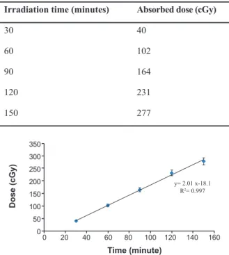
Exposure time by 131I and related absorbed dose
in TLD chips are represented in table 1.
Corre-sponding time-dose equation is shown in igure 1
Table 1: Absorbed doses in TLD chips by 131I radiation
Absorbed dose (cGy) Irradiation time (minutes)
40 30
102 60
164 90
231 120
277 150
Dose (cGy)
350
300
250
200
150
100
50
0
0 20 40 60 80 100 120 140 160
y= 2.01 x-18.1 R2= 0.997
Time (minute)
Fig 1: Absorbed doses in TLD chips by irradiation of 131I for
30, 60, 90, 120 and 150 minutes. (Error bars represent Mean ± SEM of 3 replicates).
The U87MG cell line was cultured as spheroid forms.
Growth curve of spheroids is shown in igure 2 and
this curve was used to determine the doubling time (67 hours). It lasted 11 days to have spheroids with
100 μm in diameter. After exposure of cells, induced
DNA damage in cells was evaluated as tail moments by comet assay. Figures 3A-3G represent
micropho-tographies of the 100 μm spheroid cells in control and
treated groups. Apoptosis as a result of highly dam-aged DNA was also observed. (some DNA damages such as DNA-cross link, DSB and bulky DNA ad-ducts, if not repaired, may lead to apoptosis). Irradia-tion was performed in two different places for 131I and 60Co irradiation and due to different environmental
conditions, induced base damage in control groups for 131I and 60Co irradiation were not similar.
cell/well
1000000
100000
10000
1000
0 2 4 6 8 10 12 Time (day)
Fig 2: Growth curve of U87MG cell line in the spheroid cul-tures (y=6999e0.561X). The log phase of curve is in days 2 to
5 and gradient of this phase is used to measure the volume doubling time (calculated doubling time is 67 hours). (Error bars represent Mean ± SEM of 3 replicates).
Fig 3: Comet Assay photos of U87MG cells of 100μm spheroids after irradiation with 131I and 60Co. Microphotographies are
representative of the following slides: A. control for 60Co, B. control for 131I, C. irradiated by 131I for 30 minutes, D. irradiated
by 131I for 60 minutes, E. irradiated by 131I for 90 minutes, F. irradiated by 131I for 120 minutes, G. irradiated by 200 cGy of 60Co.
B C D
A
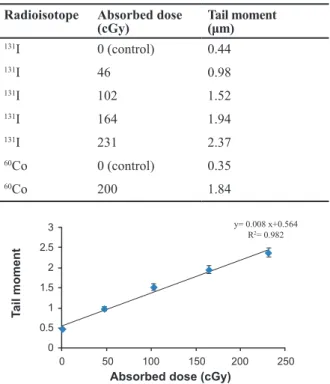
For each category of cells the average tail moment, was determined as induced DNA damage. Table 2 represents absorbed dose and corresponding DNA damage as tail moments in glioblastoma spheroid cells. Dose-response equation of 131I radiation was
Y=0.008X+0.564, where X and Y are absorbed dose in cGy and tail moment in µm as response of cells to the radiation respectively (Fig 4). In order to obtain net damage, DNA damage in control group was sub-tracted from treated groups. Resulted dose-response equation for net damage induced by 131I radiation
was Y=0.008X+0.116. With respect to this equation, 171.1 cGy of 131I radiation and 200 cGy of 60Co
diation have the same biological effect. Therefore, ra-tio of 200 cGy to 171.1 cGy, equal to 1.16, was found to be the RBE of 131I radiation to 60Co radiation. RBE
depends on many factors such as dose, dose rate, cell line and endpoint. RBE in this experiment is resulted from the dose-response equation that showed increas-ing the exposure time to 131I radiation, increases DNA
damage in glioblastoma tumor cells. Also the
corre-lation coeficient showed that there is good linearity
between absorbed dose of 131I in 100μm spheroid cells
and resulted DNA damage (R2=0.982). Comparative
chart for irradiated groups by radiations of 131I and 60Co is shown in igure 5.
Table 2: Induced tail moments in glioblastoma spheroid cells by 131I and 60Co radiations
Tail moment (μm) Absorbed dose (cGy)
Radioisotope
0.44 0 (control)
131I
0.98 46
131I
1.52 102
131I
1.94 164
131I
2.37 231
131I
0.35 0 (control)
60Co
1.84 200
60Co
T
ail moment
3
2.5
2
1.5
1
0.5
0
0 50 100 150 200 250
y= 0.008 x+0.564 R2= 0.982
Absorbed dose (cGy)
Fig 4: Resulted tail moments in U87MG cell lines of 100μm
spheroids relative to absorbed doses of 131I irradiation (Error
bars represent Mean ± SEM of 3 replicates).
T
ail moment (µm)
3 2.5 2 1.5 1 0.5 0
131I (control)131I (46 cGy)
131I (102 cGy)131I (164 cGy) 131I (231 cGy)60Co (control) 60Co (200 cGy)
Fig 5: Comparative chart for induced tail moment in irradi-ated groups by radiations of 131I and 60Co (Error bars
repre-sent SD of 3 replicates).
Discussion
RBE is a complex concept to compare a test ra-diation with reference rara-diation in a comprehen-sive way. RBE depends on dose, dose rate, LET, expose conditions, cell type and endpoint (10) and is used for radiation treatment planning and radia-tion protecradia-tion. For a good treatment planning by ion beams, in order to focus maximum RBE to the center of tumor, knowledge about dependence of RBE to depth is necessary. In radiation protection, RBE is a key factor to derive QF (6).
In this study, RBE of 131I radiation was
evalu-ated relative to 60Co gamma rays in an
experimen-tal approach (10). RBE of 1.16 indicated that 131I
radiation is more effective than 60Co radiation to
produce DNA damage in 100 µm glioblastoma spheroid cells. According to previous studies, RBE of 131I is in the range of 0.3 to 1(11). This variation
may be due to factors affecting RBE.
One of the affecting factors on RBE is energy. Experimental studies have shown that low energy photons and electrons are more effective than high energy photons or electrons to produce biological damage (6). For 60Co and 131I radiation
radiobio-logical mechanisms are different which result in different biological effects. High energy photons of 60Co as indirect ionizing radiation produce en-ergetic secondary electrons by Compton scattering and photoelectric interaction whereas 131I radiation
consists of photons and beta particles. Beta parti-cles of 131I have average range of one millimeter
therefore increase DNA damage due to the cross
ire phenomenon (12, 13). Capability of 131I beta
successful use of 131I in thyroid cancer
treat-ments, researchers have focused on treatment of other malignancies such as hepatocellular car-cinoma (HCC) and pheochromocytoma (PCC)
by radio iodine (14, 15). 131I-MIBG
(Metaiodo-benzylguanidine) is a radio labeled antibody in
target therapy of CNS tumors which uses 131I
to damage tumor cells (16). Since CNS tumors are resistant to radiation and therapies such as chemotherapy and external beam therapy are restricted because of proximity of critical or-gans to these tumors, target therapy can be an alternative.
Findings of this study can be used in radiation therapy or target therapy of glioblastoma tumors.
It is clear that comparison between biological effects of 131I radiation and 60Co gamma rays at
the DNA scale forms the basis of the information
for understanding radiobiology of 131I but more
comprehensive studies are needed in this ield.
Type of cell line is also the other factor which af-fects the RBE. Further studies are recommended to evaluate biological effects of 131I radiation on
various cell lines. Also, in this study, 131I radiation
was evaluated at low dose rate relative to 60Co
gamma rays at high dose rate. Biological effect of a radiation is affected by dose rate where at low dose and low dose rate, cellular repair mech-anism operates and reduces biological effects of radiation (6). Therefore, further studies are
rec-ommended to assess RBE of 131I at various dose
rates to ind possible relation between dose rate
and RBE of 131I radiation. Relationship between
dose rate and RBE will be important in health risks in which exposure to the radioiodine occurs at various dose rates. Moreover, it is important in
radiation therapy to ind the optimal dose rate to
achieve optimum treatment.
Conclusion
This study was an attempt to measure the RBE of 131I in a new approach with high sensitive
tech-niques at the DNA scale. It is indicated that beta particles of 131I, due to cross ire phenomenon,
have an important role in inducing considerable damage in small spheroid cells. The result of this
study (RBE of 1.16) differs from previous ind -ings, indicating dependency of RBE to the condi-tions of the experiment.
Acknowledgments
The authors wish to thank Dr. Ali Akbar Sharai
for his kind assistance during this study. This work was supported by a grant from Tehran
Uni-versity of Medical Sciences. There is no conlict
of interest in this paper.
References
1. Riad R, Kotb M, Omar W, Zaher A, Ebied E, Pitman AG, et
al. I-131 MIBG therapy for advanced stage III & IV neuro-blastoma. J Cancer Ther. 2011; 2(4): 481-489.
2. Riva P, Arista A, Sturiale C, Tison V, Lazzari S, Franceschi
G, et al. Glioblastoma therapy by direct intralesional ad
-ministration of I-131 radioiodine labeled antitenascin anti-bodies. Cell Biophys. 1994; 24(1): 37-43.
3. Task Group on Radiation Quality Effects in Radiological
Protection. Relative biological effectiveness (RBE), quality
factor (Q), and radiation weighting factor (w(R)). A report of the International Commission on Radiological Protec
-tion. Ann ICRP. 2003; 33(4): 1-117.
4. 1990 recommendations of the international commission
on radiological protection. Ann ICRP. 1991; 21(1-3): 1-201.
5. The 2007 recommendations of the international
commis-sion on radiological protection. ICRP publication 103. Ann
ICRP. 2007; 37(2-4):1-332.
6. Hunter N, Muirhead CR. Review of relative biological ef
-fectiveness dependence on linear energy transfer for low-LET radiations. J Radiol Prot. 2009; 29(1): 5-21.
7. Straume T. High-energy gamma rays in Hiroshima and
Nagasaki: implications for risk and WR. Health phys.
1995; 69(6): 954-956.
8. Collins AR. The comet assay for DNA damage and repair: principles, applications, and limitations. Mol Biotechnol.
2004; 26(3): 249-261.
9. Olive PL, Banáth JP. Multicell spheroid response to drugs predicted with the comet assay. Cancer Res. 1997; 57(24): 5528-5533.
10. Zhuang HQ, Wang JJ, Liao AY, Wang JD, Zhao Y. The
bio-logical effect of 125I seed continuous low dose rate irradia
-tion in CL187 cells. J Exp Clin Cancer Res. 2009; 28:12.
11. Hundahl SA. Perspective: National Cancer Institute sum -mary report about estimated exposures and thyroid doses received from iodine 131 in fallout after Nevada atmos
-pheric nuclear bomb tests. CA Cancer J Clin. 1998; 48(5): 285-298.
12. Enger SA, Hartman T, Carlsson J, Lundqvist H.
Cross-ire doses from beta-emitting radionuclides in targeted
radiotherapy. A theoretical study based on experimentally measured tumor characteristics. Phys Med Biol. 2008;
53(7):1909-1920.
13. Unak P, Cetinkaya B. Absorbed dose estimates at the cel
-lular level for 131I. Appl Radiat Isot. 2005; 62(6): 861-869.
14. Chen ZN, Mi L, Xu J, Song F, Zhang Q, Zhang Z, et al.
Tar-geting radioimmunotherapy of hepatocellular carcinoma with iodine (131I) metuximab injection: Clinical Phase I/II
trials. Int J Radiat Oncol Biol Phys. 2006; 65(2): 435-444.
15. Rose B, Matthay KK, Price D, Huberty J, Klencke B, Nor
-ton JA, et al. High-dose 131I-metaiodobenzylguanidine
therapy for 12 patients with malignant pheochromocyto
-ma. Cancer. 2003; 98(2): 239-248.
16. Lessig MK. The role of 131I-MIBG in high-risk