2015
U
NIVERSIDADE DEL
ISBOAF
ACULDADE DEC
IÊNCIASD
EPARTAMENTO DEB
IOLOGIAA
NIMALZebrafish as a model for toxicological studies:
is there a role for Paraoxonase?
Mestrado em Biologia Humana e Ambiente
Lara Andreia Pires Afonso
Dissertação orientada por: Ana Maria Viegas Crespo, Professora
Faculdade de Ciências, UL
Sofia de Azeredo Pereira, Professora Auxiliar NOVA Medical School, UNL
I
Agradecimentos
A realização desta dissertação só foi possível graças à participação e apoio de muitos. Em forma de lista, deixo os meus sinceros agradecimentos.
Agradeço:
À professora Ana Maria Viegas Crespo, por ter aceite ser minha orientadora. À professora Sofia de Azeredo Pereira, pela recepção calorosa, pelos ensinamentos, orientação, paciência e amizade.
À professora Judit Morello, pela partilha de conhecimentos, orientação, simpatia, solicitude e amizade e pelo muito que fica por dizer. Um bem haja.
Às minhas colegas de laboratórico Clara Dias e Aline Marinho, por toda a ajuda, partilha de saberes e técnicas e simpatia.
Aos meus pais, por me incentivarem na prossecução dos estudos. À minha irmã e amiga, Nádia Afonso, por todo o apoio e confiança que deposita em mim, mesmo quando eu a perco. És linda mana!
Aos amigos Daniela Gama, Inês Alves, Margarida Carmona, Rita Morais e Ricardo Gomes, pela ajuda preciosa que me deram a diversos e variados níveis. Sois uns fofos!
Ao Youri, companheiro de vida, por todo o amor, amparo, protecção, paciência (muita) e algumas vozes de espanto, ao longo destes longos meses. Viva eu, viva tu!
II
Abstract
Human PON1 enzyme is a multifaceted biomolecule with antioxidant, anti-atherogenic and detoxifying properties. In that way, it is considered one of the main human body endogenous free-radical scavenging system. PON1 actions are related with the three activities assigned to it: paraoxonase (POase), arylesterase (AREase) and lactonase (LACase). Due to the easy measurement of PON1 activities and to its association to several pathologies linked to oxidative stress, PON1 represents a potential biomarker of disease, in particular for renal disease.
The general goal of this exploratory and pioneer study was to explore the PON1 activities in zebrafish animal model.
Zebrafish is an emergent animal model that is becoming to be largely used as a model in human disease and toxicological studies. Zebrafish presents several characteristics that turns it in an extraordinary, even a unique, model for the toxicological study. Among these characteristics, we can identify the reduced size, the transparency of the embryo, the rapid organogenesis, the low cost of maintenance and the complete sequencing of its genome, where PON1 gene is included. The knowledge on the homology between the zebrafish and human at genetic, molecular and physiological levels is increasing. A good correlation between zebrafish and man has been found for organ toxicity as well.
In the present study, the homology between human (HGNC:9204) and zebrafish (Zgc:91887) enzymes was firstly stablished, supporting that PON1 is highly preserved on the zebrafish, presenting 49% of similitude with the man.
All experiments involving zebrafish were previously approved by the Ethical Committee at the Instituto Gulbenkian de Ciência (IGC).
With the objective of determine if human PON1 activities were present on zebrafish, in the first part of this study, the quantification of the activities POase, AREase and LACase activities were carried out in the whole animal extract, using the same spectrophotometric methods which were validated for human plasma samples. Both AREase and LACase activities were determined while POase was not quantifiable. As the homogenate of whole animal was used, the contribution of other enzymes should not be excluded. For instance, the esterases, as the
III cholinesterases and the carboxylesterase, due to their capacity of hydrolyze the phenyl acetate, might increase the AREase activity. Also, the other paraoxonases of PON family (PON2 and PON3) may contribute for the AREase and LACase activities, when whole animal extract is used, comparatively to plasma.
Aiming to exclude the contribution of the esterases, other than PON enzymes, and as anti-PON1 for the zebrafish was not available, a pharmacological approach was followed by using the paraoxon as an inhibitor of cholinesterase and carboxylesterase enzymes. A significant diminution of the AREase activity was observed when the zebrafish larvae were exposed to the xenobiotic. In attempting to clarify the contribution of PON1 for whole animal AREase and LACase activities, zebrafish larvae were exposed to acetylsalicylic acid, which is an inducer of PON1. No changes in AREase or LACase activities changes were observed upon exposition to the PON1 inducer.
There are many factors that might influence the PON1 status. However, what turns out to be more dominant is age. In that regard, the AREase, LACase and POAse activities during the zebrafish development were explored in the second part of this study. The PON1 activities were determined in zebrafish larvae from day 1 to day 7 post fertilization (dpf). The contribution of chorion – structure that evolves zebrafish embryo – for the AREase and LACase activities was also tested. At 24 hours post fertilization, the zebrafish embryos are evolved by the chorion and within 48 hours post fertilization they may or may not present that structure. The comparison of AREase and LACase activities between groups with and without chorion, at 24 and 48 hours post fertilization disclosed that only AREase activity is influenced by the presence of chorion for embryos with 24 hours post fertilization. These results suggest that proteins with AREase activity integrate the chorion. The evaluation of AREase and LACase activities until 7 dpf pointed to an increase of both activities during the zebrafish development, in particular for the AREase activity.
The zebrafish is being postulated as a good model for nephrotoxicity studies due to the similarity of its pronephro to the human kidney. As PON1 is being studied as a potential biomarker of renal disease, the third objective stablish was to evaluate the relation between PON1 activities and drugs associated to
IV nephrotoxicity. Tenofovir disoproxil fumarate (TDF) and paracetamol (PCM), as drugs with proved association to nephrotoxicity on zebrafish and man, were tested in two concentrations. For both drugs, a decrease in the AREase activity was observed.
The results herein presented show, for the first time, that the zebrafish presents AREase and LACase activities, although they are not to exclusively assign to PON1 enzyme. The POAse activity might not be present in this model. The LACase activity, and in particular the AREase activity, increase during zebrafish development. Moreover, it was for the first time showed that proteins with AREase activity are presented in zebrafish chorion.
The characterization of this enzyme and other important enzymes for xenobiotics and drug detoxification is an important step for the proposal of zebrafish as a model for translational research and to disclose its applicability to pre-clinical studies. In this regard, the present work, give new insights on the three activities involved in endogenous and exogenous compounds metabolism.
Keywords
paraoxonase-1 (PON1); paraoxonase (POase) activity; arylesterase (AREase) activity; lactonase (LAC) activity; esterases; zebrafish; drug-induced nephrotoxicity
V
Resumo
A enzima paraoxonase 1 (PON1) humana é uma biomolécula multifacetada com propriedades antioxidantes, anti-aterogénicas e destoxificantes., sendo considerada um dos principais sistemas de destoxificação de radicais livres endógenos humano. As suas acções estão relacionadas com as três actividades que lhe são associadas: paraoxonase (POase), arylesterase (AREase) e lactonase (LACase). O facto de estas actividades serem facilmente mensuráveis e de a actividade PON1 ter vindo a ser associada a diversas patologias relacionadas com stress oxidativo faz com que seja um potencial biomarcador de doença, particularmente de doenças renais.
O objectivo geral deste estudo exploratório e pioneiro, foi explorar as actividades da PON1 no modelo animal zebrafish.
O zebrafish é um modelo animal emergente que tem vindo a ser largamente utilizado como modelo em estudos de doença humana e em estudos toxicológicos. Apresenta diversas características que o tornam um modelo extraordinário, senão único, para o estudo em toxicologia. Entre elas podemos destacar o tamanho reduzido, a transparência do embrião, a rápida organogénese, o baixo custo de manutenção e a sequenciação completa do seu genoma, onde se inclui o gene PON1. O conhecimento acerca da homologia entre o zebrafish e o ser humano a nível genético, molecular e fisiológico tem vindo a aumentar. Além disso, tem vindo a ser estabelecida uma boa correlação órgão-toxicidade entre zebrafish e o homem.
No presente estudo, começou por se estabelecer a homologia entre a enzima no homem (HGNC:9204) e no zebrafish (Zgc:91887), ficou demonstrado que a PON1 está altamente conservada no zebrafish, apresentando 49% de similitude com o homem.
Todas as experiências realizadas com o zebrafish foram previamente aprovadas pelo Comité de Ética do Instituto Gulbenkian de Ciência (IGC).
Na primeira parte deste estudo e com o objectivo de determinar as actividades da PON1 humana no zebrafish, procedeu-se à quantificação das actividades Poase, AREase e LACase nos homogenatos de embriões/larvas de zebrafish. Foi usado um método espectofotométrico validado para amostras humanas.
VI Ambas as actividades AREase e LACase foram determinadas, enquanto que a Poase não foi quantificável. Contudo, pelo facto de se usar o homogenato de todo o animal, deve considerar-se a contribuição de outras enzimas. Por exemplo, as esterases, como as colinesterases e as carboxilesterases, pela sua capacidade de hidrolisar o fenilacetato, podem aumentar a actividade AREase. Também as outras paraoxonases da família PON (PON2 e PON3) podem contribuir para as actividades AREase e LACase quando o homogenato de todo o animal é usado, em comparação com o plasma.
Com vista a excluir a contribuição das esterases que não as PONs, e dada a indisponibilidade comercial de anticorpos anti-PON1 para o zebrafish, optou-se por uma abordagem farmacológica, usando o paraoxono como inibidor das actividades enzimáticas colinesterase e carboxilesterase. As larvas expostas ao xenobiótico registaram uma diminuição significativa da actividade AREase. Procurando clarificar a contribuição da PON1 nas actividades AREase e LACase foi usado o ácido acetilsalícilico como indutor da PON1 e da sua actividade. Após a exposição ao fármaco não foram registadas alterações quer na actividade AREase, quer na actividade LACase.
São conhecidos vários factores que influenciam o status da PON1 sendo o mais relevante a idade. Partindo deste princípio, na segunda parte deste estudo, quantificaram-se as actividades PON1 ao longo do desenvolvimento do zebrafish. A quantificação das actividades PON1 foi feita do dia 1 até ao dia 7 pós fertilização (dpf). A contribuição do córion – estrutura que envolve o embrião – para as actividades AREase e LACase foi também estudada.
Os embriões zebrafish às 24 hours pós fertilização (hpf) encontram-se envolvidos pelo córion, ao passo que com 48 horas podem ou não apresentar esta estrutura. A comparação das actividades AREase e LACase entre grupos com e sem córion, às 24 e às 48 hpf mostrou que apenas a actividade AREase é influenciada pela presença do córion, para os embriões com 24 hpf. Estes resultados sugerem a presença de proteínas com actividade AREase no córion. A avaliação das actividades AREase e LACase até ao 7 dpf mostrou o aumento de ambas as actividades ao longo do desenvolvimento do zebrafish, em particular para a actividade AREase.
VII O zebrafish é usado como modelo em estudos de nefrotoxicidade devido à semelhança entre o pronefro do zebrafish e do rim humano. Dado que a PON1 tem sido estudada como um potencial biomarcador de doença renal, o terceiro objectivo definido foi avaliar o efeito de fármacos associados a nefrotoxicidade nas actividades PON1. O tenofovir disoproxil fumarato (TDF) e o paracetamol (PCM), como fármacos com comprovada nefrotoxicidade no zebrafish e no homem, foram testados em duas concentrações. Para ambos os fármacos foi observada uma diminuição da actividade AREase.
Em conjunto, estes resultados demostram, pela primeira vez, que o zebrafish apresenta actividades AREase e LACase, embora não permitam atribuir exclusivamente à PON1 as actividades quantificadas. A actividade Poase parece não estar presente neste modelo. A LACase, e em particular a actividade AREase, aumenta ao longo do desenvolvimento do zebrafish. Pela primeira vez foi também demonstrada a presenta de proteínas com actividade AREase no córion do zebrafish.
A caracterização desta e de outras enzimas importantes para a destoxificação de fármacos e xenobióticos representa um passo crucial para a proposta do zebrafish como modelo para investigação de translação e para a avaliação da sua aplicabilidade em estudos pré-clínicos. Neste contexto, o presente trabalho contribui para o conhecimento de três actividades envolvidas no metabolismo de compostos endógenos e exógenos no zebrafish.
Palavras-chave:
paraoxonase-1 (PON1); actividade paraoxonase (POase); actividade arylesterase (AREase); actividade lactonase (LAC); esterases; zebrafish; nefrotoxicidade induzida por fármacos
IX
Abbreviations
AAS Acetylsalicylic acid
AchE Acetylcholinesterase AChE Acetylcholinesterase ApoA Apolipoprotein A ApoA-l Apolipoprotein Al ApoB Apolipoprotein B ApoC Apolipoprotein C ApoE Apolipoprotein E ApoJ Clusterin AREase Arylesterase CaE Carboxylesterases ChE Cholinesterases DHC Dihydrocumarin DMSO Dimethylsulphoxide
dpf Days post fertilization
H2O2 Hydrogen peroxide
Hcy Homocystein
HcyTL Homocysteine thiolactone
HDL High-density lipoproteins
hpf Hours post fertilization
IGC Instituto Gulbenkian de Ciência
LACase Lactonase
LDL Low-density lipoprotein
NAPQI N-acetyl-p-benzoquinoneimine
NSAID Non Steroidal Drug
o-HPPA 3-(o-hydroxyphenyl) proprionic acid
OP Organophosphate
oxLDL oxidized LDL
PBS Phosphate-buffered saline
PCM Paracetamol
X PON Paraoxonase PON1 Paraoxonase-1 PON2 Paraoxonase-2 PON3 Paraoxonase-3 POX Paraoxon
ROS Reactive oxygen species
TDF Tenofovir disoproxil fumarate
XI
Index
1. Introduction ... 1
1.1 Paraoxonase 1 (PON1) as a polyvalent molecule ... 1
1.2 Zebrafish: a prominent animal model ... 5
1.3 Zebrafish and paraoxonase-1 (PON1) ... 9
1.4 Rational and Objectives ... 10
2. Materials and Methods ... 11
2.1 Characterization of amino acid sequence of zebrafish PON1 ... 11
2.2. Zebrafish maintenance ... 11
2.2.1 Adult husbandry ... 11
2.2.2 Breeding and incubation ... 12
2.3. Evaluation of PON1 activities in zebrafish ... 12
2.3.1 Zebrafish euthanasia and homogenization ... 13
2.3.2 Quantification of PON1 activities in zebrafish homogenate ... 14
2.4 Acute exposure of zebrafish larvae to paraoxon, acetylsalicylic acid, tenofovir disoproxil fumarate and paracetamol ... 16
2.4.1 Acute exposure of zebrafish larvae ... 16
2.4.2 Quantification of PON1 activities ... 19
2.5 Statistical analyses ... 19
3. Results ... 20
3.1 Characterization of zebrafish PON1 ... 20
3.2 Paraoxonase activities through zebrafish development ... 20
3.3 Effect of acute exposure to drugs on PON1 activities of zebrafish larvae ... 22
4. Discussion and conclusions ... 27
5. References ... 36
XII
Index of Tables
Table 1 – Principal Landmarks in zebrafish development since 1 to 7 dpf. ... 8
Table 2 – Drugs, associated toxicity and its purpose for the study. ... 17
Table 3 – Drugs concentrations used for the acute exposure assay ... 18
Table 4 – Lethality rate ... 22
Table 5 – Potentially interfering enzymes in the measurement of the AREAse activity of PON1. ... 28
Index of Figures
Figure 1 – Diagram of zebrafish life-cycle. Zebrafish develop quickly from a one-cell zygote that sits on top of a large yolk cell.. ... 7Figure 2 – Diagram for the quantification of paraoxonase activity ... 14
Figure 3 – Diagram for the quantification of arylesterase activity. ... 15
Figure 4 – Diagram for the quantification of lactonase activity. ... 16
Figure 5 – Zebrafish (Ze) PON1 amino acid sequences aligned with the corresponding human (Hu) sequences.. ... 20
Figure 6 – Arylesterase (AREase) and Lactonase (LACase) activities in the absence or presence of zebrafish chorion. ... 21
Figure 7 – Paraoxonase 1 activities of larvae zebrafish since 2 dpf until 7dpf. ... 22
Figure 8 – Arylesterase (AREase) and Lactonase (LACase) activities in 6 dpf zebrafish larvae after paraoxon and acetylsalicylic acid. exposition. ... 24
Figure 9 – Arylesterase (AREase) and Lactonase (LACase) activities in 6 dpf zebrafish larvae after tenofovir disoproxil fumarate and paracetamol exposure. ... 26
1
1. Introduction
1.1 Paraoxonase 1 (PON1) as a polyvalent molecule
Abraham Mazur, in 1946, reported for the first time the enzymatic hydrolysis of organophosphate (OP) compounds in animal tissues (Mazur, 1946). In the early 1950s, Aldridge identified the human serum paraoxonase-1 (PON1) enzyme (Aldridge, 1953a, b).(Aldridge, 1952, 1953)PON1 owes its name to its ability to hydrolyze the OP substrate paraoxon, a toxic metabolite of the insecticide parathion (van Himbergen et al., 2006).
Human PON1 (A-esterase, EC 3.1.8.1) is an enzyme with a molecular mass of 43 kDa and 355 amino acids (Hassett et al., 1991) encoded by the PON1 gene in chromosome 7q21-22. It is a calcium-dependent esterase (Humbert et al., 1993), mostly expressed in the liver and secreted into the blood where it is associated with apolipoprotein AI (ApoA-I) and clusterin (ApoJ) in high-density lipoproteins (HDL) (Durrington et al., 2001). Although the liver is the organ where PON1 is mainly produced, the enzyme is also expressed in other tissues like kidney, brain and lungs (Rodrigo et al., 2001).
PON1 is considered a promiscuous enzyme due to its ability to hydrolyze different substrates: OP compounds such paraoxon, diazinon and chlorpyriphos (La Du et al., 1993), aromatic esters and a variety of aromatic and aliphatic lactones (Billecke et al., 2000; Jakubowski, 2000). The three identified activities of PON1 enzyme (paraoxonase – POase -, arylesterase – AREase - and lactonase - LACase) are closely associated with the mentioned substrates.
The human PON1 belongs to the same family of proteins as paraoxonase-2 (PON2) and paraoxonase-3 (PON3). The three PON genes are closely aligned on chromosome 7q21-22 (Primo-Parmo et al., 1996).
The fact that the three PON genes (PON1, PON2 and PON3) share about 65% of similarity at the amino acid level and that are highly conserved in mammals (Draganov et al., 2005; Humbert et al.,1993) suggest that they play a very important physiological role. However, the different tissue distribution of the three PON enzymes advocates that they play different physiological roles, although these still remain largely unknown (Draganov, 2007). As PON1, PON3 is mainly expressed in the liver (Reddy et al., 2001) and is associated to HDL in serum,
2 albeit to a lesser extent than PON1 (Draganov et al., 2000; Reddy et al., 2001). Regarding PON2, an intracellular enzyme, it is not detectable in serum, but it is largely expressed in a number of tissues, such as the brain, liver, kidney and testis (Mochizuki et al., 1998; Ng et al., 2001). Human PON2 do not show any POase or AREase activities and in PON3 they are very limited. However, the three enzymes show the ability to hydrolyze aromatic and long-chain aliphatic lactones. Therefore, LACase activity is a common feature of the PON enzymes (Draganov et al., 2005; Zhang et al., 2010). Of the three enzymes that form the PON family, PON1 is by far the most studied and best characterized.
The interest in PON1 enzyme was due not only to its ability of detoxification of OP derivate but also to its important physiological roles.
There are now a number of evidences of human PON1 enzyme’s antiatherogenicity, related either to its antioxidant activity, which contributes to a real protection against the oxidation of low-density lipoprotein (LDL) and HDL (Aviram et al., 1999; Navab et al., 2001) or to the hydrolysis of homocysteine thiolactone (HcyTL), a known risk factor for the development of atherosclerotic lesions (van Himbergen et al., 2006). Due to the antioxidant role of the enzyme, PON1 is considered as one of the major endogenous free-radical scavenging system in the human body (Goswami et al., 2009). PON1 protects lipids in lipoproteins (LDL and HDL), macrophages and erythrocytes from oxidation (Aviram & Rosenblat, 2004) and reduces lipid hydroperoxides to hydroxides (Ng et al., 2009). The enzyme also seems to be involved in the protection against oxidative stress. Apart from having been demonstrated that the enzyme degrades hydrogen peroxide (H2O2), a major reactive species produced under oxidative stress (Aviram et al., 1998; Yilmaz, 2012), decreased serum PON1 activity appears to be associated with increased oxidative stress (Rozenberg et al., 2003).
The physiological role of PON1 against oxidative damage, the direct influence of oxidative stress in PON1 activity and the possibility to quantify PON1 in serum, make this molecule a very attractive potential biomarker of pathologies linked to oxidative stress. In fact, decreases in PON1 activity have been associated to a number of disease states, including diabetes mellitus both type I and II (Abbott et al., 1995) familial hypercholesterolemia (Mackness et al., 1991), metabolic syndrome (Sentí et al., 2003) and Alzheimer disease (Wehr et al., 2009).
3 Furthermore, reduced POase activity has also been associated with hepatic disease (Ferré et al., 2006; Keskin et al., 2009) and markedly associated with cardiovascular complications (Shih et al., 2002). In addition, lower LACase activity has been associated with renal dysfunction, being even pointed out as a novel cardiovascular biomarker in end-stage renal disease (Sztanek et al., 2012). However, more research is needed to evaluate the specificity of PON1 as biomarker of disease.
PON1 concentration and/or hydrolytic activity vary considerably among individuals and are modelled by several factors, including environmental, pharmacological, life-style and disease state (Costa et al., 2005; Deakin & James, 2004). Additionally, one must take into account the PON polymorphisms effect on enzyme’s activities and levels. To date, a number of genetic polymorphisms in PON1 gene (Draganov et al., 2005; Humbert et al., 1993), have been described, being the most important two polymorphisms present in the PON1 coding sequence: a glutamine (Q)/arginine (R) substitution at position 192 and a leucine (L)/metionine (M) substitution at position 55 (Adkins et al., 1993; Humbert et al., 1993). The latter, has been associated with plasma PON1 protein levels, with PON1M55 linked to low plasma PON1. Separately, the 192 polymorphism do not affect the concentration of the enzyme, but the affinity and catalytic activity of its two allozymes towards several substrates (Garin et al., 1997; Mackness et al., 1998). Another factor that modelled the activity of PON1 is the age, being the major determinant of PON1 (Costa et al., 2005b)
As it was previously mentioned, PON1 has three different enzymatic activities that will be explained in the next paragraphs.
Paraoxonase activity
POase activity was the first one to be attributed to PON1 (Costa et al., 2003). The measurement of paraoxon hydrolysis, the main substrate used in tests, reflects both quantity and catalityc efficiency of the enzyme (Huen et al., 2009) Consequently, it has been widely used to refer the enzymatic activity in several species and tissues. Apart the fact that this activity is used as a biomarker for the enzyme status, and the possibility of being related to some diseases, it is important to note that this activity does not represent the physiological role of PON1 (Soyoral et al., 2011)
4 As mentioned above, this activity has a protective role against the toxicity of the metabolites of a number of specific OP insecticides and even some nerve gases like sarin and soman (Broomfield and Ford, 1991). Knowing the enzyme activity is, therefore, important to evaluate the degree of protection by PON1 in the degradation of these xenobiotics and can, therefore, be assessed the risk of toxicity to their exposure (Costa et al., 2005).
A large inter- individual variability was demonstrated in PON1 POase activity in several studies using paraoxon as substrate (Humbert et al., 1993). The majority of these variations are due genetic factors, including the occurrence of polymorphisms in PON1 gene (Rainwater et al., 2009). A lower catalytic efficiency PON1Q192 alloform has been reported when compared to PON1R192 in hydrolyzes of paraoxon (Humbert et al., 1993; Li et al., 2000; Mackness et al., 1997). Concerning L55M polymorphism, higher POase activity and mRNA levels have been associated to L allele instead of M allele (Li et al., 2000; Leviev et al., 1997). Despite de name, POase activity is absent or is very limited in PON2 and PON3 enzymes (Draganov et al., 2005) being practically exclusive of PON1.
Arylesterase activity
Recent studies point to AREase activity as the one that best reflects the antioxidant activity of PON1, despite not being directly responsible for it (Otocka-Kmiecik & Orłowska-Majdak, 2009; Rosenblat et al., 2006). Phenyl acetate is the most used substrate in determining this activity, is an artificial substrate which makes it suitable for monitoring the enzyme’s hydrolytic activity, but does not reflect a redox activity (Gur et al., 2007). In enzyme PON3 this activity is very limited, while in PON2 is totally absent (Draganov et al., 2005).
Some authors consider that this test is also a measure of PON1 enzyme quantity, since the rate of phenyl acetate hydrolysis does not differ from PON1192Q and PON1192R alloforms (Eckerson et al., 1983). Studies by Connelly and coworkers (2008) and Kujiraoka and colleagues (2000) confirm a high correlation between serum PON1 concentration and AREase activity. However, there are studies with different conclusions concerning hydrolysis rates between these polymorphisms. In 2001, Brophy and coworkers (2001) described a higher AREase activity to 192QQ genotype, while Rainwater and co-authors (2009) propose a higher AREase activity in the 192RR and 55LL genotypes.
5 Lactonase activity
LACase activity of PON1, despite being the last to be discovered, is considered as the main and native activity of the enzyme. LACase activity is the one which better reflects the physiological activities of PON1 because lactones are considered the natural and preferable substrates of PON1 (Draganov & Teiber, 2008; Khersonsky & Tawfik, 2005). One of the lactones known to be metabolized by PON1 is HcyTL, a toxic intermediate that causes protein N-homocysteinylation. As a result, the homocysteinylated proteins may lose its biological activities (Jakubowski, 2002) . PON1 hydrolyzed HcyTL in homocystein (Hcy) which is a metabolite that if in excess may be extremely toxic and that is accepted as a risk factor for several pathologies including cardiovascular, Alzheimer’s diseases and renal failure (Yilmaz, 2012). The hydrolysis of a toxic intermediate in a metabolite which causes toxicity might seem as being counterproductive and with no physiological advantages. However, some authors, such as Jakubowski (2003), suggest that HcyTL may be more toxic to human cells than Hcy itself.
LACase activity is also involved in the metabolism of some drugs containing lactones. The prodrug prulifloxacin is hydrolyzed into the active quinolone antibiotic NM394 by PON1 (Tougou et al., 1998). Also the local action glucocorticoid γ-lactones drugs relies on LACase activity for the rapid hydrolysis and inactivation of the fraction of the drug that reaches the circulation, preventing systemic side effects (Biggadike et al., 2000).
Concerning polymorphisms, the most influents appear to be 192QQ and 55LL genotypes, for which the higher activities were registered (Brophy et al., 2001; Rainwater et al., 2009).
1.2 Zebrafish: a prominent animal model
Zebrafish (Danio rerio) is a small vertebrate tropical freshwater fish, which belongs to the family Cyprinidae and is originally from the Indian continent (Spence et al., 2008).
Zebrafish has been a popular aquarium fish for decades and since it was introduced as an experiment animal model by George Streisinger in the end of the 60’s its use in biomedical research increased extensively (de Esch et al.,
6 2012; Hill et al., 2005). Zebrafish has been pointed as a prominent model organism in toxicological studies being increasingly used as an in vivo model for assessing drug toxicity and safety and ecotoxicological screening (Parng et al., 2002).
The emergence of this animal model and its increased used in investigation is due to several advantageous features of zebrafish regarding other vertebrate models (Lewis & Eisen, 2003; McGrath & Li, 2008a; Milan et al., 2006; Moens & Prince, 2002; Vascotto et al., 1997; Ward, 2002; Wilson et al., 2009). Among them are:
• The small size and low maintenance cost;
• The high fecundity (around 200-300 eggs/female/week);
• The short generation time of adult zebrafish, typically 3 to 4 months. (Figure 1);
• The transparency of the embryo;
• The remarkable homology with humans at the genetic (85% of homology in genomes), anatomical, physiological, cellular and molecular levels; • The rapid embryonic development, with precursors of all major organs
developed within 36 hours and fully developed larvae available after 48 - 72 hours post fertilization (hpf) ;
• The availability of its completed sequenced genome;
• The increasing availability of powerful genetic tools that enable in a relatively easy way the generation of transgenic and mutant zebrafish; • The ease of maintenance, zebrafish larvae can live for 7 days supported
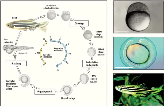
7 Figure 1 – Diagram of zebrafish life-cycle. Zebrafish develop quickly from a one-cell zygote that sits on
top of a large yolk cell. Approximately 6 h post fertilization gastrulation begins, followed by hatching between days 2 and 3, becoming a free larvae. Zebrafish reach sexual maturity around 3 months of age and can live for up to 5 years.
(Adapted from http://www.mun.ca/biology/desmid/brian/BIOL3530/DEVO_03/devo_03.html)
In addition to these characteristics, there are several other specificities of zebrafish that should be noted for toxicity studies where embryo and larvae stages are mainly used. The small size of the zebrafish embryo and larvae (1-4 mm) allows its testing in cell-culture plates or Petri dishes. The high number of offspring also allows the execution of several replicates at the same time (MacRae & Peterson, 2003). Besides, using the whole animal is extremely advantageous concerning toxicity studies, since they provide correlative information which can easily be extrapolated to humans (Wielhouwer et al., 2011). The transparency of the embryos allows for direct and real time observations of morphological alterations (Hill et al., 2005), dyes and antibodies (McGrath & Li, 2008b) under a microscope with no need of tissue sectioning. Other experimental advantages of this model include the small amount of drug that is needed and the ability to quickly absorb small molecules from the surrounding medium mainly through the skin and gills (Rubinstein, 2006).
8 Several studies have already proved the usefulness of the zebrafish model in toxicology. Moreover, there is a very good correlation of organ toxicity between zebrafish and mammals (McGrath & Li, 2008a), including humans (Milan et al., 2003; Khersonsky et al., 2003).
As previously stated, the experiments with zebrafish usually occur during the early lifestages, when the yolk is the primary source of nutrition and there is no need of complementary feeding. This includes the embryonic and larval stages of development, when the zebrafish organs, even if not totally developed, they are completely functional (Pickart & Klee, 2014).
Classically, offspring are considered embryos until they hatch, which occurs between 48 and 72 hpf. Afterwards, the larval period begins and finishes with the yolk absorption (Parichy et al., 2011).
The embryonic period is the best studied and documented of all stages of development of the zebrafish. On the other hand, little is known about the larval development of anatomy. In Table 1, are summarized some of the main landmarks of the zebrafish development between post fertilization days 1 and 7, which are the stages used in this study.
Table 1 – Principal Landmarks in zebrafish development since 1 to 7 dpf.
Dpf Developmental Landmarks References
1
• 1.9 mm embryo length
• Kidney - podocyte migration and glomerulus formation; • Heart starts beating:
• First signs of hepatocyte aggregation; • Endocrine pancreas formed
• Primary neurons differentiation; • Blood circulation start
ZFIN, 2015
Gerlach & Wingert, 2013 Ober et al., 2003 Isogai et al., 2001 Kimmel et al., 1995
2
• 3.1 mm embryo length
• Kidney- onset of glomerular filtration; functional pronephros;
• First wave of definitive hematopoiesis - erythroid myeloid
progenitors;
• Brain ventricles are formed; • Active circulation in trunk and tail;
ZFIN, 2015
Gerlach & Wingert, 2013 McGrath, 2012
Jiang et al., 1996 Isogai et al., 2001
3
• 3.5 mm total body length • Total liver vascularization;
• Establishment of the blood-brain barrier; • Digestive system is fully functional;
• Inflation of the swimbladder;
ZFIN, 2015 Ober et al., 2003 Watanabe et al., 2012 McGrath, 2012 Robertson et al., 2007
4 • 3.7 mm total body length • Hematopoiesis occurs in the thymus and pronephros;
• Liver is in the growth phase;
ZFIN, 2015 McGrath, 2012 Field et al., 2003
5
• 3.9 mm total body length
• Kidney- Proximal convoluted tubule has multiple loops and coils; • Valves between the individual heart compartments appears; • Marked expansion of the exocrine pancreas;
• Digestive system is functional;
ZFIN, 2015
Gerlach & Wingert, 2013 Wallace & Pack, 2003 Yee et al., 2005 Hu et al., 2000
9
6
• 4.2 mm total body length
• Pancreas is located asymmetrically on the right side of the body; • Skin pigmentation;
• Endocrine cells are funtional
ZFIN, 2015
Argenton et al., 2001 Li et al., 2010
7 • 4.5 mm total body length
• 8 teeth ZFIN, 2015
1.3 Zebrafish and paraoxonase-1 (PON1)
To the best of our knowledge there are no studies concerning the activities of paraoxonase (PON) enzymes in zebrafish. Thus, this is the first exploratory study that aims to evaluate the activities of PON1 in zebrafish.
Despite the lack of literature related with PON1 and zebrafish, there are some data that confirm the presence and expression of PON1 gene, as well as proteins such as HDL and ApoA-l (related to PON1 activities on humans and other species) in zebrafish. PON1 gene (zgc:91887) is located in chromosome 16 (versus the chromosome 7 in humans) and was identified by Thisse and co-workers in 2004 (http://zfin.org/ZDB-GENE-040912-6). Zebrafish PON1 protein has 356 amino acids (one more than in humans). There are no data regarding the homology between zebrafish and human PON1. However, it is expected to be high because the amino acid sequences of functionally relevant protein domains have been proven to be evolutionary conserved (de Esch et al., 2012; Reimers et al., 2004). A high similitude is demonstrated in lipid metabolism between humans and zebrafish with the identification of very low-density lipoprotein (VLDL), LDL and HDL fractions in zebrafish plasma (Babin & Vernier, 1989; Babin et al., 1997). Also, zebrafish expresses all the major classes of apolipoproteins such as apolipoprotein A (ApoA), apolipoprotein E (ApoE), apolipoprotein B (ApoB) and apolipoprotein C (ApoC), which share high homology with human apolipoproteins (Babin et al., 1997; Stoletov et al., 2010). ApoA-I is the major protein of HDL and plays an important role in reverse cholesterol transport pathway (Cuchel & Rader, 2006). Zebrafish ApoA-I sequences showed 25.6% identities to human ApoA-I sequences and its gene is largely expressed in the yolk syncytial layer, a structure involved in embryonic and larval nutrition (Babin et al., 1997).
As it is known, PON1 enzyme participates in the inhibition of LDL oxidation. The oxidized LDL (oxLDL) are directly involved in the formation and progression of atherosclerotic lesions, either in humans as in experimental animals (Glass &
10 Witztum, 2001; Miller et al., 2011) . Stoletov and co-workers (2009) demonstrated that hypercholesterolemic zebrafish shows extremely high levels of oxLDL. Although this information does not give enough factual or direct data concerning the activity of the PON1 on zebrafish, it demonstrates the high similarities of the biochemical pathways in which PON1 is involved between humans and zebrafish.
1.4 Rational and Objectives
What is known?
1. PON1 is a polyvalent biomolecule with an important role in the human endogenous free-radical scavenging system, determining susceptibility degrees and protection against insults from physiological or xenobiotics toxins (La Du et al., 2001). Furthermore, it is a potential disease status marker as well as a drug-induced organ toxicity biomarker (Yilmaz, 2012). 2. Zebrafish is an emergent animal model widely used in biomedical and
toxicological research (Parng et al., 2002) comprising nephrotoxic studies (Perner et al., 2007).
What needs to be known?
1. Does zebrafish PON1 have POase, AREase and LACase activities?
2. Do PON1 activities change throughout the embryonic and larvae development?
11
2. Materials and Methods
All experiments involving zebrafish were approved by the Ethical Committee at the Instituto Gulbenkian de Ciência (IGC), according with directives from Direcção Geral Veterinária (Portaria nº 1005/92 de 23 de Outubro).
The mutant zebrafish line mitfaw2/w2;roya9/a9, also called Casper, was specially chosen for this study due to the lack of melanocytes and iridophores. Casper zebrafish are transparent through embryogenesis and adulthood (White et al., 2008). This feature avoids interference of pigments with the colorimetric assays used for the assessment of PON1 activities. Moreover, that allows the visualization of all organs and possible changes along zebrafish development using a standard stereoscope.
2.1 Characterization of amino acid sequence of zebrafish PON1
The method used for the quantification of PON1 activities was developed for human samples. Consequently it is essential to compare the homology of PON1 between human and zebrafish.
A search for PON1 was performed at the Ensembl zebrafish database (http://www.ensembl.org/index.html) in order to check for the localization of PON1 in the zebrafish genome and to obtain the sequence of the protein. The protein sequences of zebrafish PON1 (Zgc:91887 protein) with human PON1
(HGNC:9204) were aligned using Clustal Omega
(http://www.ebi.ac.uk/Tools/msa/clustalo/).
2.2. Zebrafish maintenance
2.2.1 Adult husbandry
Adult zebrafish were grown at the zebrafish facility of Gulbenkian Institute of Science, Lisbon, Portugal.
Animals were maintained in appropriate tanks with freshwater in a 14:10 hr light/dark cycle at 28ºC according to IGC protocols.
12 2.2.2 Breeding and incubation
For each experiment, 6 to 8 pairs of male and female adult zebrafish were crossed according to the fish facility protocols. Briefly, each pair was placed in a mating container with a partition separating male from female in the afternoon of the day before eggs collection. On the next morning the partition was removed and breeding was left for 40 minutes. This breeding method allows to collect all the eggs at the same moment. This strategy decreases the variability of embryo development in the population. Following, eggs were collected using a plastic tea strainer and transferred into a petri dish with embryo medium E3 (5mM NaCl; 0,17mM KCl; 0,33mM CaCl2; 0,33mM MgSO4) and methylene blue. Finally, eggs were kept on the incubator at 28ºC. In this phase it is important to remove the unfertilized eggs because they provide a source of nutrients for bacterial and fungus growth, which rapidly spoil the medium (Brand et al., 2002).
At 24 hpf the initial E3 medium was changed and replaced by embryo medium of E3 solution plus 10 mM 4-(2-hydroxyethyl)-1-piperazineethanesulfonic acid (HEPES) buffer. In order to keep the embryo medium spotless, it was changed every two days. Dead embryos and larvae were also removed under a stereoscope (Nikon 95911) using a disposable plastic pipette.
Zebrafish embryos and/or larvae were kept at 28 ºC until the appropriate stage (1, 2, 3, 4, 5, 6 and 7 days post fertilization – dpf) to perform the experiments.
During the first two days of development zebrafish embryos are surrounded by a structure called chorion. However, at day 2 some embryos are already outside the chorion. To evaluate the influence of the chorion in PON1 activity, embryos of 1 and 2 dpf were collected in parallel with and without chorion. Pronase (Sigma) was used to dechorionate embryos with 1 dpf (Westerfield, 2007). At day 2, since some embryos were already outside the chorion, the selection was made by observation.
2.3. Evaluation of PON1 activities in zebrafish
In order to evaluate the enzymatic activity of PON1 in zebrafish embryos and larvae some preliminary processes are required to prepare the samples, which involve euthanasia and homogenization.
13 2.3.1 Zebrafish euthanasia and homogenization
The chemical methods for euthanasia of zebrafish embryos/larvae (i.e. tricaine, sodium or calcium hypochlorite) are not recommended for enzymatic studies because they can decrease and/or increase the enzyme activities. For this reason, we chose the “rapid chilling” method for zebrafish euthanasia, which works by hypothermal shock (Wilson et al., 2009). The protocol that was used is now described:
1. Prepare ice slush with embryo medium;
2. Fill an appropriate zebrafish embryo container with chilled embryo medium at 2 – 4 °C (5 parts of ice slush / 1 part of liquid);
3. Form a depression in the ice to expose the embryo medium;
4. Transfer a maximum of 20 embryos/larvae to an embryo net and remove as much embryo medium as possible (the use of embryo nets allows minimal transfer of acclimated temperature embryo media and avoids direct contact between zebrafish embryos/larvae and ice);
5. Place the embryo net (with the embryos on it) in the chilled embryo medium;
6. After loss of operculum movement, zebrafish embryos/larvae expose for 20 more minutes to chilled embryo medium in order to ensure death by hypoxia;
7. Add frozen embryo medium (ice slush) when needed during all the procedure to maintain temperature between 2 and 4 ºC.
At the end of this procedure, embryos/larvae were transferred to eppendorfs, placing 25 embryos/larvae in 110 µL of phosphate-buffered saline (PBS) 1x per eppendorf to assess AREase and LACase activities or 50 embryos/larvae in 50 µL of PBS 1x to measure POase activity. Then, embryos/larvae were homogenized using ultrasounds (VWR Ultrasonic Cleaner) between 30 min and three hours, depending of the development stage. The temperature of ultrasounds bath was kept at 4 ºC, adding ice when necessary. Finally, the homogenate was stored at –80ºC until enzymatic quantification. The storage time of the samples never exceeded 2 weeks.
14 2.3.2 Quantification of PON1 activities in zebrafish homogenate
For measurement of PON1 activities (POase, AREase and LACase) spectrophotometrics methods already validated in human samples were used. Essentially, the three activities of the enzyme catalyze the hydrolysis of distinct substrates. These reactions can be monitored spectrophotometrically by color changes which can be innate to the reaction (POase) or the result of adding the reagent phenol red (pH indicator) (AREase and LACase). Enzyme assays were performed in triplicate per sample using a microplate reader (Biotrack II plate reader, Amersham Biosciences).
2.2.2.1 Paraoxonase activity
The POase activity was measured following the method described by Batuca et al (2007). Paraoxon is a substrate of PON1 and is hydrolyzed by POase activity in diethylphosphate and p-nitrophenol (Figure 2), whose production can be monitored spectrophotometrically.
Figure 2 – Diagram for the quantification of paraoxonase activity
In short, paraoxon (1.0 mM) (Sigma-Aldrich) freshly prepared in 290 mL of 50 mM glycine buffer containing 1 mM CaCl2 (pH 10.5) was incubated with 10 µL of sample, at 37 ºC, for 10 min, in 96 well plates (Polysorp). p-nitrophenol formation was monitored at 412 nm and the activity was expressed as mmol p-nitrophenol per mL of zebrafish homogenate per min.
2.2.2.2 Arylesterase activity
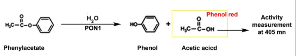
The AREase activity was assessed applying the method published by Dias et al (2014).
15 This method is based on the measurement of acetic acid production resulting from the hydrolysis of phenyl acetate, a substrate of the AREase activity of PON1 (Figure 3). A molecule of phenyl acetate is hydrolyzed into phenol and acetic acid which can be monitored spectrophotometrically by the color variation of the phenol red reagent.
Figure 3 – Diagram for the quantification of arylesterase activity.
Shortly, phenyl acetate (5.0 mM) (Fluka) freshly prepared in 190 mL of 2mM HEPES buffer containing CaCl2 (1.0 mM), BSA (0.005%) and phenol red (Fluka) (106 mM) was incubated with 10 µL of sample, at 37 ºC, for 10 min, in 96 well plates (Polysorp). Acetic acid formation was measured by reading the absorvance at 405 nm and the activity expressed as kU/L, defined as the amount of enzyme producing 1 mmol of acetic acid per minute.
2.2.2.3 Lactonase activity
To quantify LACase activity we used a similar method described above for AREase using as PON1 substrate the lactone dihydrocumarin (DHC) (1mM) (Dias et al., 2014).
This method is based on the hydrolysis reaction of the DHC, a substrate of the LACase activity (Figure 4). The product of this reaction is 3-(o-hydroxyphenyl) proprionic acid (o-HPPA) which can be monitored spectrophotometrically by the color variation of the phenol red reagent.
16 Figure 4 – Diagram for the quantification of lactonase activity.
Briefly, DHC (1.0 mM) (Sigma-Aldrich) freshly prepared in 190 mL of 2mM HEPES buffer containing CaCl2 (1.0 mM), BSA (0.005%) and phenol red (Fluka) (106 mM) was incubated with 10 µL of sample, at 37 ºC, for 10 min, in 96 well plates (Polysorp). After one minute o-HPPA formation was measured by reading the absorbance at 405 nm and the activity expressed as kU/L, defined as the amount of enzyme producing 1 mmol of o-HPPA per minute.
For all PON1 activities the specific enzyme activity was expressed as kU per mg of protein, with 1 kU defined as the amount of enzyme that hydrolyzed 1 mmol of substrate per min. Protein concentration of the samples was determined in duplicate using the spectrophotometer Nanodrop SPECTROstar Omega (BMG Labtech).
2.4 Acute exposure of zebrafish larvae to paraoxon, acetylsalicylic acid, tenofovir disoproxil fumarate and paracetamol
2.4.1 Acute exposure of zebrafish larvae
Zebrafish larvae of 5 dpf were exposed to paraoxon (POX), acetylsalicylic acid (AAS), tenofovir disoproxil fumarate (TDF) and paracetamol (PCM) during 24h at 28 ºC. The choice of the larvae at this stage of development was based on the fact that zebrafish organs are totally differentiate/developed by 120 hpf (5 dpf) (McGrath & Li, 2008a). Therefore, all organs are functional at the time of drug exposure. The features and the reasons for choosing the drugs are summarized in Table 2. Briefly, given the unspecificity of the method for differentiate PON family members in zebrafish homogenate, zebrafish larvae were exposed to POX and AAS to discriminate the activity referent to the PON1 enzyme. In order to evaluate the influence of nephrotoxic drugs in PON1 activities, zebrafish larvae were exposure to TDF and PCM.
17 Table 2 – Drugs, associated toxicity and its purpose for the study.
Drug Classification Toxicity Purpose for the study References Organ Mechanism Paraoxon (POX) • OP oxon, active metabolite of the insecticide parathion • Diethyl 4-nitrophenyl phosphate • Organophosphorous ester
• Autonomic nervous system • Somatic motor nerves • Brain
Inhibition of
acetylcholinesterase (AChE) ↓
cholinergic toxicity by stimulation of muscarinic and
nicotinic receptors Inhibitor of cholinesterases and carboxylesterases • Pubchem, 2015 • Kuster, 2005 • Mileson et al., 1998 • Watanabe, 1989 Acetylsalicylic acid (AAS)
• Non Steroidal Drug (NSAID)
• 2-Acetoxybenzoic acid • Arylester
• Gastrointestinal tract • Kidney - proximal tubule • Liver
• Others
Inhibition of prostaglandin synthesis
Mitochondrial dysfunction and oxidative stress
Increases PON1 activity and expression in HEPG2
cells and primary rat hepatocytes; Hydrolyzes by PON1 in the
plasma.
• Doi & Horie, 2010 • Patrignani et al., 2011 • Jaichander et al., 2008 • Santanam & Parthasarathy,
2007b Tenofovir disoproxil fumarate(TDF) • Nucleotide reverse transcriptase inhibitor • fumaric acid salt of
bis-isoproxycarbonyloxymet hyl ester derivative of tenofovir
• Kidney - proximal tubule • Nephrotoxic in zebrafish
larvae
↑ROS production and ↓ antioxidants and antioxidant
enzymes ↓ Mitochondrial toxicity Nephrotoxic in zebrafish larvae PON1 susceptible to oxidative inactivation
• Van Gelder et al., 2002b • Ramamoorthy et al., 2011 • Unpublish data for our group
Paracetamol (PCM) • Analgesic and antipyretic • N-acetyl-p-aminophenol • Liver
• Kidney - proximal tubule • Nephrotoxic in zebrafish larvae Reactive intermediate NAPQI(*) ↑ROS(**) production Nephrotoxic in zebrafish larvae PON1 susceptible to oxidative inactivation
• Davis & Hanumegowda, 2009 • Karadas et al., 2014
• Lorz et al., 2004 • Peng et al., 2010
18 Two different concentrations were selected for each drug (Table 3).
The rational for the choice of the two concentrations was the following:
1. Paraoxon: inhibitory doses of cholinesterases and carboxylesterases based in literature (toxicity studies with zebrafish larvae) (Küster & Altenburger, 2006; Yozzo et al.; 2013) and in preliminary experiences with several concentrations (supplementary figures).
2. AAS: preliminary experiences with several concentrations (Table in Suplementary figures); maximum concentration achieved by limiting the solubility. As second concentration half of a the maximum concentration was select;
3. TDF and PCM: empirically adjusted from the experience of our group with those drugs.
Table 3 – Drugs concentrations used for the acute exposure assay
Drug Concentration
POX 1 µM 2 µM
AAS 500 µM 1000 µM
TDF 800 µM 3200 µM
PCM 7000 µM 20 000 µM
Stock solutions were prepared in 100% dimethylsulphoxide (DMSO) for POX (100 000 µM) and AAS (4700 µM), or ultrapure water for TDF (9500 µM) or PCM (53 000 µM). Before each experiment serial dilutions were made to obtain the test concentrations. The maximum DMSO concentration was 1%.
Negative controls were exposed to the drug vehicle, DMSO 1% or water.
Zebrafish larvae of 5 dpf were transferred to six well plates (Sarstedt). In order to ensure that in the end of the experiment we had a minimum of 25 larvae, a total of 34 larvae were transferred in a total volume of 6 mL of embryo media per well.
The tested drugs were delivered by soaking to be absorbed mainly through the skin and gills (Rubinstein, 2006).
After 24 hours of drug exposure, zebrafish larvae were subjected to visual observation under a stereoscope (Leika MZ6) to record the lethality. Lethality was defined as absence of a heartbeat or the presence of necrosis.
Four independent exposure experiments were performed. In all cases, at least two replicates were used for each drug concentration. The totals of samples obtained in these experiments were: 7 samples of AAS 1000 µM, TDF 800 µM, PCM 7000 µM
19 and PCM 20 000 µM; 6 samples of POX 1 µM; 5 samples of TDF 3200 µM and 4 samples of POX 2 µM and AAS 500 µM.
2.4.2 Quantification of PON1 activities
PON1 activities in zebrafish homogenate samples were quantified according with the procedures described in 2.3 sections.
2.5 Statistical analyses
Statistical analyses were performed with GraphPad Prism ® version 5.0. One-way ANOVA followed by Dunnett’s post-test or Two-way ANOVA followed by Bonferroni post-test were used to compare differences among the groups. Data were expressed as mean ± standard deviation (SD) and P values < 0.05 were considered statistically significant.
20
3. Results
3.1 Characterization of zebrafish PON1
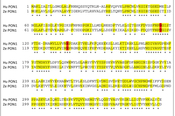
PON1 is highly conserved in mammals (Draganov et al., 2005) and PON1-like proteins can be found in several animal species (Primo-Parmo et al., 1996). Bearing this in mind the human (HGNC:9204) and the zebrafish PON1 (Zgc:91887 protein) protein sequence were aligned. The alignment showed an identity of 49% with an overlap of 356 amino acids (Figure 5).
Hu PON1 1 MAKLIALTLLGMGLALFRNHQSSYQTRLN-ALREVQPVELPNCNLVKGIETGSEDMEILP Ze PON1 1 MGKLAVLSLAVVALAVFIGERLVTLRHVALSYRELTQNYLPNCHLIKGIECGSEDITIID * ** * * ** * ** **** * **** **** * Hu PON1 60 NGLAFISSGLKYPGIKSFNPNSPGKILLMDLNEEDPTVLELGITGSKFDVSSFNPHGIST Ze PON1 61 DGLAFLSTGVKAPGLP-FCSDDPGKIYTLNLLDSEPKIKALSIKGD-FDQDTFNPHGISV **** * * * ** * **** * * * * * ** ******* Hu PON1 120 FTDE-DNAMYLLVVNHPDAKSTVELFKFQEEEKSLLHLKTIRHKLLPNLNDIVAVGPEHF Ze PON1 119 YTDDKDGTMYLFVINHPRGNSQVEIFEFVKDEHALKYIKTIEHELLHSVNDIVAVGTESF ** * *** * *** * ** * * * * *** * ** ******* * * Hu PON1 179 YGTNDHYFLDPYLQSWEMYLGLAWSYVVYYSPSEVRVVAEGFDFANGINISPDGKYVYIA Ze PON1 179 YATNDHYFSNEILKIVENFFTLPWCDVIYYSPETVQVVADGFLLANGINLSLDKRYLYVS * ****** * * * * * **** * *** ** ***** * * * * Hu PON1 239 ELLAHKIHVYEKHANWTLTPLKSLDFNTLVDNISVDPETGDLWVGCHPNGMKIFFYDSEN Ze PON1 239 DVLKHTVTVLEIKKNTVLSRVKEINVGSLADNIELDRESGDLWIGCHPNGFKFMLGDPND * * * * * * * * *** * * **** ****** * * Hu PON1 299 PPASEVLRIQNILTEEPKVTQVYAENGTVLQGSTVASVYKGKLLIGTVFHKALYCE Ze PON1 299 PPGSEVIKIENIHSEKPLVTQVYSDNGSVIIGSSVAAPYRGKVLIGTVYHKVLLCD ** *** * ** * * ***** ** * ** ** * ** ***** ** * *
Figure 5 – Zebrafish (Ze) PON1 amino acid sequences aligned with the corresponding human (Hu) sequences. Zebrafish PON1 (Ze PON1) was aligned with human PON1 (HuPON1). The
sequences have 49% identity in 356 aa overlap. The asterisks indicate the overlap, also highlighted in yellow for better viewing. H115 and H134, residues that mediate AREase and LACase activities of PON1 in humans, are highlighted in red.
3.2 Paraoxonase activities through zebrafish development
Contrarly to AREase and LACase the POase activity was absent in all tested samples. The AREase and LACase activities in zebrafish embryos of 24 and 48 hpf with or without corion are presented on Figure 6.The AREase activity, in 24 hpf
21 samples, was higher in samples with chorion (W/C) (0.86 ± 0.12) with those without chorion (WO/C) (0.79 ± 0.15). This difference was not observed at 48 hpf. The LACase activity was not affected by the chorion in the two tested time-points.
Figure 6 – Arylesterase (AREase) and Lactonase (LACase) activities in the absence or presence of zebrafish chorion. WO/C: without chorion; W/C: with chorion; hpf: hours post fertilization A) AREase activity
measured at 24 and 48 hpf. AREase activity is significantly higher in the zebrafish samples with chorion at 24 hpf.
B) LACase activity measured at 24 and 48 hpf. No significant differences in the activity. Each bar represents
mean ± SD. P values were calculated using Two-way ANOVA and Bonferroni post test. *p<0.05
Since a higher AREase activity was observed in samples of zebrafish embryos at 24hpf, a set of experiments using embryos with 48hpf to evaluate PON1 activity in post-chorion stages was performed.
PON1 activities were measured from 2 up to 7 days of development of zebrafish. Both AREase and LACase activities are shown in Figure 7. There was a gradual increase for the AREase activity throughout the days, experiencing the large increase from day 2 until day 7 (0.44 ± 0.11 versus 0.92 ± 0.04, p<0.001. For the LACase activity, there was only a significant increase in day 7 when compared with day 2 in comparison with day 2 (0.20 ± 0.01 versus 0.27 ± 0.02, p<0.01).
24 hpf 48 hpf 0.0 0.2 0.4 0.6 0.8 1.0 WO/C * A W/C n=6 n=8 n=6 n=6 A R E as e A c tiv ity ( kU /m g pr ot ) 24 hpf 48 hpf 0.0 0.1 0.2 0.3 0.4 0.5 WO/C B W/C n=7 n=10 n=5 n=10 LA C as e A c tiv ity ( kU /m g pr ot )
22 Figure 7 – Paraoxonase 1 activities of larvae zebrafish since 2 dpf until 7dpf: A) Arylesterase activity
(AREase) with significant increase in the activity in 4, 5, 6 and 7 dpf versus 2 dpf. B) Lactonase activity (LACase) with significant increase in the activity in 7 dpf versus 2 dpf. Each bar represents mean ± SD. P values were calculated using One-way ANOVA with Dunnett post test. *p<0.05, **p<0.01 and ***p<0.001.
The POase activity was undetectable in all samples tested since 2 dpf until 7 dpf.
3.3 Effect of acute exposure to drugs on PON1 activities of zebrafish larvae
Zebrafish larvae of 5 dpf were exposed to two different concentrations of POX and AAS (Table 3). The Table 4 contains the lethality observed after 24 hours of drug exposure.
Table 4 – Lethality rate
Drug % Lethality S.D. a Control water 0.59 1.32 Control DMSO 1% 0.59 1.32 POX 1 µM 2.5 2.21 POX 2 µM 61.1 39.6 AAS 500 µM 2.35 1.32 AAS 1000 µM 1.47 3.60 TDF 800 µM 3.0 3.14 TDF 3200 µM 7.35 5.81 PCM 7000 µM 5.15 4.08 PCM 20 000 µM 7.35 5.88
POX had the highest mortality rate (61.1% to POX 2 µM), while AAS caused the lowest mortality rate (1.47% to AAS 1000 µM). Death larvae were removed and AREase and LACase activities were quantified in 25 larvae of each well. Due to high lethality rate of POX 2 µM animals of different wells were collected to equal 25 larvae.
2 3 4 5 6 7 0.0 0.2 0.4 0.6 0.8 1.0 * * * ** *** A n=5 n=5 n=5 n=6 n=4 n=6 Days post-fertilization (dpf) A R E as e (k U /m g pr ot ) 2 3 4 5 6 7 0.0 0.1 0.2 0.3 * B n=5 n=5 n=6 n=6 n=5 n=6 Days post-fertilization (dpf) LA C as e (k U /m g pr ot )
23 The results are presented in Figure 8. AREase activity decreased significantly in zebrafish larvae exposed to POX 1 µM and to POX 2 µM when compared to controls (0.7 ± 0.07 in controls; 0.53 ± 0.05 in POX 1 µM and 0.54 ± 0.03 in POX 2 µM, p < 0.01). AAS did not affect the AREase activity. Concerning LACase activity, there was no impact of POX or AAS concentrations.
24 Figure 8 – Arylesterase (AREase) and Lactonase (LACase) activities in 6 dpf zebrafish larvae after paraoxon and acetylsalicylic acid exposition. POX: paraoxon; AAS:
acetylsalicylic acid. A) AREase activity in zebrafish exposed to POX 1 and 2 µM. Significant decreased in the activity in both POX concentrations versus DMSO 1%. B) AREase activity in zebrafish exposed to AAS 500 and 1000 µM. There was no significant difference in the AREase activity. C) LACase activity in zebrafish exposed to POX 1 and 2 µM
D) LACase activity in zebrafish exposed to AAS 500 and 1000 µM. LACase activity was not affected by POX or AAS at the tested cncentrations. Each bar represents mean ± SD.
P values were calculated using One-way ANOVA and Dunnette post test . **p<0.01
0.0 0.2 0.4 0.6 0.8 Control DMSO 1% (v/V) POX 1 µM POX 2 µM ** ** A n=4 n=5 n=4 A R E as e (k U /m g pr ot ) 0.0 0.2 0.4 0.6 0.8 1.0 AAS 1000 µM Control DMSO 1% (v/V) B n=4 n=4 n=5 AAS 500 µM A R E as e (k U /m g pr ot ) 0.0 0.1 0.2 0.3 Control DMSO 1% (v/V) POX 1 µM POX 2 µM C n=5 n=6 n=3 LAC as e (k U /m g pr ot ) 0.0 0.1 0.2 0.3 Control DMSO 1% (v/V) AAS 500 µM D n=5 n=4 n=7 AAS 1000 µM LAC as e (k U /m g pr ot )
25 Figure 9 shows the results of the quantification of the AREase and LACase activities in 6 dpf zebrafish larvae after acute exposure to TDF or PCM. The lowest concentration of TDF significantly decreased the AREase activity when compared with controls (0.81 ± 0.09 versus 0.69 ± 0.06). Both 7000 µM and 20 000 µM PCM concentrations showed a significant influence in AREase activity, leading to an even higher decrease (0.81 ± 0.09 in controls versus 0.66 ± 0.07 and 0.61 ± 0.08, respectively). Once more, LACase activity was not influenced by any of the drugs at the tested concentrations.
26 Figure 9 – Arylesterase (AREase) and Lactonase (LACase) activities in 6 dpf zebrafish larvae after tenofovir disoproxil fumarate and paracetamol exposure. TDF:
tenofovir disoproxil fumarate; PCM: paracetamol. A) AREase activity in zebrafish exposed to TDF 800 and 3200 µM. Significant decreased in AREase activity for TDF 800 µM versus control. B) AREase activity in zebrafish exposed to PCM 7000 and 20000 µM. Both concentrations induced a significant reduction of the AREase activity. C) LACase activity in zebrafish exposed to TDF 800 and 3200 µM. D) LACase activity in zebrafish exposed PCM 7000 and 20000 µM. LACase activity was not affected either by TDF or Paracetamol at the tested concentrations. Each bar represents mean ± SD. P values were calculated using One-way ANOVA and Dunnette post test . *p<0.05, **p<0.01.
0.0 0.1 0.2 0.3 Control water TDF 800 µM TDF 3200 µM C n=5 n=7 n=5 LA C as e ( k U /m g pr ot ) 0.0 0.1 0.2 0.3 Control water PCM 7000 µM PCM 20000 µM D n=5 n=7 n=7 LA C as e ( k U /m g pr ot ) 0.0 0.2 0.4 0.6 0.8 1.0 Control water TDF 800 µM TDF 3200 µM * A n=5 n=6 n=3 A R E as e ( k U /m g pr ot ) 0.0 0.2 0.4 0.6 0.8 1.0 Control water PCM 7000 µM ** ** B n=5 n=7 n=6 PCM 20 000 µM A R E as e (k U /m g pr ot )
27
4. Discussion and conclusions
The high homology between zebrafish and human has been identified at different levels, of which it can be highlighted the genetic (85%), the morphologic and the molecular levels (Lewis & Eisen, 2003; Milan et al., 2006; Moens & Prince, 2002). In this work we found a high homology between human and zebrafish PON1 protein sequencs. This high homology is supported by: a) the protein alignment of human and zebrafish PON1sequences overlaps at 49% of the amino acids; b) the residues of His 115 and His134, which mediated the AREase and LACase activity in humans (Khersonsky & Tawfik, 2006), are in the same position in both human and zebrafish PON1 proteins, as it can be seen in Figure 5. These evidences allowed us to conclude that zebrafish PON1 constitutes a closer homologue to human PON1.
The POase activity was undetectable in all samples tested from 2 dpf until 7 dpf (data not show) suggesting that zebrafish is unable to hydrolyze ethyl paraoxon and, consequently, does not present POase activity. These results are in agreement with the absence of paraoxonase domain in the PON1 zebrafish protein sequence (supplementary figures). It is also important to emphasize that the POase activity is also absent in other species of fish and some birds, like turkey (Brealey et al., 1980). Even though the POase activity has been the first of the PON1 activities to be found (Costa et al., 2003), the POase activiy does not reflect a physiologic function. It was demonstrated by evolutionary studies that the LACase is the native activity of the enzyme while the POase came as a promiscuous activity across its evolution (Aharoni et al., 2005). These evidences suggest that the POAse activity was not integrated in the PON1 of the zebrafish across its evolutionary process.
The method used for the measurement of PON1 activities allowed quantifying both AREase and LACase. However, considering that our sample, represents the whole zebrafish embryo/larvae, it is important to take into account the unspecificity of the method to discern for the different enzymes with AREase and LACase activity. In human, besides PON1, there are other enzymes with esterase capability such as albumin, acetylcholinesterase, butyrilcholinesterase, carboxylesterase (Li et al., 2005; Taylor et al., 2010), which haves the ability to hydrolyze ester substrates like
28 phenyl acetate. While it is disclosed that the hydrolysis of phenyl acetate in the plasma (primary fluid where articles evaluate the enzymatic activity of PON1) is due almost exclusively to the PON1, because the contribution of other esterases is just residual (Ceron et al., 2014), the same principle cannot be stated when a whole organism is used as a sample. There are no studies that allow us an exhaustive knowledge of esterase enzymes in zebrafish. Table 5 shows some relevant enzymes with esterase activity that are known to be present or absent in the zebrafish. Acetylcholinesterase and carboxylesterase have esterase activity and both are present in zebrafish larvae and thus they can be potential interfering enzymes when measuring AREAse activity.
Table 5 – Potentially interfering enzymes in the measurement of the AREAse activity of PON1. Enzymes Presence in zebrafish References
Albumin No Noël et al., 2010
Acetylcholinesterase Yes Bertrand et al., 2001
Butyrilcholinesterase No Bertrand et al., 2001
Carboxylesterase Yes De Lima et al., 2013
Apart from the esterases enzymes above mentioned, it is necessary to evaluate the possibility of a contribution for other members of the PON family (PON2 and PON3). According to Draganov and co-authors, the three human PONs hydrolyzed aromatic esters in a very different way. As far as the phenyl acetate is concerned, the PON1 hydrolyze it at a higher rate than PON2 and PON3. PON3 present a limited AREase activity and in PON2 it is absent (Draganov et al., 2005). If we assume that the same happens in zebrafish, we can say that we are quantifying PON1 at the expense of PON2 and PON3. However, we emphasize that there is insufficient data to support this extrapolation. Similarly, when measuring the LACase activity, we were confronted with the lack of specificity of the dihydrocoumarin as it is a common substract to the three paraoxonases, even though the PON2 presents a reduced capability to hydrolyze it (Draganov et al., 2005). Noteworthy that this question is due to the use of homogenate of all animal and the lack of data on the enzymatic constitution of zebrafish. The application of this quantification method of the LACase activity in the plasma is specific of the PON1 once the serum PON3 is less abundant than the PON1 and the PON2 is undetectable (Draganov et al., 2000).
29 The use of anti-PON1 zebrafish antibodies would have allowed the specific identification and quantification of the PON1. However, since there are no available anti-PON1 zebrafish antibodies in the market we cannot conclude that the AREase or LACase activities that we quantified proceed exclusively from PON1 enzyme. The AREase activity that we are measuring can be due to other enzyme with ability to hydrolyze the phenyl acetate. For LACase activity, we have to take into account the possible contribution of the three PONs. In humans, LACase activity is much more restricted in PON2 than in PON1 and 3 (Draganov et al., 2005). If we assume a similar behavior for zebrafish PONs, possible we are quantifying PON1 and PON3.
In the absence of antibodies, POX was used in an attempt to isolate AREase PON activity from other esterases and the AAS was used with the aim of clarifying the role of PON1 in AREase and LACase activities.
In accordance with Aldridge (1953) the esterases can be divided in A-esterases (paraoxonase), with the capability to hydrolyze OP compound and B-esterases (cholinesterases - ChE- and carboxylesterases – CaE), inhibited by the latter. The POX, active metabolite of the OP parathion, was used as an inhibitor of any esterases but PONs.
After the 24h exposure of zebrafish larvae to POX it was verified a significant reduction of AREase activity for both tested concentrations, suggesting the complete inhibition of the B-esterases (Figure 8). There are several studies with results pointing in this direction, allowing to assume that the paraoxon concentrations that were used were enough for the enzyme inhibition. In a study performed by Küster (2005) with zebrafish embryos, both enzymes where inhibited with 0.4 µM paraoxon-methyl. Also, Yozzo and its colaborators (2012) demonstrated an 85% reduction on the acetylcholinesterase (AchE) activity in 96hpf zebrafish larvae, when exposed to a concentration of 500nM paraoxon, compared with the vehicle controls.
With the inhibition of the B-esterases, we can conclude that the quantified Arease activity probably can be attributed to PONs (1, 2 or 3). As mentioned before, based on the experiences in human PONs, the PON1 is likely the responsible for this activity as phenylacetate is slowly hydrolyzed by the PON2 and modestly hydrolyzed by PON3 (Draganov et al., 2005). However, it cannot be excluded the possibility of the existence of nonspecific esterases which somehow are not inhibited by the POX and may interfere in the measurement of the AREase activity, increasing its signal.