CLINICAL SCIENCE
Hemodynamic responses and upper airway
morbidity following tracheal intubation in patients
with hypertension: Conventional laryngoscopy
versus an intubating laryngeal mask airway
Elif Bengi Sener, Emre Ustun, Burcu Ustun, Binnur Sarihasan
Ondokuz Mayis University, Faculty of Medicine, Department of Anesthesiology, Kurupelit/Samsun, Turkey.
OBJECTIVES:We compared hemodynamic responses and upper airway morbidity following tracheal intubation via conventional laryngoscopy or intubating laryngeal mask airway in hypertensive patients.
METHODS: Forty-two hypertensive patients received a conventional laryngoscopy or were intubated with a intubating laryngeal mask airway. Anesthesia was induced with propofol, fentanyl, and cis-atracurium. Measurements of systolic and diastolic blood pressures, heart rate, rate pressure product, and ST segment changes were made at baseline, preintubation, and every minute for the first 5 min following intubation. The number of intubation attempts, the duration of intubation, and airway complications were recorded.
RESULTS:The intubation time was shorter in the conventional laryngoscopy group than in the intubating laryngeal mask airway group (16.33¡10.8 vs. 43.04¡19.8 s, respectively) (p,0.001). The systolic and diastolic blood pressures in the intubating laryngeal mask airway group were higher than those in the conventional laryngoscopy group at 1 and 2 min following intubation (p,0.05). The rate pressure product values (heart rate x systolic blood pressure) at 1
and 2 min following intubation in the intubating laryngeal mask airway group (15970.90¡3750 and
13936.76¡2729, respectively) were higher than those in the conventional laryngoscopy group (13237.61¡3413 and 11937.52¡3160, respectively) (p,0.05). There were no differences in ST depression or elevation between the groups. The maximum ST changes compared with baseline values were not significant between the groups (conventional laryngoscopy group: 0.328 mm versus intubating laryngeal mask airway group: 0.357 mm;p= 0.754). The number and type of airway complications were similar between the groups.
CONCLUSION: The intense and repeated oropharyngeal and tracheal stimulation resulting from intubating laryngeal mask airway induces greater pressor responses than does stimulation resulting from conventional laryngoscopy in hypertensive patients. As ST changes and upper airway morbidity are similar between the two techniques, conventional laryngoscopy, which is rapid and safe to perform, may be preferred in hypertensive patients with normal airways.
KEYWORDS: Intubating laryngeal mask airway; Conventional laryngoscopy; Hemodynamic responses; Airway morbidity; Hypertensive patients.
Sener EB, Ustun E, Ustun B, Sarihasan B. Hemodynamic responses and upper airway morbidity following tracheal intubation in patients with hypertension: Conventional laryngoscopy versus an intubating laryngeal mask airway. Clinics. 2012;67(1):49-54.
Received for publication onJuly 7, 2011;First review completed onAugust 23, 2011;Accepted for publication onSeptember 20, 2011 E-mail: [email protected]
Tel.: 00 90 362 3121919/3350
INTRODUCTION
The pressor response to laryngoscopy and intubation is a sympathetic reflex that is provoked by stimulation of the oro-laryngopharynx. Although the corresponding increases in blood pressure and heart rate are transitory and variable,
they are more pronounced and unpredictable in hypertensive patients following laryngoscopy. Consequently, life-threaten-ing complications may develop in these patients, such as pulmonary edema, cerebrovascular hemorrhage, and myo-cardial infarction (1). Hypertensive patients have increased activity of the sympathetic nervous system and may exhibit an exaggerated hemodynamic response to the induction of anesthesia compared with normotensive patients (2,3). Marked increases in catecholamine concentration and in the sensitivity of peripheral vessels to catecholamines in these patients have been reported (4). Thus, undesirable hemody-namic responses to intubation should be reduced via different intubation techniques or pharmacological agents. Copyrightß2012CLINICS– This is an Open Access article distributed under
the terms of the Creative Commons Attribution Non-Commercial License (http:// creativecommons.org/licenses/by-nc/3.0/) which permits unrestricted non-commercial use, distribution, and reproduction in any medium, provided the original work is properly cited.
The intubating laryngeal mask airway (ILMA) is a device used for blindly introducing a tracheal tube. Because it does not require direct exposure of the larynx, tracheal intubation via an ILMA may be less stimulating than conventional laryngoscopy (CL). The cardiovascular effect of inserting a laryngeal mask airway (LMA) has been shown to be similar to that of establishing an oropharyngeal airway and to be less than the effect resulting from a tracheal intubation (5,6). However, the ILMA may exert pressure against the pharyngeal mucosa and may increase airway morbidity because of its rigid silicone-coated steel tube.
To our knowledge, especially in hypertensive patients, no study has been performed to compare the ILMA and CL techniques with respect to the rate pressure product (RPP), ST segment changes and upper airway morbidity following tracheal intubation. As the RPP is an index of myocardial oxygen consumption, higher RPP values together with ST changes may be early warning signals for myocardial ischemia or infarction in hypertensive patients. Airway tissues may be more vulnerable to mechanical damage and pressure from endotracheal intubation in hypertensive patients (7). Hypertension is associated with atherosclerotic changes in the arterial vasculature and microcirculatory insufficiency of the laryngeal nerves (7).
Therefore, we conducted a prospective, randomized study to assess hemodynamic responses to tracheal intuba-tion using CL or an ILMA in hypertensive patients. Included in the study were analyses of the RPP index, the ST segment changes and upper airway morbidity.
MATERIALS AND METHODS
We obtained institutional ethics committee approval and written informed consent from the study participants. We examined 42 patients with controlled hypertension (ASA physical status II) who were scheduled for elective ophthalmic surgery under general anesthesia requiring tracheal intubation. The patients were divided into two groups using a sealed envelope technique. The groups consisted of ILMA (n = 21) and CL (n = 21) groups. The general exclusion criteria were ages of,18 yr or.65 yr; a history of serious pulmonary, coronary artery, or cervical spine disease; a history of difficult intubation; gastroeso-phageal reflux; oto-laryngologic surgery or neurosurgery. Prior to the surgery, all of the patients were evaluated by cardiologists to optimize their antihypertensive treatment and to determine whether other cardiac problems existed. All of the patients received their antihypertensive medica-tion, including diuretics, beta-blockers, calcium channel blockers, or angiotensin-converting enzyme inhibitors (ACEIs) approximately 2 h prior to anesthesia induction. In addition, diazepam (10 mg) PO and famotidine (40 mg) PO were administered 2 h prior to the surgery.
Mallampati scores, thyromental distances, and sternomen-tal distances were measured prior to the surgery, and the type of antihypertensive medications used by the patients were recorded. An electrocardiograph (ECG) capable of automatic ST-segment analysis, a pulse oximeter, and a non-invasive blood pressure monitor were used in the operating room. The baseline values of the aforementioned hemody-namic parameters were recorded following a stabilization period of 3-5 minutes. The patients were in the supine position. Oxygen was administered via a face-mask for 3 min. Lidocaine (0.5 mg.kg-1) was given i.v. to reduce
propofol injection pain. Anesthesia was induced with 2-3 mg.kg-1 propofol and 1
mg.kg-1 fentanyl and was
main-tained with 2% sevoflurane in oxygen and 66% nitrous oxide. Muscle relaxation was achieved using 0.2 mg.kg-1 cis-atracur-ium administered via i.v. The orotracheal intubation was performed when the train-of-four (TOF) count was zero.
A standardized hemodynamic management protocol was used during induction. Any hypotension (systolic blood pressure,80 mmHg) was treated with volume replacement and ephedrine as indicated; persistent hypertension (sys-tolic blood pressure.160 mmHg lasting more than one minute) was treated with i.v. nitroglycerin; tachycardia (HR.120 beats.min–1) was treated with boluses of 30 mg esmolol iv; and bradycardia (HR,50 beats?min–1) was treated with 0.5 mg atropine i.v.
All of the tracheal intubations were performed by skilled anesthesiologists using one of the two intubation techni-ques. Well-lubricated silicone tracheal tubes with internal diameters of 7.5 and 7.0 mm were used for male and female patients, respectively. In the CL group, tracheal intubation was performed with a size 3 Macintosh laryngoscope. In the ILMA group, an ILMA was inserted using a single-handed rotational technique. A size 3 ILM was used for patients
,60 kg, a size 4 ILM was used for patients 60–80 kg, and a size 5 ILM was used for patients.80 kg in weight. The cuff was inflated with air (size 3, 20 mL; size 4, 30 mL; and size 5, 40 mL), and an anesthesia circuit was connected. The position of the ILMA was adjusted until optimal ventilation was obtained. This position was maintained by firmly holding the handle. The tracheal tube was inserted through the ILMA and advanced to 8-9 cm beyond the epiglottic elevating bar if no resistance was felt. If resistance was felt through the tracheal tube, the ILM was readjusted in the patient’s mouth prior to the second attempt of tracheal tube insertion. If tracheal intubation was unsuccessful during the second attempt, either the ILMA was withdrawn 1.5–2.0 cm or the size of the ILMA was changed. The ILMA was removed from the pharynx following successful intubation. In both groups, failed intubation was defined as a procedure lasting more than 3 min. Successful intubation was con-firmed using capnography. In the CL group, face-mask ventilation was permitted between attempts if required. In the ILMA group, ventilation using the ILMA was permitted between attempts if required.
The following data were recorded by an unblinded observer: grade of face mask ventilation (easy, moderate, difficult, failed); number of intubation attempts (a failed attempt was defined as removal of the tracheal tube from the oral cavity or the ILM); intubation time (from insertion of the intubation device to capnographic confirmation); and intrao-perative complications, namely: a - esophageal intubation (lack of a capnography trace following tracheal tube inser-tion), b - mucosal bleeding (blood detected on the intubation device following use), c - lip or dental injury (laceration), and d - episodes of hypoxia during intubation (SpO2,95%).
memory and were verified by other anesthesiologists, thereby reducing bias and error.
Routine monitoring was performed using a Siemens SC 7000 Monitor (Siemens Medical Systems, Denvers, MA, US) and by observing the II and V5 ECG leads. ECG-detected episodes of ischemia were defined as reversible ST segment changes lasting at least 1 min and involving a shift from baseline of either a 1-mm ST depression or a 1-mm ST elevation at the J point. ST segment depression was measured 60 ms following the J point. The baseline level of the ST segment was defined as its position during a stable preoperative period, and the maximal ST change from baseline was determined for each episode.
Postoperative airway complications (sore throat, hoarse-ness, dysphagia, and cough) were assessed by a blinded investigator 24 h following the surgery. All of the complica-tions were graded on an established four-point scale (8).
Statistical Analysis
A power analysis based on a previous article (2) indicated that a sample size of 21 patients per group was required to achieve a power of 80%, to obtain an alpha of 0.05 and to detect a 2300 mmHg.beat.min-1 difference with a standard deviation of 2600 mmHg.beat.min21in the RPP.
All of the data were analyzed using SPSS 16.0 software (SPSS Inc, Chicago, Ill, USA). The descriptive data were analyzed using a factorial analysis of variance. Heart rate, blood pressure, and RPP values were examined using analysis of variance repeated measures. Pair-wise comparisons of the mean values were assessed using the Bonferroni-Dunn test. The Kruskal-Wallis test was used for the scored data. The Pearson correlation and the Spearman rank correlation were used to determine the relationship between the degree of change of the hemodynamic variables and the intubation time and number of intubation attempts, respectively. The data are presented as the mean¡SD unless otherwise noted.p,0.05 was considered statistically significant.
RESULTS
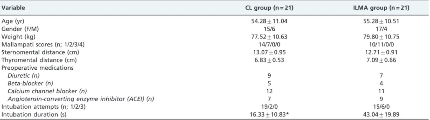
Forty-two hypertensive patients were enrolled in this study. There were no significant differences between the groups with respect to demographic characteristics, airway features or concurrent medications (Table 1). Face-mask ventilation was graded as easy and moderate for all patients.
There was no difference in the number of intubation attempts between the groups, but intubation time was shorter in the CL group than in the ILMA group (p,0.001, Table 1).
The hemodynamic data are presented in Table 2. No significant differences were observed between the groups with respect to heart rate (HR) or blood pressure (BP) values prior to anesthetic induction and insertion of the device. Following the induction of anesthesia, the systolic blood pressure (SBP) and diastolic blood pressure (DBP) decreased to a similar extent in both groups, but HR did not change relative to baseline values. Significant increases in HR were observed only at 1 min following intubation in the CL group relative to baseline values (p,0.05). In the ILMA group, the HR values at 1, 2, and 3 min following intubation were significantly increased compared to base-line values (p,0.05). There were no differences in HR measurements between the groups.
SBP measurements in the ILMA group were significantly higher than for the CL group at 1 and 2 min following intubation (p,0.05). DBP measurements in the ILMA group were also significantly higher than in the CL group at 1 and 2 min following intubation (p,0.01 andp,0.05, respectively). The RPP values in the ILMA group were greater than those for the CL group at 1 and 2 min following intubation (p,0.05). There were no differences in SpO2values between the groups at any time point. None of the patients developed hypoxia (SpO2#95%).
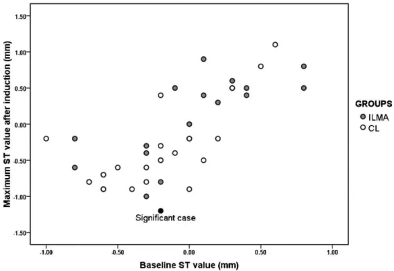
A clinically significant ST change (-1.0-mm depression) was observed in only 1 patient in the CL group, whereas no significant ST changes were observed in the ILMA group. The patient who exhibited a significant ST change was 42 years old and had been treated with a diuretic and a calcium channel blocker preoperatively.
For eight patients in the CL group and 16 patients in the ILMA group, automated ST segment trend analyses revealed ST depression. Nine patients in the CL group and five patients of the ILMA group exhibited ST elevations. There were no differences between the groups with respect to the number of cases of ST depression or elevation (p= 0.094). Four patients in the CL group exhibited no ST changes. The maximum ST changes compared with baseline values were not significant between the groups (CL group: 0.328 mm, ILMA group: 0.357 mm;p= 0.754). The baseline values and the maximum ST values following induction for each case are plotted in Figure 1.
Table 1 -Demographic characteristics, airway assessments, and concurrent medications of the groups.
Variable CL group (n = 21) ILMA group (n = 21)
Age (yr) 54.28¡11.04 55.28¡10.51
Gender (F/M) 15/6 17/4
Weight (kg) 77.52¡10.63 79.80¡10.75
Mallampati scores (n; 1/2/3/4) 14/7/0/0 10/11/0/0
Sternomental distance (cm) 13.07¡0.95 12.71¡0.91
Thyromental distance (cm) 6.83¡0.53 7.09¡0.66
Preoperative medications
Diuretic (n) 9 7
Beta-blocker (n) 5 4
Calcium channel blocker (n) 12 11
Angiotensin-converting enzyme inhibitor (ACEI) (n) 7 9
Intubation attempts (n; 1/2/3) 19/2/0 15/6/0
Intubation duration (s) 16.33¡10.83* 43.04¡19.89
The presented data are the mean¡SD.
CL = Conventional laryngoscopy group, ILMA = Intubating laryngeal mask airway group. *:p
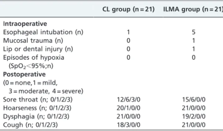
The number and type of intraoperative and postoperative airway complications were similar between the groups (Table 3).
Ten patients in the ILMA group and five patients in the CL group were treated for hypertension with i.v. nitrogly-cerin. None of the patients developed severe bradycardia (HR#50 bpm), hypotension (SBP#80 mmHg), or adverse events (persistent, significant myocardial ischemia, or infarction) during the observation period.
DISCUSSION
The results of this study are important for hypertensive patients with normal airways. Our study demonstrated that hemodynamic responses to intubation with CL are accom-panied by a smaller increase in blood pressure and RPP at 1 and 2 min following intubation than with the ILMA method. Although blood pressures and RPPs increased following intubation in our study, the maximum ST changes compared with baseline values were not significant between the groups. Our results demonstrate that intense and
repeated oropharyngeal and tracheal stimulation by an ILMA induces greater pressor responses than does stimula-tion by laryngoscopy in hypertensive patients.
Normally, orotracheal intubation using a laryngoscope requires elevation of the epiglottis and exposure of the glottis. These maneuvers can cause significant pressor and tachycar-diac responses by enhancing sympathetic activity. Although these are only transient cardiovascular stress responses, they may lead to pulmonary edema, intracranial hemorrhage, and myocardial infarction in hypertensive patients (1).
ILMA-guided orotracheal intubation has some advantages because this procedure does not distort the base of the tongue or directly stimulate the receptors in the larynx. Theoretically, ILMA-guided orotracheal intubation may produce less adverse cardiovascular stress responses. However, there is a controversy as to whether an ILMA significantly attenuates hemodynamic changes following tracheal intubation when compared with the CL technique (9,10).
In a trial by Kihara et al., (9) the hemodynamic response to direct laryngoscopy (DL) was compared with an ILMA and a lightwand device in groups of normotensive and hypertensive patients. The authors concluded that the ILMA attenuated the stress response to tracheal intubation compared with the DL in the hypertensive group. Another study comparing hemodynamic responses to CL and ILMA by Choyce et al. (11) demonstrated that delayed removal of the ILMA was associated with a second pressor response and did not support the use of an ILMA over a direct laryngoscopy purely to decrease the response to intubation. Increases in hemodynamic parameters may be associated with direct tracheal stimulation by a tracheal tube rather than oropharyngeal stimulation.
In contrast to our expectations and theoretical knowledge, the ILMA-guided intubation failed to attenuate the increases in blood pressure and RPP following intubation in our study. Moreover, this technique resulted in higher pressor responses, which is inconsistent with the results of other studies (9,12).
It is likely that our contrasting results are related to factors such as the duration and applied force during intubation. The ILMA technique required a significantly longer dura-tion (approximately three-fold) for intubadura-tion than CL, which is associated with the three-stage process of ILMA placement, intubation, and removal. The longer apnea duration and repeated airway manipulation may have enhanced the hemodynamic responses. In addition, when compared with CL, ILMA-guided orotracheal intubation may impose a greater pressure on the oropharyngeal structures, even exceeding the capillary perfusion pressure of the pharyngeal structures, thus resulting in greater stimuli to the local structures (13). During intubation, moving the ILMA and lifting the epiglottic elevating bar of the ILMA may cause additional stimuli to the orophar-yngeal structures. A mechanical stimulus to the supralar-yngeal area, which is rich in nociceptive receptors, can cause strong hemodynamic responses (14). Finally, removal of the ILMA produces more significant hemodynamic responses, compared to CL, and using the stabilizing rod to advance the tracheal tube may result in greater stimulation of the tracheal wall (15). As we removed the ILMA from the pharynx immediately following successful intubation, the removal of the ILMA may have induced a greater pressor response than CL.
It has been reported that a single 1-min episode of myocardial ischemia, as detected by a 1-mm ST depression
Table 2 -Hemodynamic parameters of the groups.
Variable CL group (n = 21) ILMA group (n = 21)
Heart rate (bpm)
Baseline 81.00¡19.19 83.66¡13.96
Preintubation 77.95¡22.53 81.42¡11.56
Postintubation (min)
1 87.90¡16.65* 93.19¡14.85*
2 86.00¡13.04 89.76¡11.92*
3 85.00¡14.01 90.14¡14.24*
4 81.57¡14.57 86.85¡12.26
5 80.71¡15.40 84.47¡13.06
Systolic BP (mmHg)
Baseline 152.19¡17.46 149.85¡21.03
Preintubation 113.85¡24.57* 116.80¡30.10*
Postintubation (min)
1 150.90¡28.90 171.28¡27.25*{
2 137.90¡22.21* 155.00¡20.15{
3 141.76¡22.36 146.90¡21.90
4 132.76¡22.27* 135.95¡19.69*
5 129.23¡22.02* 128.09¡16.88*
Diastolic BP (mmHg)
Baseline 87.09¡11.37 88.09¡10.57
Preintubation 71.71¡16.27* 71.09¡16.89*
Postintubation (min)
1 91.04¡17.31 105.04¡13.55*{
2 87.00¡15.22 96.19¡11.99*{
3 84.66¡17.51 91.76¡13.83
4 84.00¡16.25 87.28¡9.98
5 79.28¡14.70* 81.90¡9.87*
Rate pressure product (RPP) (mmHg.beat.min-1)
Baseline 12364.90¡3524.73 12659.80¡3342.04
Preintubation 9030.19¡3565.33* 9567.95¡2959.23*
Postintubation (min)
1 13237.61¡3413.47 15970.90¡3750.56*{
2 11937.52¡3160.65 13936.76¡2729.84{
3 12095.76¡3083.06 13308.00¡3290.13
4 10943.66¡3205.21 11865.23¡2768.96
5 10475.80¡2992.56 10889.80¡2494.24*
All values are expressed as the mean¡SD.
CL = Conventional laryngoscopy group, ILMA = Intubating laryngeal mask airway group.
BP = Blood pressure. *:p
,0.05 versus baseline values,
{
or elevation on the ECG, increases the risk for cardiac events 10-fold and the risk of death 2-fold (16). Laryngoscopy and intubation may trigger ST changes due to pressor responses. In our study, a clinically significant ST change was observed in only one patient, who was in the CL group. The maximum ST changes compared with baseline values were not significant between the groups. Theodoraki (17) noted that hemodynamic responses to laryngoscopy and tracheal intubation were not associated with ischemia as assessed by ST-segment monitoring. Our results are consistent with those of her study. We believe that our commercial ST software has moderate sensitivity and specificity in terms of accurately detecting ST segment changes when compared with Holter monitors. Considering this degree of accuracy, ST segment changes in our study were not accompanied by significant increases in SBP or RPP. Hypertensive patients may develop left ventricular hypertrophy (LVH) as target-organ damage. The ST-T configuration seen in the ECG of patients with LVH has a typical pattern of ST depression and asymmetrical T wave inversion (left ventricular strain pattern) (18). The cause of ST segment depression in hypertensive patients is usually a secondary disturbance of the repolarization processes related to LVH development. During anesthesia, ST segment changes may be related to LVH. However, none of our patients exhibited significant clinical or electrocardiographic symptoms of LVH.
RPP is the product of systolic blood pressure and the heart rate and normally is less than 12000; a RPP exceeding 20000 is commonly associated with myocardial ischemia and angina (2,19). An increase in blood pressure without a change in heart rate may be better for myocardial oxygenation than an increase in heart rate along with an increase in blood pressure (20). Kanaide et al. (21) compared the hemodynamic responses, including the RPP and ST-segment changes, to intubation using a lightwand device and a laryngoscope in elderly hypertensive patients. They observed no significant
difference in the RPP and ST-segment changes between the two techniques. Their mean RPP values were less than 15000 following intubation in both groups. They concluded that the major cause of hemodynamic responses to tracheal intuba-tion by the different techniques was direct stimulaintuba-tion of the trachea by the tube. Our results demonstrate that the RPP increases following intubation compared with baseline values in both groups due to increased SBP levels rather than increased HR values. However, increases in the RPP were higher at 1 and 2 min following intubation in the ILMA group than in the CL group. Fortunately, RPPs exceeding 16000 were not observed in any of the patients. The differences observed in the RPP were primarily due to higher arterial pressures in the patients intubated using an ILMA. In contrast, heart rates were not affected by the intubation technique. This situation may be due to preoperative beta-blocker or calcium channel beta-blocker treatment. Therefore, we recommend continuing beta-blocker treatment for hyperten-sive patients during the perioperative period.
We observed no differences in pharyngolaryngeal com-plaints or intraoperative airway complications between the groups. It has been noted that the use of manometry following insertion of an LMA reduces pharyngolaryngeal complications by 70% (22). Thus, the LMA cuff pressure should be routinely measured using manometry and by deflating the intracuff pressure to less than 44 mmHg or 60 cm H2O. Pharyngeal mucosal pressures are generally higher for the ILMA compared with the LMA over the range of cuff volumes (0-40 ml) and are always greater than 157 cm H2O in the distal oropharynx where the curved metal tube is pressed into the posterior oropharyngeal wall. This is much higher than the perfusion pressure of the posterior phar-yngeal wall, which is between 34 and 80 cm H2O (23). An intracuff ILMA pressure that is higher than the recom-mended limits may be attributed to excessive hemodynamic responses due to oropharyngeal stimulation.
There are some possible limitations of this study worth noting. First, we conducted our study on patients with normal airways. In difficult airways, longer intubation durations may lead to different results between CLs and ILMAs. ILMAs may be life-saving for difficult airway management. Therefore, our results may not be applicable to patients with difficult airways. Second, we were not able to use a double-blinded technique to collect hemodynamic parameters. However, the hemodynamic data were stored in the memory of the monitor and were verified by other anesthesiologists. Third, our patients could not perform routine treadmill tests or echocardiograms prior to the surgery. However, all of the patients had controlled hypertension, had no target-organ damage and had been evaluated by cardiologists in terms of coronary artery disease and LVH prior to the surgery. Fourth, our results are specific to the administered anesthetic agents and may not be applicable to other anesthesia regimes, such as the administration of large doses of opioids.
We found that intense and repeated oropharyngeal and tracheal stimulation using an ILMA induces greater pressor responses than does stimulation by CL in hypertensive patients. In conclusion, tracheal intubation with an ILMA does not attenuate the hemodynamic responses to intuba-tion compared with CL in hypertensive patients. As ST changes and upper airway morbidity are similar between the two techniques, the rapid and safe CL technique may be preferable in hypertensive patients with normal airways. In addition, the adjuvant drug choice may be more important than the technique in this population in terms of controlling the hemodynamic responses associated with intubation.
AUTHOR CONTRIBUTIONS
Sener EB conceived, designed and executed the study and was also responsible for the writing of the manuscript. Ustun E was responsible for the data analysis and writing of the manuscript. Ustun B was responsible for the design and execution of the study. Sarihasan B wrote the manuscript.
REFERENCES
1. Fox EJ, Slar CS, Hill CH, Villanueva R, King BD. Complications related to the pressor response to endotracheal intubation. Anesthesiology. 1977;47:524-25, doi: 10.1097/00000542-197712000-00013.
2. FujiiY, Tanaka H, Toyooka H. Circulatory responses to laryngeal mask airway insertion or tracheal intubation in normotensive and hyperten-sive patients. Can J Anaesth. 1995;42:32-36, doi: 10.1007/BF03010568. 3. Stone JG, Foe¨x P, Sear JW, Johnson LL, Khambatta HJ, Triner L. Risk of
myocardial ischaemia during anaesthesia in treated and untreated hypertensive patients. Br J Anaesth. 1988;61:675–9, doi: 10.1093/bja/61. 6.675.
4. Low JM, Harvey JT, Prys-Roberts C, Dagnino J. Studies of anaesthesia in relation to hypertension. VII. Adrenergic responses to laryngoscopy. Br J Anaesth. 1986;58:471–7, doi: 10.1093/bja/58.5.471.
5. Hickey S, Cameron AE, Asbury AJ. Cardiovascular response to insertion of Brain’s laryngeal mask. Anaesthesia. 1990;45:629–33, doi: 10.1111/j. 1365-2044.1990.tb14384.x.
6. Braude N, Clements EA, Hodges UM, Andrews BP. The pressor response and laryngeal mask insertion. A comparison with tracheal intubation. Anaesthesia 1989;44:551–54, doi: 10.1111/j.1365-2044.1989. tb11439.x.
7. Kikura M, Suzuki K, Itagaki T, Takada T, Sato S. Age and comorbidity as risk factors for vocal cord paralysis associated with tracheal intubation. Br J Anaesth. 2007;98(4):524-30, doi: 10.1093/bja/aem005.
8. Stout DM, Bishop MS, Dwersteg JF, Cullen BF. Correlation of endotracheal tube size with sore throat and hoarseness following general anesthesia. Anesthesiology. 1987;76:419–21, doi: 10.1097/00000542-198709000-00025.
9. Kihara S, Brimacombe J, Yaguchi Y, Watanabe S, Taguchi N, Komatsuzaki T. Hemodynamic responses among three tracheal intuba-tion devices in normotensive and hypertensive patients. Anesth Analg. 2003;96(3):890-95, doi: 10.1213/01.ANE.0000048706.15720.C9.
10. Kihara S, Watanabe S, Taguchi N, Suga A, Brimacombe JR. Tracheal intubation with the Macintosh laryngoscope versus intubating laryngeal mask airway in adults with normal airways. Anaesth Intensive Care. 2000;28(3):281-6.
11. Choyce A, Avidan MS, Harvey A, Patel C, Timberlake C, Sarang K, Tilbrook L. The cardiovascular response to insertion of the intubating laryngeal mask airway. Anaesthesia. 2002;57(4):330-3, doi: 10.1046/j. 1365-2044.2002.02463.x.
12. Kahl M, Eberhart LH, Behnke H, Sa¨nger S, Schwarz U, Vogt S, et al. Stress response to tracheal intubation in patients undergoing coronary artery surgery: direct laryngoscopy versus an intubating laryngeal mask airway. J Cardiothorac Vasc Anesth. 2004;18(3):275-80, doi: 10.1053/j. jvca.2004.03.005.
13. Keller C, Brimacombe J. Pharyngeal mucosal pressures, airway sealing pressures, and fiberoptic position with the intubating versus the standard laryngeal mask airway. Anesthesiology. 1999;90:1001-1006, doi: 10.1097/00000542-199904000-00012.
14. Hamaya Y, Dohi S. Differences in cardiovascular response to airway stimulation at different sites and blockade of the responses by lidocaine. Anesthesiology. 2000;93:95-103, doi: 10.1097/00000542-200007000-00018. 15. Shimoda O, Yoshitake A, Abe E, Koga T. Reflex responses to insertion of the intubating laryngeal mask airway, intubation and removal of the ILMA. Anaesth Intensive Care. 2002;30:766-70.
16. Wallace A. Cardiovascular Disease. Chapter 25. In: Stoelting RK, Miller RD (Eds). Basics of anesthesia. 5th ed. Philadelphia: Churchill Living-stone. 2007;pp.363-92.
17. Theodoraki K, Fassoulaki A. Cardiovascular responses to laryngoscopy and tracheal intubation are not accompanied by ST-segment changes. Eur J Anaesth. 2009;26:520–36, doi: 10.1097/EJA.0b013e32831a468d. 18. Huwez FU, Pringle SD, Macfarlane PW. Variable patterns of ST-T
abnormalities in patients with left ventricular hypertrophy and normal coronary arteries. Br Heart J. 1992;67(4):304-7, doi: 10.1136/hrt.67.4.304. 19. Gobel FL, Nordstrom LA, Nelson RR, Jorgensen CR, Wang Y. The
rate-pressure product as an index of myocardial oxygen consumption during exercise in patients with angina pectoris. Circulation. 1978;57:549-56. 20. Kissin I, Reves JG, Mardis M. Is the rate-pressure product a misleading
guide? Anesthesiology. 1980;52:373-4, doi: 10.1097/00000542-198004000-00024.
21. Kanaide M, Fukusaki M, Tamura S, Takada M, Miyako M, Sumikawa K. Hemodynamic and catecholamine responses during tracheal intubation using a lightwand device (Trachlight) in elderly hypertensive patients. J Anesth. 2003;17(3):161-5, doi: 10.1007/s00540-003-0166-8.
22. Seet E, Yousaf F, Gupta S, Subramanyam R, Wong DT, Chung F. Use of manometry for laryngeal mask airway reduces postoperative pharyngo-laryngeal adverse events: a prospective, randomized trial. Anesthesio-logy. 2010;112(3):652-7, doi: 10.1097/ALN.0b013e3181cf4346.
23. Kihara S, Yaguchi Y, Brimacombe J, Watanabe S, Taguchi N. Routine use of the intubating laryngeal mask airway results in increased upper airway morbidity. Can J Anaesth. 2001;48(6):604-8.
Table 3 -Intraoperative and postoperative airway complications.
CL group (n = 21) ILMA group (n = 21)
Intraoperative
Esophageal intubation (n) 1 5
Mucosal trauma (n) 0 1
Lip or dental injury (n) 0 1
Episodes of hypoxia (SpO2,95%;n)
0 0
Postoperative
(0 = none,1 = mild, 3 = moderate, 4 = severe)
Sore throat (n; 0/1/2/3) 12/6/3/0 15/6/0/0 Hoarseness (n; 0/1/2/3) 20/1/0/0 21/0/0/0
Dysphagia (n; 0/1/2/3) 21/0/0/0 19/2/0/0
Cough (n; 0/1/2/3) 18/3/0/0 21/0/0/0