Association between gingivitis and anterior
gingival enlargement in subjects undergoing
fixed orthodontic treatment
Fabricio Batistin Zanatta1, Thiago Machado Ardenghi1, Raquel Pippi Antoniazzi2, Tatiana Militz Perrone Pinto2, Cassiano Kuchenbecker Rösing3
» The authors report no commercial, proprietary or financial interest in the products or companies described in this article.
How to cite this article: Zanatta FB, Ardenghi TM, Antoniazzi RP, Pinto TMP, Rösing CK. Association between gingivitis and anterior gingival enlargement in subjects undergoing fixed orthodontic treatment. Dental Press J Orthod. 2014 May-June;19(3):59-66. DOI: http://dx.doi.org/10.1590/2176-9451.19.3.059-066.oar
Submitted: September 22, 2012 - Revised and accepted: April 08, 2013
Contact address: Tatiana Militz Perrone Pinto
Rua Roberto Holtermann, 314 – Medianeira – Santa Maria/RS — Brazil. CEP: 97.015-570 – E-mail: [email protected]
1 Associate professor, Department of Stomatology, Federal University of Santa
Maria (UFSM).
2 Assistant professor, Franciscano University Center (UNIFRA).
3 Postdoc in Periodontics, Adjunct professor, Federal University of Rio Grande
do Sul, (UFRGS).
» Patients displayed in this article previously approved the use of their facial and in-traoral photographs.
Objective:The aim of this study was to investigate the association among gingival enlargement (GE),
periodon-tal conditions and socio-demographic characteristics in subjects undergoing fixed orthodontic treatment. Meth-ods: A sample of 330 patients undergoing fixed orthodontic treatment for at least 6 months were examined by a single
calibrated examiner for plaque and gingival indexes, probing pocket depth, clinical attachment loss and gingival en-largement. Socio-economic background, orthodontic treatment duration and use of dental floss were assessed by oral interviews. Associations were assessed by means of unadjusted and adjusted Poisson’s regression models. Results: The
presence of gingival bleeding (RR 1.01; 95% CI 1.00-1.01) and excess resin around brackets (RR 1.02; 95% CI 1.02-1.03) were associated with an increase in GE. No associations were found between socio-demographic characteristics and GE. Conclusion: Proximal anterior gingival bleeding and excess resin around brackets are associated with higher
levels of anterior gingival enlargement in subjects under orthodontic treatment.
Keywords:Epidemiology. Orthodontics. Gingivitis. Gingival enlargement. DOI: http://dx.doi.org/10.1590/2176-9451.19.3.059-066.oar
Objetivo:o objetivo desse estudo foi verificar a associação entre volume gengival (AG) com condições periodontais
e características sócio-demográficas em sujeitos com aparelho ortodônticos fixo. Métodos: uma amostra, de 330
participantes com aparelho ortodôntico fixo, por pelo menos seis meses, foi examinada, por um único examinador calibrado, para os índices de placa e gengivais, profundidade de sondagem, nível de inserção clínico e aumento de vo-lume gengival. O status socioeconômico, tempo com aparelho ortodôntico fixo e uso de fio dental foram verificados por entrevista oral. A verificação das associações foi realizada por meio de modelos de regressão de Poisson sem ajuste e ajustados. Resultados: a presença de sangramento gengival (RR 1.01; 95% IC 1.00-1.01) e o excesso de resina em
torno dos braquetes (RR 1.02; 95% IC 1.02-1.03) foram associadas a um aumento do AG. Não foram encontradas as-sociações entre características sócio-demográficas e AG. Conclusão: sangramento gengival proximal na região anterior
e excesso de resina no entorno dos braquetes estão associados a níveis mais altos de aumento de volume gengival na região anterior em sujeitos com aparelho ortodôntico fixo.
INTRODUCTION
The balance of health-disease processes in Periodon-tics depends on adequate supra and subgingival plaque control achieved by patient and professional’s combined eforts. Accumulation of supragingival plaque results in inlammatory alterations of gingival tissues. However, interindividual diferences might explain the diferent patterns of response and time needed for evident clini-cal responses. It is possible that these variations may be associated with diferent plaque growth patterns, local and systemic individual resistance or even a speciic mi-crobial challenge.1,2
Clinical studies suggest that orthodontic treat-ment may be associated with a decrease in periodon-tal health.3,4,5 One of the adverse periodontal
altera-tions is a hypertrophic form of gingivitis.5,6,7 The exact
mechanism for the development of gingival enlarge-ment (GE) is not yet completely understood, but it probably involves increased production by ibroblasts of amorphous ground substance with a high level of glycosaminoglycans. Increases in mRNA expression of type I collagen and up-regulation of keratinocyte growth factor receptor could play an important role in excessive proliferation of epithelial cells and develop-ment of GE.8 In some studies, poor oral hygiene
in-creased GE.9,10 Other clinical studies concluded that
overall gingival changes during orthodontic treatment are transient with no permanent damage to the peri-odontal supporting tissues.11,12
The presence of orthodontic brackets also increases the skills and efort required to maintain good levels of oral hygiene, especially on the proximal surfaces.11
Microbiological studies demonstrate that when ixed orthodontic appliances are placed, the potential for quantitative13,14 and qualitative15,16 changes in the
micro-bial composition of these areas enhances. Thus, peri-odontal reaction might be elicited by a change in the microbiological environment.
To the best of our knowledge, there are no studies assessing GE and associated factors in individuals un-dergoing orthodontic therapy. Thus, this study aimed at assessing the prevalence of GE and associated factors in a group of orthodontic patients.
MATERIAL AND METHODS
This cross-sectional study examined subjects who were undergoing orthodontic treatment in an
orth-odontic graduate program in Santa Maria, Brazil. Ethi-cal approval was obtained from the Franciscan Univer-sity Center Institutional Review Board prior to the start of the study (protocol registration number 1246 in the National Ethics Committee). Subjects who agreed to participate signed an informed consent form. Patients diagnosed with oral pathological conditions were ad-vised to seek consultation and treatment.
Eligibility criteria
To be eligible for the study, individuals should have been undergoing ixed orthodontic treatment for at least 6 months. Exclusion criteria comprised presence of diseases and conditions that could pose health risks to the participant or that could interfere in clinical exami-nation, for instance, users of nifedipine, cyclosporine or phenytoin, contraceptive and decompensated diabet-ics. Individuals who had undergone antibiotic therapy within three months prior to examination were also excluded. Female subjects were excluded if they were pregnant or breastfeeding. Additionally, individuals who required a prophylactic regimen of antibiotics for clinical examination were excluded.
Study sample
The orthodontic dental clinic of Ingá College (UNINGÁ) was treating, during data collection period, an estimated population of seven hundred patients, six hundred of which were regularly under treatment and, therefore, were assessed for eligibility criteria. This pro-cess resulted in four hundred eligible individuals, 330 of which were assessed, resulting in a non-response rate of less than 20%. Clinical examinations were performed between September/2009 and July/2010.
Examinations
A single, calibrated examiner performed all clini-cal examinations with the aid of a dental assistant who took the records. All permanent and fully erupted teeth, except for third molars, were examined by means of a manual periodontal probe (Neumar®, São Paulo,SP,
Brazil). Six sites (mesio-buccal, mid-buccal, disto-buc-cal, disto-lingual, mid-lingual, and mesio-lingual) were assessed for each tooth.
depth (PPD), attachment loss (CAL) and bleeding on probing were assessed. Thereater, gingival enlarge-ment19 was assessed. The degree of gingival thickening
in both labial and lingual aspects was scored as follows: 0 = normal; 1 = thickening up to 2 mm; 2 = thickening greater than 2 mm. The extent of gingival tissues en-croachment onto the adjacent crowns was also graded, using 0, 1, 2 and 3 on the labial and lingual surfaces. The sum of both scores (thickening and gingival en-croachment) resulted in an enlargement score for each gingival unit. The maximum score obtainable using this method is 5. Additionally, excess resin was dichoto-mously assessed by inspection with a probe around the bracket on the buccal surfaces of each bonded bracket. For this purpose, the buccal surface was divided into distal, mesial and cervical. Excess resin, located at less than 1 mm from the gingival margin was present.
Ater clinical examination, socioeconomic and de-mographic data were collected using a structured writ-ten questionnaire. Race was scored as white or non-white. Socioeconomic status was scored by the individ-ual’s level of education (≤ 11 years / > 11 years) which, in Brazil, corresponds to those who have completed high school or those educated beyond high school level. Household income information was measured in terms of the Brazilian minimum wage, which corresponded to approximately US$290 (US dollars) during the pe-riod of data collection. Income information was mea-sured dichotomously (≤ 5 national minimum wages / > 5 national minimum wages). PII and GI were dichot-omized as visible plaque (present / absent) and gingi-val bleeding20(present / absent), respectively. The
per-centages of sites with visible plaque, gingival bleeding and bleeding on probing were calculated individually. The questionnaire also reported the declared frequency dental loss use. Regular interdental hygiene was deined as the use of dental loss at least once a day. Non-users of dental-loss were deined as subjects who did not use interdental oral hygiene devices every day or who did not perform interdental hygiene.
Measurement reproducibility
The examiner was trained and calibrated to per-form the clinical measurements before the study started. Assessment of measurement reproducibility was con-ducted with 15 subjects who were divided into three groups of 5 subjects each. In each one of the groups,
replicate measurements were made by the examiner on two occasions, with a two-day interval. At the site level, reproducibility was assessed by means of the weighted Kappa (± 1 mm) and resulted in values of 0.73 for prob-ing pocket depth and of 0.68 for clinical attachment loss. Gingival enlargement reproducibility was assessed by means of Intraclass Correlation coeicient at the site level in 15 subjects, resulting in an ICC of 0.86.
Data analysis
Most participants presented low mean values for PPD and CAL. Thus, these data were not used in the present analysis. For this exploratory analysis, data pat-tern distribution was analyzed and non-parametric (Mann-Whitney and Kruskal-Wallis) tests were used. Ater descriptive analysis, the frequencies of gingival enlargement scores (full mouth mean) were compared for diferences between demographic characteristics, so-cioeconomic indicators and clinical status.
Unadjusted Poisson regression analysis with robust variance was performed to correlate the overall mean of the anterior gingival enlargement (AGE) score with each demographic, socioeconomic and clinical indicator. AGE was considered for anterior teeth located in es-thetic regions, only. This region was chosen because a recent publication21 emphasized the impact of AGE in
oral health related quality of life (OHRQoL) in sub-jects undergoing ixed orthodontic treatment. In this analysis, the outcome was considered as continuous. Additionally, rate ratios (RR), which correspond to the quotient between average scores of each comparison group, and 95% conidence intervals (95% CI) were calculated. A multivariate model was then run with the covariates. These covariates were selected using a back-ward stepwise procedure. Only variables with p ≤ 0.20 or those that presented a conceptual association with the primary outcome were included in the model. In order to be retained in the inal multivariate model, the vari-ables should present p ≤ 0.05. The statistical sotware STATA 9.0 (Stata Corp, College Station, USA) was used for all analyses.
RESULTS
The unadjusted Poisson regression assessment of as-sociations revealed use of dental loss (RR 1.25; 95% CI 1.08-1.43), percentage of proximal anterior gingival bleeding (RR 1.01; 95% CI 1.0-1.01) and percentage of sites with excess resin around brackets (RR 1.02; 95% CI 1.02-1.03) as the main covariates associated with higher levels of anterior gingival enlargement. In the multivariate regression model, the percentage of proxi-mal anterior gingival bleeding remained associated, in which higher frequencies of anterior gingival bleed-ing were associated with a 1.01 fold increase in average AGE scores (RR 1.01; 95% CI 1.0-1.01). Additionally, the percentage of sites with excess resin around brack-ets also remained associated, with higher frequencies of sites with excess resin around brackets being associated with a 1.02 fold increase in average AGE scores (RR 1.02; 95% CI 1.02-1.03).
DISCUSSION
The present study aimed at investigating poten-tial associations among gingival enlargement, socio-demographic and clinical characteristics. When GE was assessed by frequency of scores, statistically signiicant diferences between nearly all independent variables and frequencies of GE were observed. While the clinical rel-evance of these diferences could be dubious for some of these variables, others such as household income, use of dental loss and time with orthodontic appliances pre-sented substantial clinical diferences for the prevalence of gingival enlargement score two. Ater regression analysis had been performed, proximal anterior gingival bleeding and excessive resin around brackets were re-vealed as independent variables associated with the level of anterior gingival enlargement.
Since hyperplasic gingival response is a common re-sponse to plaque accumulation in subjects undergoing ixed orthodontic therapy. this study used two indexes to assess gingival inlammatory status: color alteration and/or swelling and bleeding ater marginal probing. Gingival bleeding IS shown by clinical and histological studies to be an earlier and more sensitive sign of gingi-val inlammation in comparison to cardinal signs such as redness and swelling, which may be rather subjective and not very reliable.23,24 Furthermore, a single
calibrat-ed examiner, unaware of dental loss use habits, assesscalibrat-ed all parameters. These methods probably increased the quality of data collection as well as results reliability.
Variables n (%)
Demographic characteristics
Sex
Male 159 (48.1%)
Female 171 (51.8%)
Ethnicity
White 263 (79.7%)
Non-White 67 (20.3%)
Age (years)
14 - 19 162 (49.0%)
20 - 24 121 (36.6%)
25 - 30 47 (14.2%)
Socioeconomic status
Household Income
≤ 5 270 (81.8%)
> 5 60 (18.1%)
Educational level
> 11 years 159 (48.1%)
≤ 11 years 171 (51.8%)
Clinical status
TUFOT (Months)
6 - 12 185 (56.06%)
> 12 145 (43.93%)
Dental floss
Users 81 (24.5%)
Non-users 249 (75.5%)
TUFOT: Time under fixed orthodontic treatment.
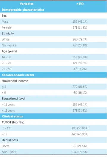
Table 1 - Research subjects’ clinical and socio-demographic characteristics.
study sample comprised 330 individuals aged between 14 and 30 years old, 171 (51.8%) female and 159 (48.2%) male, 263 (79.7%) white and 67 (20.3%) non-white. As for periodontal diagnosis, based on patient’s age and periodontal data explored in another study22 (2.06 mm
± 0.18 and 1.6 ± 0.11 for mean probing depth and clini-cal attachment levels in proximal sites, respectively) most patients had periodontal diagnosis of gingivitis, only.
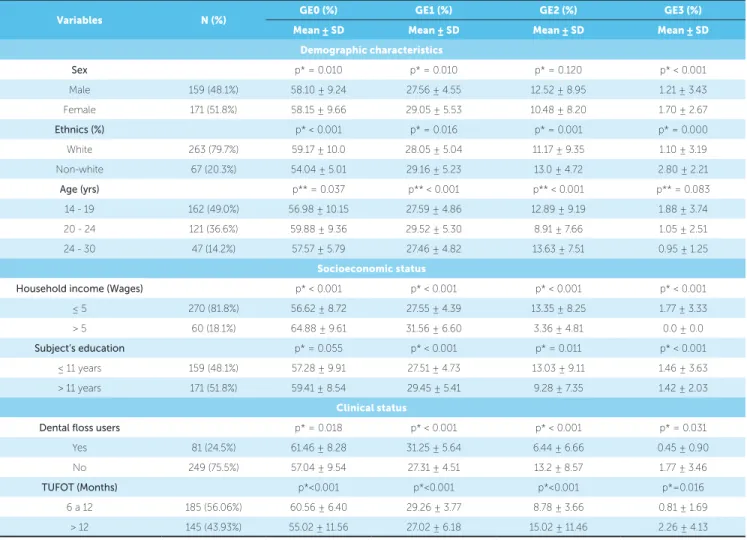
Table 2 - Univariate analysis between socioeconomic factors and oral clinical conditions related to frequencies of different scores of gingival enlarge-ment index.
Caption :GE0=Gingival enlargement score 0; GE1=Gingival enlargement score 1; GE2=Gingival enlargement score 2; GE3=Gingival enlargement score 3; TU-FOT: Time under fixed orthodontic treatment; p*= Mann Whitney test; p**= Kruskall Wallis test
Variables N (%) GE0 (%) GE1 (%) GE2 (%) GE3 (%)
Mean ± SD Mean ± SD Mean ± SD Mean ± SD
Demographic characteristics
Sex p* = 0.010 p* = 0.010 p* = 0.120 p* < 0.001
Male 159 (48.1%) 58.10 ± 9.24 27.56 ± 4.55 12.52 ± 8.95 1.21 ± 3.43
Female 171 (51.8%) 58.15 ± 9.66 29.05 ± 5.53 10.48 ± 8.20 1.70 ± 2.67
Ethnics (%) p* < 0.001 p* = 0.016 p* = 0.001 p* = 0.000
White 263 (79.7%) 59.17 ± 10.0 28.05 ± 5.04 11.17 ± 9.35 1.10 ± 3.19
Non-white 67 (20.3%) 54.04 ± 5.01 29.16 ± 5.23 13.0 ± 4.72 2.80 ± 2.21
Age (yrs) p** = 0.037 p** < 0.001 p** < 0.001 p** = 0.083
14 - 19 162 (49.0%) 56.98 ± 10.15 27.59 ± 4.86 12.89 ± 9.19 1.88 ± 3.74
20 - 24 121 (36.6%) 59.88 ± 9.36 29.52 ± 5.30 8.91 ± 7.66 1.05 ± 2.51
24 - 30 47 (14.2%) 57.57 ± 5.79 27.46 ± 4.82 13.63 ± 7.51 0.95 ± 1.25
Socioeconomic status
Household income (Wages) p* < 0.001 p* < 0.001 p* < 0.001 p* < 0.001
≤ 5 270 (81.8%) 56.62 ± 8.72 27.55 ± 4.39 13.35 ± 8.25 1.77 ± 3.33
> 5 60 (18.1%) 64.88 ± 9.61 31.56 ± 6.60 3.36 ± 4.81 0.0 ± 0.0
Subject’s education p* = 0.055 p* < 0.001 p* = 0.011 p* < 0.001
≤ 11 years 159 (48.1%) 57.28 ± 9.91 27.51 ± 4.73 13.03 ± 9.11 1.46 ± 3.63
> 11 years 171 (51.8%) 59.41 ± 8.54 29.45 ± 5.41 9.28 ± 7.35 1.42 ± 2.03
Clinical status
Dental floss users p* = 0.018 p* < 0.001 p* < 0.001 p* = 0.031
Yes 81 (24.5%) 61.46 ± 8.28 31.25 ± 5.64 6.44 ± 6.66 0.45 ± 0.90
No 249 (75.5%) 57.04 ± 9.54 27.31 ± 4.51 13.2 ± 8.57 1.77 ± 3.46
TUFOT (Months) p*<0.001 p*<0.001 p*<0.001 p*=0.016
6 a 12 185 (56.06%) 60.56 ± 6.40 29.26 ± 3.77 8.78 ± 3.66 0.81 ± 1.69
> 12 145 (43.93%) 55.02 ± 11.56 27.02 ± 6.18 15.02 ± 11.46 2.26 ± 4.13
The use of dental loss was dichotomized into sub-jects who use it every day and individuals who do not have this habit. This cutof was made based on evidence that demonstrates reduction in inlammatory parameters associated with gingivitis when lossing is performed ev-ery day.25 The use of dental loss was not an independent
predictor for AGE. There is no evidence in population basis evaluating the association between dental loss and GE. However, short-term clinical studies with subjects without orthodontic appliances have demonstrated a signiicant improvement in the interproximal gingival condition with the correct use of dental loss.25,26,27 It is
important to highlight that some studies conducted with highly motivated individuals who eiciently brush their teeth revealed that the incorporation of lossing in oral hygiene routines does not signiicantly contribute to improve interproximal gingival conditions.28 Thus, the
lack of association between dental loss and AGE may be
due to the possibility of toothbrushing alone resulting in low levels of plaque accumulation in anterior segments. These results should be interpreted with care. Although no statistically signiicant diferences were observed in AGE in users and non-users of dental loss, this habit is also recommended for other purposes, such as preventing attachment loss, halitosis, carious lesions, etc.
The positive correlation found between sites with ex-cess resin around brackets and levels of AGE conirmed the hypothesis that oversight in the bonding of brackets might inluence gingival enlargement. The standard of inishing/polishing techniques and surface roughness proved to be important factors for bacterial adhesion with diferent types of dental materials.29,30 Thus, our
Variables n (%) AGE AGE AGE
Mean ± SD RR (95%IC) RR* (95% IC)
Demographic characteristics
Sex p = 0,142 **
Male 159 (48.1%) 0.72 ± 0.33 1.08 (0.97-1.20)
Female 171 (51.8%) 0.66 ± 0.34 1
Ethnics (%) 0.188 **
White 263 (79.7%) 0.7 ± 0.34 1
Non-white 67 (20.3%) 0.64 ± 0.32 0.91 (0.79-1.04)
Age (yrs) p = 0.123 **
14-19 162 (49.0%) 0.73 ± 0.35 1.11 (0.96-1.29)
20-24 121 (36.6%) 0.66 ± 0.34 1.00 (0.85-1.17)
24-30 47 (14.2%) 0.65 ± 0.29 1
Socioeconomic status
Household income (Wages) p = 0.213 **
≤ 5 270 (81.8%) 0.70 ± 0.33 1.10 (0.94-1.28)
> 5 60 (18.1%) 0.64 ± 0.35 1
Subject’s education p = 0.14 **
< 11 years 159 (48.1%) 0.73 ± 0.35 1.08 (0.97 – 1.20)
≥ 11 years 171 (51.8%) 0.63 ± 0.31 1
Clinical status
Dental floss users (%) p = 0.000 p = 0.239
Yes 81 (24.5%) 0.58 ± 0.34 1 1
No 249 (75.5%) 0.73 ± 0.33 1.25 (1.08-1.43) 1.07 (0.95-1.22)
TUFOT (Months) p = 0.18 **
6 a 12 185 (56.06%) 0.64 ± 0.32 1
> 12 145 (43.93%) 0.75 ± 0.35 0.91 (0.79-1.04)
PAGB (%) - p = 0.000 p = 0.000
1.01(1.0 – 1.01) 1.01 (1.00-1.01)
Excessive resin (% of sites) - p = 0.000 p = 0.000
1.02 (1.02-1.03) 1.02 (1.02-1.03)
Table 3 - Unadjusted (RR) and Adjusted assessment (RR*) for association between socioeconomic factors and oral clinical conditions related to anterior gingival
enlargement average. Robust poisson regression analysis.
RR: Ratios rate; AGE: Anterior gingival Enlargement; TUFOT: Time under fixed orthodontic treatment; PAGB: Proximal anterior gingival bleeding. *Adjusted by sex, ethnics, age, Household income, subjects´s education, TUFOT, dental floss, GAE and excessive resin ** Variables not included in the final multiple model after adjustment.
In this study, variables related to socio-demographic characteristics, such as sex, ethnicity, household in-come and subjects’ level of education, were not asso-ciated with anterior gingival enlargement. However, it has been established that individuals from diferent socioeconomic backgrounds are exposed to diferent risk factors that afect oral health. Individuals with lower socioeconomic status are subjected to material deprivation which could inluence their engaging in riskier behaviors, thereby resulting in worse oral health conditions.31 Furthermore, low educational level may
lead to reduced income, unemployment and poor
occupational status, all of which could inluence oral health.31 Our contradictory results could be explained
Studies assessing the association between levels of anterior gingival enlargement and socioeconomic as well as clinical conditions in orthodontic subjects (us-ing multiple regression analyses controlled for other socio-demographic and clinical variables which may act as confounders) are inexistent. From this perspec-tive, the present study provides new information. In addition, Poisson regression with robust variance was used in order to provide PR estimates which are eas-ier to interpret than odd ratios. In a situation of an-terior gingival enlargement, with prevalence higher than 50%, odd ratios would strongly overestimate PRs.32 It is important to highlight that this study had
a cross-sectional design, which hypothesizes relations between the outcome and predictor variables without establishing causal relationships. This is a limitation of this study. However, conclusions from cross-sectional studies are important to identify indicators that may be included in longitudinal or, even, experimental stud-ies. The present study comprised 330 orthodontic pa-tients attending a private orthodontic specialist train-ing program. This sample limited the extent to which these indings can be generalized to a larger popula-tion. Nevertheless, analyses were performed with suf-icient power and the analytical results strengthen the hypotheses of this study.
Evidence shows that gingival enlargement is as-sociated with esthetic impairment and, in more se-vere cases, with phonetic alterations and masticatory problems.7 A recent publication21 conducted with
an orthodontic sample emphasized the impact of anterior gingival enlargement in oral health related quality of life (OHRQoL). Thus, prevention and/ or treatment of gingival enlargement may contribute to improve the OHRQoL of orthodontic patients. According to our results, proximal anterior bleed-ing and excess resin around brackets were associated with higher levels of anterior gingival enlargement. However, further studies are required to understand whethwe it is possible that prevention and treatment of gingivitis and careful bonding of brackets may re-sult in decrease in the prevalence or even the severity of GE in orthodontic subjects.
CONCLUSIONS
Anterior gingival enlargement is associated with gingival inlammation and excess resin around brackets.
Acknowledgements
1. Trombelli L, Scapoli C, Tatakis DN, Grassi L. Modulation of clinical expression of plaque-induced gingivitis: efects of personality traits, social support and stress. J Clin Periodontol. 2005;32(11):1143-50.
2. Antoniazzi RP, Miranda LA, Zanatta FB, Islabao AG, Gustafsson A, Chiapinotto GA, et al. Periodontal conditions of individuals with Sjogren’s syndrome. J Periodontol. 2009;80(3):429-35.
3. Levin L, Samorodnitzky-Naveh GR, Machtei EE. The association of orthodontic treatment and ixed retainers with gingival health. J Periodontol. 2008;79(11):2087-92.
4. Polson AM, Subtelny JD, Meitner SW, Polson AP, Sommers EW, Iker HP, et al. Long-term periodontal status after orthodontic treatment. Am J Orthod Dentofacial Orthop. 1988;93(1):51-8.
5. Zachrisson S, Zachrisson BU. Gingival condition associated with orthodontic treatment. Angle Orthod. 1972;42(1):26-34. 6. Kloehn JS, Pfeifer JS. The efect of orthodontic treatment on the
periodontium. Angle Orthod. 1974;44(2):127-34.
7. Kouraki E, Bissada NF, Palomo JM, Ficara AJ. Gingival enlargement and resolution during and after orthodontic treatment. N Y State Dent J. 2005;71(4):34-7.
8. Trackman PC, Kantarci A. Connective tissue metabolism and gingival overgrowth. Crit Rev Oral Biol Med. 2004;15(3):165-75.
9. Reali L, Zuliani E, Gabutti L, Schonholzer C, Marone C. Poor oral hygiene enhances gingival overgrowth caused by calcineurin inhibitors. J Clin Pharm Ther. 2009;34(3):255-60.
10. Somacarrera ML, Lucas M, Scully C, Barrios C. Efectiveness of periodontal treatments on cyclosporine-induced gingival overgrowth in transplant patients. Br Dent J. 1997;183(3):89-94.
11. Gomes SC, Varela CC, Veiga SL, Rosing CK, Oppermann RV. Periodontal conditions in subjects following orthodontic therapy. A preliminary study. Eur J Orthod. 2007;29(5):477-81.
12. Sadowsky C, Begole EA. Long-term efects of orthodontic treatment on periodontal health. Am J Orthod Dentofacial Orthop. 1981;80(2):156-72. 13. Diamanti-Kipioti A, Gusberti FA, Lang NP. Clinical and microbiological
efects of ixed orthodontic appliances. J Clin Periodontol. 1987;14(6):326-33.
14. Paolantonio M, Festa F, di Placido G, D’Attilio M, Catamo G, Piccolomini R. Site-speciic subgingival colonization by Actinobacillus actinomycetemcomitans in orthodontic patients. Am J Orthod Dentofacial Orthop. 1999;115(4):423-8.
15. Lee SM, Yoo SY, Kim HS, Kim KW, Yoon YJ, Lim SH, et al. Prevalence of putative periodontopathogens in subgingival dental plaques from gingivitis lesions in Korean orthodontic patients. J Microbiol. 2005;43(3):260-5.
16. Lo BA, Di Marco R, Milazzo I, Nicolosi D, Cali G, Rossetti B, et al. Microbiological and clinical periodontal efects of ixed orthodontic appliances in pediatric patients. New Microbiol. 2008;31(2):299-302.
REFERENCES
17. Loe H, Silness J. Periodontal disease in pregnancy. I. Prevalence and severity. Acta Odontol Scand. 1963;21:533-51.
18. Loe H. The gingival index, the plaque index and the retention index systems. J Periodontol. 1967;38(6 Suppl.):s610-6.
19. Seymour RA, Smith DG, Turnbull DN. The efects of phenytoin and sodium valproate on the periodontal health of adult epileptic patients. J Clin Periodontol. 1985;12(6):413-9.
20. Ainamo J, Bay I. Problems and proposals for recording gingivitis and plaque. Int Dent J. 1975;25(4):229-35.
21. Zanatta FB, Ardenghi TM, Antoniazzi RP, Pinto TM, Rosing CK. Association between gingival bleeding and gingival enlargement and Oral health-related quality of life (OHRQoL) of subjects under ixed orthodontic treatment: a cross-sectional study. BMC Oral Health. 2012;12(1):53. 22. Zanatta FB, Moreira CH, Rosing CK. Association between dental loss use
and gingival conditions in orthodontic patients. Am J Orthod Dentofacial Orthop. 2011;140(6):812-21.
23. Greenstein G, Caton J, Polson AM. Histologic characteristics associated with bleeding after probing and visual signs of inlammation. J Periodontol. 1981;52(8):420-5.
24. Polson AM, Greenstein G, Caton J. Relationships between epithelium and connective tissue in inlamed gingiva. J Periodontol. 1981;52(12):743-6. 25. Sharma NC, Charles CH, Qaqish JG, Galustians HJ, Zhao Q, Kumar LD. Comparative efectiveness of an essential oil mouthrinse and dental loss in controlling interproximal gingivitis and plaque. Am J Dent. 2002;15(6):351-5.
26. Finkelstein P, Grossman E. The efectiveness of dental loss in reducing gingival inlammation. J Dent Res. 1979;58(3):1034-9.
27. Lobene RR, Soparkar PM, Newman MB. Use of dental loss. Efect on plaque and gingivitis. Clin Prev Dent. 1982;4(1):5-8.
28. Reitman WR, Whiteley RT, Robertson PB. Proximal surface cleaning by dental loss. Clin Prev Dent. 1980;2(3):7-10.
29. Al-Marzok MI, Al-Azzawi HJ. The efect of the surface roughness of porcelain on the adhesion of oral Streptococcus mutans. J Contemp Dent Pract. 2009;10(6):E017-24.
30. Aykent F, Yondem I, Ozyesil AG, Gunal SK, Avunduk MC, Ozkan S. Efect of diferent inishing techniques for restorative materials on surface roughness and bacterial adhesion. J Prosthet Dent. 2010;103(4):221-7. 31. Sisson KL. Theoretical explanations for social inequalities in oral health.
Community Dent Oral Epidemiol. 2007;35(2):81-8.