Cephalometric effects of the Jones Jig appliance followed by
fixed appliances in Class II malocclusion treatment
Mayara Paim Patel1, José Fernando Castanha Henriques2,
Karina Maria Salvatore de Freitas3, Roberto Henrique da Costa Grec4
Objective:The aim of this study was to cephalometrically assess the skeletal and dentoalveolar effects of Class II maloc-clusion treatment performed with the Jones Jig appliance followed by fixed appliances. Methods: The sample comprised 25 patients with Class II malocclusion treated with the Jones Jig appliance followed by fixed appliances, at a mean initial age of 12.90 years old. The mean time of the entire orthodontic treatment was 3.89 years. The distalization phase lasted for 0.85 years, after which the fixed appliance was used for 3.04 years. Cephalograms were used at initial (T1), post-distalization (T2) and final phases of treatment (T3). For intragroup comparison of the three phases evaluated, dependent
ANOVA and Tukey tests were used. Results: Jones Jig appliance did not interfere in the maxillary and mandibular
component and did not change maxillomandibular relationship. Jones Jig appliance promoted distalization of first molars with anchorage loss, mesialization and significant extrusion of first and second premolars, as well as a significant increase in anterior face height at the end of treatment. The majority of adverse effects that occur during intraoral distalization are subsequently corrected during corrective mechanics. Buccal inclination and protrusion of mandibular incisors were identified. By the end of treatment, correction of overjet and overbite was observed. Conclusions: Jones Jig appliance promoted distalization of first molars with anchorage loss represented by significant mesial movement and extrusion of first and second premolars, in addition to a significant increase in anterior face height.
Keywords:Malocclusion. Angle Class II. Corrective Orthodontics. Tooth movement.
How to cite this article: Patel MP, Henriques JFC, Freitas KMS, Grec RHC. Cephalometric effects of the Jones Jig appliance followed by fixed appliances in Class II malocclusion treatment. Dental Press J Orthod. 2014 May-June;19(3):44-51. DOI: http://dx.doi.org/10.1590/2176-9451.19.3.044-051.oar
Submitted: December 08, 2012 - Revised and accepted: May 26, 2013
Contact address: Karina Maria Salvatore de Freitas
Faculdade de Odontologia de Bauru, Universidade de São Paulo. Rua Jamil Gebara 1-25, Apto 111 – CEP: 17017-150 — Brazil E-mail: [email protected]
1 PhD in Orthodontics, School of Dentistry — University of São Paulo/Bauru. 2 Full professor, University of São Paulo, USP.
3 Adjunct professor, Masters program in Orthodontics, Ingá College, UNINGÁ. 4 PhD resident in Orthodontics, School of Dentistry — University of São Paulo/
Bauru.
» The authors report no commercial, proprietary or financial interest in the products or companies described in this article.
DOI: http://dx.doi.org/10.1590/2176-9451.19.3.044-051.oar
Objetivo:a proposta desse estudo foi avaliar cefalometricamente os efeitos esqueléticos e dentoalveolares do tratamento da má oclusão de Classe II com o distalizador Jones jig, seguido do uso do aparelho fixo corretivo. Métodos: a amostra constituiu de 25 pacientes com má oclusão de Classe II, tratados com o distalizador Jones Jig, seguido do uso de aparelho fixo corretivo, com média de idade inicial de 12,90 anos. O tempo médio de tratamento ortodôntico total foi de 3,89 anos. A fase de distalização durou 0,85 anos, e a fase de aparelho fixo pós-distalização foi de 3,04 anos. Foram utilizadas as telerradiografias nos tempos inicial (T1), pós-distalização (T2) e final do aparelho fixo (T3). Para comparação intragrupo
nos três tempos avaliados, foram utilizados os testes ANOVA dependente e de Tukey. Resultados: os resultados
de-monstraram que o Jones Jig não interferiu no componente maxilar e mandibular, e não promoveu alterações na relação maxilomandibular. O Jones Jig promoveu distalização dos primeiros molares, com perda de ancoragem, mesialização e extrusão significativa dos primeiros e segundos pré-molares, e aumento significativo da altura facial anteroinferior ao final do tratamento. A maioria dos efeitos adversos ocorridos na fase de distalização intrabucal são posteriormente corrigidos durante a mecânica corretiva. Verificou-se vestibularização e protrusão dos incisivos inferiores. Ao final do tratamento, foi observada a correção dos trespasses horizontal e vertical. Conclusões: o distalizador Jones Jig promoveu a distalização dos primeiros molares, com perda de ancoragem, representada pela mesialização e extrusão significativa dos primeiros e segundos pré-molares, e aumento significativo da altura facial anteroinferior.
INTRODUCTION
Class II malocclusion is an anteroposterior dis-crepancy characterized by dentoalveolar or skeletal change or a combination of both, of which mandibu-lar retrusion is the predominant etiologic factor.1
There are several methods used to treat this antero-posterior discrepancy, in which case treatment is cer-tainly diversiied by patients’ etiologic factor, growth pattern, age, degrees of cooperation and, specially, their chief complaint. They may opt for a treatment with or without extractions, with the use of headgear, intermaxillary elastics, functional or mechanical or-thopedics removable appliances, ixed intraoral appli-ances, and even surgical-orthodontic treatment.
Patient’s cooperation is a determinant factor for successful orthodontic treatment, thus, protocols that require minimal collaboration are of great value in orthodontic practice. Intraoral distalizers fulfill this function, i.e., they correct Class II malocclusion without entirely depending on the patient to achieve satisfactory results by the end of treatment.2-5
Intraoral distalizers difer in the site of action, ei-ther buccal or palatal, and promote diferent results during distalization.6 Another important factor is the type of anchorage which can be performed in decidu-ous molars or pre-molars, supported by two or four teeth.7 Anchorage reinforcement can be currently ac-complished by miniscrews ixed on the palate, thereby promoting skeletal anchorage and reducing adverse ef-fects that are characteristic of intraoral distalizers.8
However, intraoral distalization through intraoral fixed appliances is only the first phase of treatment that will be finalized with fixed corrective mechan-ics. In the literature, there are only a few studies sci-entifically assessing the results of the two treatment phases;9-12 most researches only assess the results of distalization.2,4,7,13-17 Therefore, it is of paramount importance to conduct a study that assesses distal-ization phase and post-distaldistal-ization fixed appliance phase, separately.
Thus, the aim of this study was to cephalomet-rically assess the skeletal and dentoalveolar changes of young subjects with Class II malocclusion treated with the Jones Jig appliance followed by corrective fixed appliances, comparing the changes caused by the distalization phase with the changes of the cor-rective fixed appliances phase.
MATERIAL AND METHODS
Material
This study was approved by Ingá College Institution-al Review Board. The prospective sample comprised 75 lateral cephalograms of 25 subjects treated with the Jones Jig appliance followed by ixed appliances.
The criteria for sample selection were based on the following characteristics: Presence of Class II, division 1 malocclusion; mild to moderate crowding; absence of previous orthodontic treatment; absence of supernumerary teeth or agenesis.
Fourteen out of 25 patients were male, while 11 were female, all presenting Class II, division 1 mal-occlusion, 4 full-cusp Class II, 3 ¾-cusp Class II, 7 ½-cusp and 11 ¼-cusp Class II.
These patients were part of a prospective sample treated by a single student of Masters in Orthodon-tics at Ingá College. Class II molar relationship was initially corrected by means of the Jones Jig appli-ance and maintained as a result of the nightly use of headgear during the entire treatment. Fixed correc-tive appliances were also installed.
The mean initial age was 13.10 years (SD 1.40; minimum 10.83, maximum 16.24), the mean post-distalization age was 13.95 (SD 1.48; minimum 11.33, maximum 17.23), and the mean final age was 16.99 years (SD 1.87; minimum 15.03, maximum 23.15). The mean time of total orthodontic treatment was 3.89 years (SD 0.99). The distalization phase lasted for 0.85 years (SD 0.30; minimum 0.41, maximum 1.95) whereas the phase of fixed appliance after dis-talization lasted for 3.04 years (SD 0.97; minimum 1.73, maximum 5.93).
Methods
Orthodontic treatment with Jones Jig appliance
modified Nance button. The compression spring cor-responded to a distance of 5 mm, which promoted a dissipation of 120 grams (0.12N) of force, in average.
By the end of distalization and correction of mo-lar relationship, an average overcorrection of 2 mm beyond normal molar relationship was endeavored. After removal of the Jones Jig, a modified Nance but-ton was installed on the distalized molars in associa-tion with a headgear with middle-high tracassocia-tion (jeans helmet). This phase was followed by bonding of fixed orthodontic appliances (Morelli, Roth, slot 0.022 x 0.028-in). In the maxillary anterior retraction phase, the Nance button was removed and in addition to the night use of the headgear, (with a force of 250 grams, 0.25 N), 3/16-in Class II elastics were used, releas-ing an average force of 200 g/side (0.2 N), between 12 and 20 h/day. After the fixed orthodontic appli-ance was removed, a maxillary Hawley plate and a mandibular 3x3 were installed for retention.
Lateral cephalograms
Three lateral cephalograms of each patient were taken at three different times: T1 (initial), T2 (post-distalization) and T3 (final).
Each radiograph was traced with landmarks set in a darkened room. Through a Numonics A-30TL digitizing table, attached to an AMD K-6 II 500MHz microcomputer, the location of the cephalometric landmarks was transferred to the Dentofacial Plan-ner 7.02 software (Dentofacial PlanPlan-ner Software Inc., Toronto, Ontario, Canada) in which measurements involving planes and lines were processed. Magnifica-tion factors were set at 6% and 9.8%.
Error of the method
Intra-examiner error was assessed by retracing and obtaining new measurements of 20 randomly selected cephalometric radiographs. The irst and second mea-surements were performed within a month interval. The formula proposed by Dahlberg18 (Se2 = Sd2/2n) was applied to estimate the magnitude of casual errors, while systematic errors were analyzed by paired t-tests.19
Statistical analysis
Descriptive statistics was performed to ob-tain means and standard deviations of age and treatment times.
For intragroup comparisons of the three times evaluated, dependent ANOVA as well as Tukey tests were used whenever necessary.
STATISTICA for Windows (7.0 version, StatSoft. Inc.) software was used for analysis. Significance level was set at 5% (P < 0.05).
RESULTS
Two systematic errors were observed (PTVI-A and NAP). Casual error ranged from 0.26 mm of overjet to 1.75 degrees of NAP variable.
There were significant changes in almost all components of the three assessed phases (Table 1). The Jones Jig appliance did not affect the maxil-lary and mandibular component and did not change maxillomandibular relationship. There was distaliza-tion of first molars with loss of anchorage, extrusion and mesial movement of first and second premolars. Anterior face height was increased after treatment. Mandibular incisors were uprighted and protruded. By the end of treatment, overjet and overbite were corrected (Table 1).
DISCUSSION
The use of a control group could have added more data to this research by allowing the differentiation of changes produced by the Jones Jig and the fixed appliances from changes that occur with individu-als’ normal growth. However, the main objective of this study was to observe, separately, the changes in the period of use of the Jones Jig and in the period of use of fixed appliances, headgear and intermaxil-lary elastics. With this variation of time, there was no compatible control group that could have been used for comparison. Nevertheless, in no way, the lack of a control group invalidates the results obtained herein.
Maxillary component
Distalization with the Jones Jig followed by cor-rective fixed appliances did not produce statistically significant changes in the effective length of the max-illa (Fig 1, Table 1). This result was already expected, since intraoral distalizers do not promote skeletal changes, as reported in other studies.9,14-16
Variables T1 T2 T3 p
Mean ± SD Mean ± SD Mean ± SD
Maxillary component
SNA (degrees) 81.91 ± 3.96AB 82.84 ± 4.20A 81.72 ± 4.84B 0.022*
Co-A (mm) 81.85 ± 5.21A 82.97 ± 5.65A 82.58 ± 5.48A 0.216
PTVI-A (mm) 47.85 ± 4.51A 48.06 ± 4.23A 48.77 ± 4.72A 0.148
Mandibular component
SNB (degrees) 78.50 ± 3.00A 79.31 ± 3.26A 79.18 ± 4.37A 0.210
Co-Gn (mm) 104.41 ± 5.02A 106.30 ± 6.50B 109.67 ± 6.90C 0.000*
P-NB (mm) 1.40 ± 1.11A 1.47 ± 1.17A 1.66 ± 1.30A 0.284
PTVI-B (mm) 47.28 ± 5.43A 47.00 ± 5.11A 49.36 ± 5.98B 0.000*
Maxillomandibular relationship
ANB (degrees) 3.42 ± 2.63A 3.57 ± 2.41A 2.54 ± 2.62A 0.055
NAP (degrees) 5.44 ± 5.89A 5.69 ± 5.77A 3.34 ± 5.58A 0.051
Vertical component
FMA (degrees) 29.84 ± 4.17A 31.99 ± 4.88B 31.76 ± 4.04B 0.005*
SN.PP (degrees) 6.40 ± 4.07A 5.77 ± 3.70A 6.84 ± 3.34A 0.285
SN.GoGn (degrees) 31.80 ± 4.11A 31.86 ± 4.98A 32.25 ± 4.59A 0.650
SN.GoMe (degrees) 34.95 ± 4.18A 35.33 ± 4.46A 35.40 ± 4.63A 0.563
NS.Gn (degrees) 66.50 ± 3.58A 66.32 ± 3.89A 67.35 ± 4.34A 0.059
SN.Ocl (degrees) 9.77 ± 4.05A 10.04 ± 4.45AB 11.85 ± 3.40B 0.015*
LAFH (mm) 61.75 ± 5.54A 63.77 ± 5.71B 66.72 ± 6.95C 0.000*
Maxillary dentoalveolar component
SN.1 (degrees) 107.26 ± 5.52A 111.77 ± 6.73B 106.16 ± 6.29A 0.000*
PTVI-1 (mm) 55.71 ± 5.23A 56.82 ± 5.06A 57.04 ± 5.35A 0.086
PP-1 (mm) 26.90 ± 2.82A 27.09 ± 2.95A 28.58 ± 3.38B 0.000*
1.NA (degrees) 25.33 ± 6.19AB 28.73 ± 6.93A 24.45 ± 6.95B 0.015*
1-NA (mm) 5.05 ± 2.72 A 6.33 ± 2.67A 5.48 ± 3.36A 0.087
SN.4 (degrees) 82.20 ± 4.57A 94.60 ± 5.55B 80.66 ± 5.61A 0.000*
PTVI-4 (mm) 36.41 ± 3.87A 38.87 ± 3.61B 38.60 ± 4.21B 0.000*
PP-4 (mm) 19.20 ± 2.47A 20.61 ± 2.49B 21.20 ± 2.81C 0.000*
SN.5 (degrees) 78.24 ± 5.24A 88.78 ± 5.53B 79.80 ± 5.69A 0.000*
PTVI-5 (mm) 29.99 ± 3.76A 32.72 ± 3.73B 32.14 ± 4.33B 0.000*
PP-5 (mm) 18.69 ± 2.50A 20.64 ± 2.44B 20.69 ± 2.86B 0.000*
SN.6 (degrees) 65.48 ± 4.82A 55.30 ± 6.13B 66.80 ± 5.21A 0.000*
PTVI-6 (mm) 21.83 ± 3.61A 19.66 ± 3.34B 23.58 ± 4.23C 0.000*
PP-6 (mm) 16.90 ± 2.29A 16.46 ± 2.44A 19.15 ± 3.04B 0.000*
SN.7 (degrees) 50.26 ± 6.44A 48.57 ± 6.08A 55.42 ± 6.82B 0.000*
PTVI-7 (mm) 12.14 ± 3.14A 11.34 ± 3.36A 13.53 ± 3.98B 0.000*
PP-7 (mm) 11.48 ± 3.64A 11.82 ± 3.24A 15.68 ± 3.18B 0.000*
Mandibular dentoalveolar component
1.NB degrees) 25.79 ± 5.70A 24.62 ± 5.75A 28.48 ± 5.17B 0.001*
1-NB (mm) 4.54 ± 2.21A 4.73 ± 2.04A 5.90 ± 1.93B 0.000*
PTVI-6i (mm) 20.86 ± 4.51A 21.04 ± 4.47A 23.25 ± 4.34 B 0.000*
GoMe-6i (mm) 27.72 ± 2.78A 28.29 ± 2.76A 30.92 ± 3.62 B 0.000*
Dental relationships
Molar relationship (mm) -0.35 ± 1.09A -4.54 ± 1.07B -2.76 ± 0.58C 0.000*
Overjet (mm) 4.59 ± 1.59A 5.71 ± 1.98B 2.80 ± 0.57C 0.000*
Overbite (mm) 3.82 ± 1.52A 3.40 ± 1.62A 2.35 ± 0.51B 0.000*
Table 1 - Intragroup comparison of the three evaluated phases: Initial (T1), post-distalization (T2) and final (T3) (dependent ANOVA and Tukey tests).
* statistically significant difference (p < 0.05)
phase, vertical changes occurred to correct overbite, which decreased in 1.47 mm from the initial to the final stage.9 Moreover, increased LAFH may also be credited to normal vertical facial growth.
Maxillary dentoalveolar component Maxillary incisors
Maxillary incisors showed statistically significant buccal inclination in the post-distalization phase. This accentuated inclination is a characteristic of anchor-age loss during intraoral distalization.9,15-17 However, in the final phase of orthodontic treatment, maxillary of an extraoral headgear, since after distalization
per-formed by means of the Jones Jig the headgear was used as anchorage so as to maintain molars distalized as well as to verticalize their roots.9,20
Mandibular component
The efective length of the mandible was gradually and signiicantly increased, showing changes in the end of the distalization phase and at the end of cor-rective orthodontic treatment (Fig 1). Assessment of PTVI-B demonstrated statistically signiicant man-dibular increase in the inal phase of corrective treat-ment (Table 1). This change was probably related to craniofacial growth and development, which proves mandibular growth in the long-term.21
Maxillomandibular relationship
Maxillomandibular relationship was improved by the end of corrective orthodontic treatment; how-ever, these changes were not statistically significant in all three stages (Table 1). This result was already expected, specially at the end of distalization of max-illary molars, since intraoral distalizers do not signifi-cantly interfere in changes of bone bases.9,14-16
Vertical component
The variables related to the vertical component showed a slight tendency towards angular increase in the phase of fixed corrective treatment; howev-er, only FMA showed a statistically significant in-crease during distalization, a change that remained in the final stage. Likewise, the occlusal plane angle showed a statistically significant increase at the end of corrective treatment compared to the initial stage (Table 1). There was also a statistically significant in-crease in lower anterior face height in the post-distal-ization stage and at the end of corrective orthodontic treatment (Table 1).
Changes in cephalometric variables related to the vertical component certainly occurred due to clock-wise rotation of the mandible probably caused by significant extrusion of first and second premolars during distalization.9,16
Changes in the occlusal plane and LAFH were also observed at the end of corrective orthodontic treat-ment and were related to extrusion of first premolars and first and second molars (Fig 2). However, in this
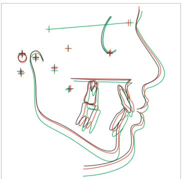
Figure 1 - Mean cephalometric landmarks in the three observation phases: (T1, black; T2, red; and T3, green) (overlap on line SN).
incisors showed significant palatal inclination, there-by reversing anchorage loss. Protrusion of maxillary incisors was not statistically significant (Table 1).
In the inal phase of corrective orthodontic treat-ment, maxillary incisors also showed signiicant extru-sion related to correction of buccal inclination, verti-cal displacement of the maxilla due to the craniofacial growth22 and the use of Class II elastics.
First and second maxillary premolars
In the post-distalization phase, irst premolars dem-onstrated efects of anchorage loss related to distal move-ment through intraoral distalizers.7,10-13,20 There was sig-niicant linear and angular mesial movement associated with extrusion in relation to the palatal plane. Mesial angulation was corrected at the end of orthodontic treat-ment; however, mesial positioning remained in the inal stage (Table 1), probably due to anterior displacement of the maxilla in an attempt to accompany mandibular dis-placement.10,20 First premolars showed signiicant extru-sion at the end of distalization and orthodontic treatment. Previous researches9,16 also assessed the efects of the Jones Jig appliance, but focused only on second premo-lars, which are the anchorage teeth selected for placement of the Nance button during intraoral distalization.
Second premolars showed similar behavior to irst premolars, also characterizing anchorage loss due to in-traoral distalization.2,4,7,9-13,15-17,20,23,24 There was statistical-ly signiicant angular and linear mesial movement in the post-distalization phase. Conversely, in the inal stage, mesial angulation was corrected, but mesial positioning also remained accompanying the displacement of the maxillary base in relation to the mandible.10,20
There was statistically signiicant extrusion during distalization in comparison to the initial position. This vertical change was related to mesial angulation and me-sial movement, i.e., relecting anchorage loss.9,16
Longitudinal observations show that extrusion, mesial angulation of premolars and anterior inclination of max-illary incisors are reversed efects that occur during active treatment with maxillary ixed appliances.9,15 They may even occur spontaneously in the period of verticaliza-tion and stabilizaverticaliza-tion with the Nance button posiverticaliza-tioned in distalized molars. However, the vertical positioning of second premolars remained at the end of corrective treat-ment, similarly to the post-distalization phase. In other words, it is probable that the accentuated curve of Spee
preserved extrusion of second premolars to correct over-bite, which was greater at the beginning of treatment.
First and second maxillary molars
Maxillary irst molars demonstrated signiicant distal angulation, as already anticipated in treatment of Class II with intraoral devices.2-4,7,9,11-17,20
Accentuated distal angulation is observed in all re-searches that assess distalization with the Jones Jig ap-pliance,5,9,14-17,25 including cases of absolute anchorage.26 However, this angulation was reversed and, by the end of corrective orthodontic treatment, maxillary irst molars showed a verticalized position in relation to the cranial base (Table 1 and Fig 2). This correction was already ex-pected, since patients, by the end of the distalization pro-cess, used the extraoral headgear in order to anchor and verticalize distalized molars.16,20
Maxillary irst molars were signiicantly distalized in the post-distalization phase; however, ater corrective orthodontic treatment, signiicant angular and linear me-sial movement was observed, probably due to correction of accentuated distal angulation and displacement of the maxilla of which goal is to maintain normal maxilloman-dibular relationship10 (Fig 2).
By the end of corrective orthodontic treatment, mo-lar extrusion was statistically signiicant, probably due to correction of overbite (Fig 2). Extrusion of maxillary irst molars can also be related to appositional maxillary growth and vertical lotation process.22 In this group, the use of headgear may not have promoted extrusion of irst molars, since the headgear was used with me-dium-high traction (jeans helmet), thereby preventing tooth extrusion.
Maxillary second molars showed no statistically sig-niicant changes in the post-distalization phase; however, at the end of corrective orthodontic treatment, second molars showed mesial angulation in relation to the cra-nial base, as well as statistically signiicant mesialization. Statistically signiicant extrusion was also observed at the end of corrective orthodontic treatment. Changes caused to second molars were not only due to eruption, but also to movement of maxillary irst molars in the corrective phase.9
Mandibular dentoalveolar component Mandibular incisors
With regard to mandibular incisors, there was statistically significant buccal inclination and pro-trusion at the end of corrective orthodontic treat-ment (Table 1 and Fig 3). This change was related to correction of overjet which was greater at the be-ginning of treatment. Therefore, changes observed in mandibular incisors occurred to decrease overjet and were due to the use of Class II elastics.
In the literature, only the study conducted by Brickman, Sinha and Nanda9 assessed the effect of Class II treatment with the Jones Jig appliance fol-lowed by corrective fixed appliances. Nevertheless, the authors did not report the behavior of mandib-ular incisors. However, studies that assessed other distalizers observed buccal inclination during orth-odontic treatment.10,11,20 According to Angelieri et al,20 mandibular incisors are buccally inclined due to anterior mandibular displacement and the use of Class II elastics.
Mandibular first molars
Mandibular molars showed statistically signifi-cant mesialization during corrective treatment when initial and post-distalization phases were compared (Table 1). Class II molar relationship was corrected not only by maxillary molars distalization, but also by mesial movement of mandibular molars.10,20
As for the vertical component, at the end of cor-rective treatment, mandibular molars showed signifi-cant extrusion when the initial and post-distalization phases were compared (Fig 3). Changes in mandibu-lar momandibu-lars are not reported by studies assessing dis-talization with the Jones Jig 5,15-17 followed by fixed appliances;9 however, extrusion was observed in stud-ies that use the Pendulum appliance.10,11 Therefore, it is assumed that mandibular molars extrusion may be related to the use of Class II elastics, vertical displace-ment due to appositional growth of the lower base of the mandible and to vertical floating.22
Dental relationships
Molar relationship showed statistically signifi-cant changes in the three phases of assessment. In the initial phase, patients showed Class II relationship, whereas in the post-distalization phase, there was
overcorrection with molars ending up in a “super” Class I relationship, as suggested by previous stud-ies.9,11,12 However, as expected, maxillary molars me-sially moved during corrective treatment to establish normal molar relationship, (Table 1).
At the beginning of treatment, both overjet and overbite were increased. In the post-distalization phase, there was an increase in overjet due to buccal tipping of maxillary incisors, thereby characterizing anchorage loss related to distalization with the Jones Jig appliance.16 By the end of corrective orthodontic treatment, both overjet and overbite were reduced, thus demonstrating that orthodontic treatment goals were achieved. Correction of overjet was related to uprighting of maxillary incisors and buccal tipping of mandibular incisors, as described above. Correction of overbite, on the other hand, was probably related to extrusion of first and second premolars and first and second molars, as previously reported.
Brickman, Sinha and Nanda9 observed an increase of 0.45 mm in overjet and a decrease of 1.28 mm in overbite during distalization of maxillary molars. According to the authors, changes in overjet were re-lated to mesial movement of maxillary premolars and buccal inclination of maxillary incisors, as reported by other studies assessing intraoral distalization.12,13,16,20
CONCLUSION
The Jones Jig appliance did not interfere in maxil-lary and mandibular components and did not change maxillomandibular relationship. The Jones Jig appli-ance promoted distalization of irst molars with an-chorage loss, signiicant mesialization and extrusion of irst and second premolars, and a signiicant increase in
anterior face height at the end of treatment. Most ad-verse efects occurred during the intraoral distalization phase and were subsequently corrected with corrective mechanics. There was buccal inclination and protru-sion of mandibular incisors. At the end of treatment, overjet and overbite correction was observed.
1. McNamara J. Components of class II malocclusion in children 8-10 years of age. Angle Orthod. 1981;51(3):177-202.
2. Fortini A, Lupoli M, Giuntoli F, Franchi L. Dentoskeletal efects induced by rapid molar distalization with the irst class appliance. Am J Orthod Dentofacial Orthop. 2004;125(6):697-704.
3. Gianelly AA, Vaitas AS, Thomas WM. The use of magnets to move molars distally. Am J Orthod Dentofacial Orthop. 1989;96(2):161-7.
4. Hilgers J. The pendulum appliance for Class II non-compliance therapy. J Clin Orthod. 1992;26(11):706-14.
5. Jones RD, White JM. Rapid Class II molar correction with an open-coil jig. J Clin Orthod. 1992;26(10):661-4.
6. Antonarakis GS, Kiliaridis S. Maxillary molar distalization with noncompliance intramaxillary appliances in Class II malocclusion. A systematic review. Angle Orthod. 2008;78(6):1133-40.
7. Kinzinger GSM, Wehrbein H, Diedrich PR. Molar distalization with a modiied pendulum appliance--in vitro analysis of the force systems and in vivo study in children and adolescents. Angle Orthod. 2005;75(4):558-67.
8. Oncag G, Seckin O, Dincer B, Arikan F. Osseointegrated implants with pendulum springs for maxillary molar distalization: a cephalometric study. Am J Orthod Dentofacial Orthop. 2007;131(1):16-26. 9. Brickman CD, Sinha PK, Nanda RS. Evaluation of the Jones jig
appliance for distal molar movement. Am J Orthod Dentofacial Orthop. 2000;118(5):526-34.
10. Burkhardt DR, McNamara JA Jr, Baccetti T. Maxillary molar distalization or mandibular enhancement: a cephalometric comparison of comprehensive orthodontic treatment including the pendulum and the Herbst appliances. Am J Orthod Dentofacial Orthop. 2003;123(2):108-16. 11. Chiu PP, McNamara JA, Franchi L. A comparison of two intraoral molar
distalization appliances: distal jet versus pendulum. Am J Orthod Dentofacial Orthop. 2005;128(3):353-65.
12. Ngantung V, Nanda RS, Bowman SJ. Posttreatment evaluation of the distal jet appliance. Am J Orthod Dentofacial Orthop. 2001;120(2):178-85. 13. Ghosh J, Nanda RS. Evaluation of an intraoral maxillary molar distalization
technique. Am J Orthod Dentofacial Orthop. 1996;110(6):639-46. 14. Gulati S, Kharbanda OP, Parkash H. Dental and skeletal changes after
intraoral molar distalization with sectional jig assembly. Am J Orthod Dentofacial Orthop. 1998;114(3):319-27.
REFEREnCES
15. Haydar S, Uner O. Comparison of Jones jig molar distalization appliance with extraoral traction. Am J Orthod Dentofacial Orthop. 2000;117(1):49-53.
16. Patel MP, Janson G, Henriques JF, Almeida RR, Freitas MR, Pinzan A, et al. Comparative distalization efects of Jones jig and pendulum appliances. Am J Orthod Dentofacial Orthop. 2009;135(3):336-42.
17. Runge ME, Martin JT, Bukai F. Analysis of rapid maxillary molar distal movement without patient cooperation. Am J Orthod Dentofacial Orthop. 1999;115(2):153-7.
18. Dahlberg G. Statistical methods for medical and biological students. New York: Interscience; 1940.
19. Houston WJ. The analysis of errors in orthodontic measurements. Am J Orthod. 1983;83(5):382-90.
20. Angelieri F, Almeida RR, Almeida MR, Fuziy A. Dentoalveolar and skeletal changes associated with the pendulum appliance followed by ixed orthodontic treatment. Am J Orthod Dentofacial Orthop. 2006;129(4):520-7.
21. Martins DR, Janson G, Almeida RR, Pinzan A, Henriques JFC, Freitas MR. Atlas de crescimento craniofacial. São Paulo: Ed. Santos; 1998. 22. Enlow DH, Kuroda T, Lewis AB. The morphological and morphogenetic
basis for craniofacial form and pattern. Angle Orthod. 1971;41(3):161-88. 23. Bolla E, Muratore F, Carano A, Bowman SJ. Evaluation of maxillary molar
distalization with the distal jet: a comparison with other contemporary methods. Angle Orthod. 2002;72(5):481-94.
24. Bussick T, McNamara J Jr. Dentoalveolar and skeletal changes associated with the pendulum appliance. Am J Orthod Dentofacial Orthop. 2000;117(3):333-43.
25. Mavropoulos A, Karamouzos A, Kiliaridis S, Papadopoulos MA. Eiciency of noncompliance simultaneous irst and second upper molar distalization: a three-dimensional tooth movement analysis. Angle Orthod. 2005;75(4):532-9.