An interview with
How to cite this interview: Hans MG. An interview with Mark G. Hans. Dental Press J Orthod. 2014 May-June;19(3):26-35. DOI: http://dx.doi.org/10.1590/2176-9451.19.3.026-035.int - Submitted: February 05, 2014 - Revised and accepted: February 27, 2014.
Mark G. Hans
It is a great honor to conduct an interview with Professor Mark G. Hans, ater following his outstanding work ahead of the Bolton-Brush Growth Study Center and the Department of Orthodontics at the prestigious Case Western Reserve School of Dental Medicine (CWRU) in Cleveland, Ohio. Born in Berea, Ohio, Professor Mark Hans attended Yale University in New Haven, CT, and earned his Bachelor of Science Degree in Chemistry. Upon graduation, Dr. Hans received his DDS and Masters Degree of Science in Dentistry with specialty certiication in Orthodontics at Case Western Reserve University. During his education, Dr. Hans’ Master’s Thesis won the Harry Sicher Award for Best Research by an Orth-odontic Student and being granted a Presidential Teaching Fellowship. As one of the youngest doctors ever certiied by the American Board of Orthodontics, Dr. Hans continues to maintain his board certiication. He has worked through aca-demics on a variety of research interests, that includes the demographics of orthodontic practice, digital radiographic data, dental and craniofacial genetics, as obstructive sleep apnea syndrome, with selected publications in these ields. One of his noteworthy contributions to the orthodontic literature came along with Dr. Donald Enlow on the pages of “Essentials of Facial Growth”, being reference on the study of craniofacial growth and development. Dr. Mark Hans’s academic career is linked to CWRU, recognized as the renowned birthplace of research on craniofacial growth and development, where the classic Bolton-Brush Growth Study was historically set. Today, Dr. Hans is the Director of The Bolton-Brush Growth Study Center, performing, with great skill and dedication, the handling of the larger longitudinal sample of bone growth study. He is Associate Dean for Graduate Studies, Professor and Chairman of the Department of Orthodontics, working in clinical and theoretical activities with students of the Undergraduate Course from the School of Dental Medicine and residents in the Department of Orthodontics at CWRU. Part of his clinical practice at the university is devoted to the treatment of craniofacial anomalies and to special needs patients. Prof. Mark Hans has been wisely conducting the Joint Cephalometric Experts Group (JCEG) since 2008, held at the School of Dental Medicine (CWRU). He coordinates a team composed of American, Asian, Brazilian and European researchers and clinicians, working on the transition from 2D cephalometrics to 3D cone beam imaging as well as 3D models for diagnosis, treatment planning and assessment of orth-odontic outcomes. Dr. Hans travels to diferent countries to give lectures on his ields of interest. Besides, he still maintains a clinical orthodontic practice at his private oice. In every respect, Dr. Hans coordinates all activities with particular skill and performance. Married to Susan, they have two sons, Thomas and Jack and one daughter, Sarah and he enjoys playing jazz guitar for family and friends.
Matilde da Cunha Gonçalves Nojima » DDS, School of Dental Medicine, Case Western Reserve University,
Cleveland, OH, USA.
» MSD, Department of Orthodontics, Case Western Reserve University, Cleveland, OH, USA.
» Chairman, Department of Orthodontics, Case Western Reserve University, Cleveland, OH, USA.
» Director, The Bolton-Brush Growth Study Center, Case Western Reserve University, Cleveland, OH, USA.
» Diplomate, American Board of Orthodontics.
1. As director of the Bolton-Brush Growth Study Center, could you tell us some points about this renowned research center?
(Lincoln Nojima)
Located on third loor of the Bolton Dental Build-ing, the Bolton-Brush Growth Study Center houses the world’s largest longitudinal radiographic collections of cranial and post cranial skeleton. The Bolton Study was started in 1930 by the inventor of the cephalostat, B. Holly Broadbent, with the goal of increasing our un-derstanding of the normal growth of the human face. A total of 4,309 subjects were enrolled. Lateral and frontal cephalograms as well as hand wrist radiographs and dental study casts were taken yearly on these chil-dren, usually on or around their birthday. Under the direction of T. Wingate Todd, the Brush Study began at the same time with the goal of radiographically doc-umenting the normal growth and development of the appendicular skeleton. All of Bolton subjects were also enrolled in the Brush Study. The Brush Study ended in 1950 and the Bolton study ended in 2001. To be part of the Bolton-Brush subject population, individuals had to be in excellent health and free from any major illness or inirmity. Oten, these children were enrolled because they had won “Healthy Child” contests at their schools. Landmark publications resulted from these legendary studies. Some examples are the Greulich and Pyle Hand Wrist Atlas used by pediatricians around the world to assess skeletal development in growing children, the Bolton Standards for Dentofacial Growth and Develop-ment used by craniofacial practitioners to assess facial proportions and skeletal balance, as well as the classic work by Rolf Behrents on Adult Craniofacial Growth and Development.
Although we stopped enrolling new subjects in these studies, the collections are still used by researchers from around the globe to answer important questions pertaining to human health. One of the main activi-ties for center staf is to convert the fragile radiographs to digital format. A small portion of the Bolton Col-lection is available online by visiting the American As-sociation of Orthodontist Legacy Collection website: http://www.aaolegacycollection.org/aaof_collection. html?id=CASEBolton
Many more radiographs are available in digital for-mat and can be accessed at the center or by ordering digital copies of the radiographs. A complete index to
the radiographs in the Bolton and Brush collections is available by sending an email to [email protected] with the subject line “Bolton-Brush Index”. The index is in Filemaker Pro format and a demo version of the Filemaker Pro program can be downloaded for free from the Filemaker Pro website.
2. In your opinion, which theories of Facial Growth should be taught in an orthodontic program?
(Juan Martin Palomo)
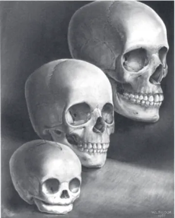
In my opinion, we do not understand the switches that turn bone growth on and of. However, the mor-phologic changes that occur as a result of the growth process have been well described (Fig 1). Therefore, orthodontic education should focus on giving students a thorough understanding of morphologic changes that occur as well as the biological processes that result in
Figure 1 - Morphologic changes of the face. The infant and young child are char-acterized by a wide-appearing face but vertically short. During later childhood and into adolescence, vertical nasal enlargement keeps pace with growing body and lung size, dental and other oral components have approached adult sizes and configuration. Overall, the early wide face has become altered in propor-tion by the later vertical changes. (In Enlow, D. H. and Hans, M. G.: Essentials of Facial Growth. 2nd ed. Ann Arbor: Needham Press, 2008. Courtesy of William L.
those changes. For example, orthodontic programs must teach students about bone remodeling and dis-placement. Remodeling activity includes the deposition of new bone on both periosteal and endosteal surfaces by osteoblasts concomitant with the resorption of bone on these surfaces mediated by osteoclasts. The areas of bone deposition and resorption were mapped for the human face as part of the life work of Donald H. Enlow, PhD. Dr. Enlow was my predecessor as department chair at CWRU and I was very fortunate when he asked me to coauthor the textbook Essentials of Facial Growth in 1996. Now, in its second edition, Essentials describes in detail, the remodeling process. As second equally important concept is that of bony displacement. Dis-placement is the movement in space of an entire bone en masse. All morphologic changes we see in the bony skeleton are the result of these two basic processes. And since all orthodontic and orthopedic treatments must afect changes in one or both of these processes, they must be taught in all orthodontic training programs.
In terms of theories of facial growth, I think it is im-portant for students to know something about Moss’ Functional Matrix hypothesis as well as Scott’s Nasal Septum Theory. Each of these theories adds something of value to students understanding of facial growth. In the case of the functional matrix hypothesis, it leads the student to think about the relationship between hard and sot tissues of the head and neck. The theory is par-ticularly strong when applied to the growth of the neu-rocranium. In this area, growth of brain tissue clearly paces enlargement of the lat bones of the skull. As you move into the area of the cranial base, Scott’s cartilage theory becomes attractive because of the ability of car-tilage to grow interstitially and the histologic similarity between synchondrosis and epiphyseal growth plates. As we approach areas closer to the occlusal plane, things get more confusing. For example, what is the most logical theory regarding the tissue separating force that drives midfacial growth? The nasal septal cartilage sits in the midline and the septopremaxillary ligament at-taches to the anterior nasal spine of the maxilla allowing expansile growth of the cartilaginous septum to pull the maxilla downward and forward. This is consistent with the observed displacement of the maxilla downward and forward. The functional matrix theory holds that it is respiration and in particular nasal respiration that drives growth of the midface downward and forward. It is most
likely that both of these processes (i.e. septal growth and respiration) inluence maxillary development, and further, that a deiciency in one driving force is likely compensated by an excess in the other. That there exists a certain amount of redundancy in critical growth sys-tems seems plausible. However, the complexity of hu-man maxillary growth will likely not allow the relative contributions of the functional matrix compared to the nasal septum to be tested by hypothesis driven research methods. Finally, neither theory provides a compelling rationale for mandibular growth. Clearly, the man-dibular condyle does not function as a locus of control. Rather, the most important role for this cartilage is in establishing and maintaining the integrity of the tem-poromandibular joint. It is also diicult to comprehend how mastication and/or deglutition inluence mandibu-lar growth. I would say that both theories fail to provide a compelling argument for the driving force behind the downward and forward displacement of the mandible.
3. How do you focus on the importance of knowl-edge on Craniofacial Biology for orthodontic treatment planning and achievement of better treatment outcomes? (Matilde Nojima)
Orthodontists need to be very efective at moving teeth in all three dimensions (vertical, lateral, and horizontal) within the alveolar process. We need to pay attention to tooth movements and control the movements. To do this efectively, we should know the normal way teeth move during growth and development. We are all fa-miliar with the term eruption of teeth. I want to clarify an important diference between eruption of teeth and drit of teeth. Eruption is a biologic process whereby the tooth moves towards the occlusal plane until it con-tacts its opposing tooth and is said to be “in occlusion”. Once a tooth is in occlusion, it is inished with the eruption process. However, the face continues to grow downward and forward and to maintain this occlusion of the teeth, they must drit towards the occlusal plane. This means maxillary teeth drit inferiorly and man-dibular teeth drit superiorly. In addition to the verti-cal drit of teeth, the teeth drit mesially with normal growth and development. Thus, drit of teeth naturally occurs toward the occlusal plane and mesially. So, what does this mean for the average orthodontist and their patients? We all know it is easier to move teeth in the direction they naturally want to go. So, if you do not pay any attention to your treatment mechanics, you will naturally accelerate the movement of teeth toward the occlusal plane and mesially. This means that when you are inished with your treatment, the face will be lon-ger (vertical dimension will be increased), and the teeth will be more forward in the face (i.e. more bimaxillary protrusive). If these movements are outside of the physi-ologic boundaries of stability, then you will have created an unstable result. Not what we are looking for. So, to achieve better treatment outcomes, you need to pay at-tention to your mechanics if you do not want a longer face or a more protrusive denture. The skilled ortho-dontist can control the teeth in all three dimensions so it is critical that we do so.
4. In order to better understand growth and de-velopment of the face, do you believe that estab-lishing VTOs on growing patients can be helpful? If so, which method do you recommend us to use?
(Kunihiko Miyashita)
To be an excellent orthodontist, you need to have two goals in mind. First, the alignment of the teeth and second the placement of the teeth within the face. The alignment of teeth is almost always dictated by the
contact points of adjacent teeth. This results in little disagreement among dentists as to the ideal position of the teeth relative to each other. Likewise, there is general agreement among dentists about the proper occlusion of the teeth, that is, axial loading of posterior teeth with anterior guidance provided by the cuspids and incisors. However, there is not universal agree-ment as to the ideal place for the teeth within the face. It is in this area that I believe a VTO is helpful. A VTO gives the orthodontist a target to shoot for and he or she should aim for the bull’s eye. Without a target you cannot measure the accuracy of treatment plan for the position of teeth within the face.
So, I believe you should have a goal for the posi-tion of the dentiposi-tion within the face. I prefer to start my VTO by asking the question, “Where should the upper central incisor be located in this patient’s face?” There are several ways to determine this position. I prefer to use the Bolton Standards as a guide for upper incisor location. In addition, I conirm the use of the Bolton Standard position by using a Nasion Vertical and placing the maxillary central incisor 5 mm in front of the line and vertically about 2-3 mm below the inferior border of the upper lip in the relaxed state. Once I have estab-lished the position of the upper incisor, I then decide on how best to establish anterior guidance and an accept-able interincisal angle. In terms of growth prediction, I prefer to use a mean change expansion as described by Johnston et al.1 In general, this method assumes that the
maxilla will grow 1 mm forward at A Point per year and 1 mm vertically at ANS per year. Mandibular growth will exceed maxillary growth by 1 mm per year in both vertical and horizontal directions.
5. What are the biological indicators that should be observed in order to predict the amount of fu-ture growth in a speciic bone of the face?
(Ana Maria Bolognese)
appliance used ater the anterior crossbite is corrected. If you correct Class III any sooner, you have the prob-lem of retaining correction during the transition of the incisor dentition. It is important to me that there be contact of natural dentition during the post treatment period. I think proprioceptive feedback during func-tion is an important factor in the success of early Class III treatment. Ideally, if I treat a patient early for Class III, I like to wait to begin full ixed orthodontic treat-ment until ater the pubertal growth spurt is complete.
If you miss the orthopedic treatment window, then I think it is best to wait to begin full ixed treatment until ater the pubertal growth spurt. I think this is best because at this point you have only two options remain-ing, camoulage or surgery. And, in one case (camou-lage) you will be adding dental compensations to es-tablish the best possible occlusion while in the other (surgery) you will be removing dental compensations to allow correction for skeletal disharmony. It is impos-sible for the orthodontist to do both at the same time. So, you are forced to choose. And delaying the choice as long as possible gives you the best chance to make the correct decision between these two options.
7. What are your views on growth modiication through the use of functional appliances?
(Juan Martin Palomo)
I believe the data on modiication of maxillary growth both forward using a protraction facemask or backward using cervical pull headgear is compelling and I recommend these treatment options to my younger patients who would beneit from such therapy. I rec-ommend that these orthopedic devices be worn only at night because we know that humans only grow at night and that teeth only erupt at night. There seem to be two major factors that predict treatment success. The irst is patient compliance. To see any efect with the headgear, it must be worn almost every night. Since orthopedic treatment seeks to inluence facial growth, we have to allow time for growth to occur. This means that a mini-mum of 6 months of compliant wear must be achieved before you can assess the second factor that inluences treatment success. That second factor is the genetic sus-ceptibility of the patient to growth modiication, i.e. is the patient a “responder” or a “non-responder” to or-thopedic therapy? I think most orthodontists tend to the amount of growth, one can use chronological age,
hormonal indicators of maturity (acne, onset of men-struation in girls, facial hair in boys) to help estimate the amount of growth potential remaining. However, I still believe that the only way to determine when facial growth has slowed is to use serial cephalometric ra-diographs taken a minimum of 6 months apart. When the two ilms shows less than 0.5 mm of change over a six month period prior, then the clinician can safely assume adolescent facial growth has been completed.
6. What are your thoughts on the best tim-ing to treat skeletal disharmonies as Class III malocclusions? (Matilde Nojima)
think all non-responders are non-compliant, but I dis-agree. I think you can get most patients to comply with treatment for a few months. The ones that see results are encouraged and continue to wear their device at night, the ones that do not respond get discouraged and stop wearing the device. I never blame the patient for not re-sponding, in most cases it is not their fault. As my friend Gerry Samson says: “You can’t ask them to take another dip in the gene pool”. It would be great if we could ind a way to determine responders from non-responders without having to wait 3-6 months, but I do not see that as an option in the near future.
8. Since you are a great expert on craniofacial growth, what is your opinion on the historical debate between functional appliances and head-gear traction for the correction of Class II maloc-clusions? (Lincoln Nojima)
I take an approach to the use of headgear traction in the correction of Class II that is very similar to Dr. Robert Ricketts. I like to use a cervical pull facebow in combination with a lower utility arch. The head-gear is effective in correcting maxillary skeletal prog-nathism and maxillary dental protrusion, and the util-ity arch uncouples the upper and lower anterior teeth by intrusion of lower incisors. Ricketts’s theory, and I agree, was that by uncoupling the anterior teeth you allow the mandible to be displaced downward and forward. Since the effect of orthodontic treatment on mandibular growth is non-specific and highly vari-able, I do not find a big difference in mandibular growth response between functional appliances that advance the mandible and headgear/utility arch me-chanics. And, since headgear is a fixed device, it is much more effective at addressing maxillary protru-sions that often accompany Class II maloccluprotru-sions.
By the way, I use the cervical pull headgear exclu-sively for facebow type headgear traction. I ind the pos-terior high pull headgear not as efective as an anpos-terior J-Hook headgear in controlling vertical. Plus, you need to add the Transpalatal Arch to the Posterior high pull to negate the buccal rolling of molars. I used to apply straight pull facebows but found that patient compli-ance was much better with the simpler cervical pull. And, since I do not use facebow headgear in high angle cases, the small diference between the angle of pull for straight pull and cervical was not clinically signiicant.
9.Do you think that using temporary anchorage devices (TAD) can help us control craniofacial growth patterns? (Kunihiko Miyashita)
I think that TADs can help control vertical dental drit in growing patients. As mentioned earlier, verti-cal drit of dentition occurs towards the occlusal plane. So, controlling this natural movement could be helpful in patients with increased lower vertical facial height. Of course, to achieve this goal it would be necessary to control both the inferior drit of the maxillary buccal segments, as well as the superior drit of the mandibular buccal segments. In addition, because we are trying to limit vertical facial development by modifying the den-tition and alveolus, these mechanics would likely need to be continued until vertical facial growth was com-pleted. This type of growth modiication will face the same challenges as we faced when we used chin cups to limit mandibular growth, i.e. achieving long-term sta-bility will require a long-term retention strategy. I have used miniplates to intrude posterior segments in several patients including one that we published in the Journal of Plastic Surgery. TADs are an exciting addition to our mechanical systems and their efective use will require orthodontists to apply bone biology in their TAD place-ment planning. For example, long-term placeplace-ment of a TAD in an area of bone that is undergoing bone re-sorption as part of remodeling process will likely fail. Whereas, placement of a TAD in an area of natural bone deposition has a higher chance of success based on biol-ogy. Applying this concept to vertical control of dental drit would mean that maxillary TADs should be placed in the palate, and not on the buccal surface. In the man-dible, TADs could be placed on the buccal cortices ad-jacent to the molars. TADs placed in alveolar bone are likely to fail sooner than those placed in cortical bone. Since you need long-term TAD success to modify cra-niofacial growth, the resorptive and depository remod-elling patterns are important to understand.
10. Is there any diferent clinical response in the sutural tissues of growing patients using mini-plates or headgear traction considering growth and displacement of facial bones?
(Ana Maria Bolognese)
the growing craniofacial complex is to change the dis-placement and remodelling of the bones. For example, cervical pull headgear can be used in a variety of ways to change facial growth depending on how the force is ap-plied. Dr. Andy Haas, Robert Ricketts and others have documented the efects of cervical pull headgear applied solely to maxillary irst molars. When the headgear is attached only to maxillary irst molars you are using the teeth via the periodontal ligament (PDL) as a transducer to send mechanical signals to the periosteum and sutural systems. The sutural system can be inluenced by this mechanical system including the intermaxillary suture, the circummaxillary suture system, and, to a lesser ex-tent, the circumfacial sutures. Cervical headgear applied to maxillary irst molars combined with a lower util-ity arch to intrude the lower incisors can also inluence mandibular growth. The most logical explanation of the efect of cervical headgear on mandibular growth is that by disengaging the dentition with the intrusion arch, the mandible outgrows the nasomaxillary complex. In contrast to the biological impact of orthopedic force ap-plication, miniplates are just devices that can be rigidly attached to bone. There is no biologic rationale to think that miniplates are anything like headgear.
11. Considering the important concepts includ-ed in your classic “Essentials of Facial Growth”, how do you feel about research on craniofacial growth and airway? And how such informa-tion can be correlated to clinical interveninforma-tion?
(Matilde Nojima)
Probable, one of the most classic experiments that demonstrated the impact of forced oral respiration on facial growth was conducted by Egil Harvold when he plugged the noses of growing rhesus monkeys. What ev-eryone remembers about this experiment is that Harvold was able to cause open bite malocclusions in these mon-keys. And these open bite malocclusions were character-ized by increased lower vertical facial height, maxillary transverse deiciency and dental crowding. What most people forget is that not all of Harvold’s monkeys de-veloped malocclusions. This variability in response to airway obstruction has not been talked about very much in our literature. In my opinion, if we have such vari-ability in a genetically homogeneous population of ex-perimental animals I would expect even greater variation in human populations. And, this is in fact what we have
found with any large study on the impact of airway on facial growth. It is impossible to show a simple cause and efect relationship between mouth breathing and maloc-clusion. It makes sense that there should be some inlu-ence since the roof of the mouth and the loor of the nose are the same bone, but I do not think we will ever be able to prove such a relationship. In terms of clinical intervention, I think we do know that rapid palatal ex-pansion reduces nasal airway resistance. And, we know that moving the mandible forward with functional ap-pliances increases the oral pharyngeal airway. These are anatomic facts. Therefore, if you have a patient that has nasal obstruction and is a mouth breather you could con-sider palatal expansion as one mode of treatment. If the pediatrician asks whether removal of adenoid tissue would be beneicial for facial growth I would answer the following way: If the child needs to have adenoid tissue removed for medical reasons (i.e. recurrent infection), then I would support the operation and indicate that there could be a positive efect on facial development. In contrast, if a pediatrician asks me if I would recommend removal of adenoid tissue for improving facial growth I would say that the evidence is not strong enough to sup-port such a recommendation from the orthodontist.
12. What changes can be achieved, after growth, to correct the morphology of the nasomaxil-lary complex in cases of open bite and mouth breathers ? (Ana Maria Bolognese)
caused a monumental paradigm shift in Orthodontics. Prior to these publications, orthodontists considered stability of orthodontic correction to be one of the main goals of treatment. And to achieve this goal, the removal of permanent teeth was often prescribed so that expansion of dental arches in the anterior and lateral dimensions could be avoided. After these pub-lications, lifetime retention was de rigueur for all pa-tients. I think that this change in our approach to sta-bility is very dangerous to our profession. The main reason for my thinking is that if we give up on stabil-ity as a treatment goal, we run the risk of diminishing the specialty to the level of a cosmetic rather than a functional service. This, in turn, will make it much easier for the public to view orthodontic treatment as a commodity. When the public purchases a com-modity, they focus on value. If stability is not part of the value equation, we are left with price and ment time. Patients will assume that quality of treat-ment results is equal among providers. Our specialty is much more than aligning teeth. Fit and function as well as lifetime dental health should be important goals of treatment. Making beautiful smiles is fine, but we cannot do so at the expense of dental health.
My main concern about lifetime retention is that there is a real possibility that significant chang-es in the supporting periodontium could rchang-esult if teeth retained for a lifetime in an unstable position. We know from our studies of bone biology that un-der pressure bone resorbs. So, if an orthodontist moves teeth beyond their physiologic boundary, pa-thology will ensue. The critical question is “What are those boundaries?” Identifying these boundar-ies should be the focus of orthodontic research for the coming decade. Until we have a better idea of the limits of treatment, we should be wary of life-time retention. I think a more biologic approach would be to tell our patients that we can produce an orthodontic treatment result as good as some-one who was born with straight teeth. However, we know from the Bolton Brush Studies, that naturally straight teeth do not stay that way for a lifetime. The idea that a single orthodontic intervention will lead to a lifetime of perfectly straight teeth is not realis-tic. We should inform patients that additional treat-ment may be needed in the future due to naturally occurring growth of the adult craniofacial skeleton. extraction of permanent teeth has severely limited
what we can achieve for our patients ater growth. For the non-growing patient, the need for extraction of permanent teeth must be carefully evaluated.
13. How do you address the relationship between Craniofacial Growth and stability of orthodontic treatment outcomes?
(Lincoln Nojima)
This is an interesting question, especially as it pertains to craniofacial growth that occurs after orthodontic treatment. During treatment, the or-thodontist is constantly monitoring craniofacial growth, treatment response, and the progress to-wards completion of therapy. Treatment decision can be made on a monthly basis to compensate or decompensate the dentition in response to cranio-facial growth. Once the treatment goals of dental alignment, anterior guidance, axial loading of pos-terior teeth, proper smile arch, pleasing smile, etc., have been achieved and braces are removed, the game changes. Now, the patient no longer has the skilled orthodontist to help maintain equilibrium among all of components of the craniofacial com-plex that are involved in maintaining the dental oc-clusion. Although there are no easy answers to this dilemma, I can offer one suggestion. Do not try to finish all of your cases with centric relation (the liga-mentous position of the mandibular condyle in the glenoid fossa) coincident with the position of the condyle in the glenoid fossa dictated by the maxi-mum intercuspation of the teeth. Allow at least 1-2 mm of difference between these two condylar posi-tions. That way, if you get a couple millimeters of late mandibular growth, a change in CR-MIC rela-tionship can occur and compensate for this growth, thereby keeping teeth in proper occlusion.
14. In a constantly changing craniofacial com-plex, what are your views on the changes that occur during adulthood, the appearance of late dental misalignment and the concept of retainers for life? (Juan Martin Palomo)
15. Which bone is your favorite? And why?
(Kunihiko Miyashita)
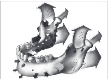
Without question, the mandible is my favorite bone in the craniofacial regions. My attraction to the study of the mandible is based on three factors. First, the historic orthodontic controversies involving this bone. These begin with the myth of the condylar cartilage as a growth center that magically determined the size and shape of this complex bone. This was followed by the functional appliance craze in the United States with the outrageous claims of mandibular protrusive devices being capable of stimulating mandibular corpus growth in the six to seven millimeter range. Then, came the false claims that malocclusions caused temporomandibular joint dis-ease. And inally, the most recent controversy over the role of the size and shape of the mandible in Obstruc-tive Sleep Apnea Syndrome. The second reason I am fond of the mandible is its anatomic complexity (Fig 2).
When I teach students about the mandible I like to di-vide the bone into ive areas based on function. Area one, the condyle with the primary function of articula-tion. Area two, the coronoid process, primary function, attachment of the temporalis muscle. Area three, the corpus, primary function to connect the right and let halves for the mandible as a rigid strut. Area four, the al-veolus, primary function to support the dentition. Area ive, the ramus, primary functiont to provide compen-sations in both the vertical and horizontal dimensions to insure occlusion of molar teeth within 6 mm among the entire human race. Finally, I like the mandible be-cause we have much to learn about the control processes involved in its growth. Right now, I like to say to my students that all orthodontic/orthopedic treatments of growing patients increase mandibular growth a little bit, I refer to this non-speciic stimulation of mandibular growth as the “fertilizer efect”.
1. Johnston LE. A simplified approach to prediction. Am J Orthod. 1975;67(3):253-7.
2. Greulich W, Pyle S. Radiographic atlas of skeletal development of the hand and wrist. 1. ed. Redwood City: Stanford University Press; 1999.
REFERENCES
Ana Maria Bolognese
» DDS, Federal University of Rio Grande do Sul, Brazil. » MSc and Phd in Orthodontics, Federal University of Rio
de Janeiro, Brazil.
» Postdoctoral in Oral Biology, Northwestern University, Chicago, USA.
» Chairman, Department of Orthodontics, Federal University of Rio de Janeiro, Brazil.
Juan Martin Palomo
» DDS, State University of Ponta Grossa, Brazil.
» MSc in Orthodontics, Case Western Reserve University, Cleveland, OH, USA.
» Director of Orthodontics Residency — Case Western Reserve University, Cleveland, OH, USA.
» Director of Craniofacial Imaging Center - Case Western Reserve University, Cleveland, OH, USA.
» Diplomate of the American Board of Orthodontics. » Director of Craniofacial Biology Group - International
Association of Dental Research (IADR).
Kunihiko Miyashita
» DDS, School of Dentistry, Nihon University, Tokyo, Japan. » Certificate of Oral Surgeon, Department of Oral Surgery,
School of Dentistry, Nihon University, Tokyo, Japan. » Certificate of Orthodontics, Department of Orthodontics,
University of California, Los Angeles, USA.
» PhD, Department of Anatomy, School of Dentistry, Nihon University, Tokyo, Japan.
» Visiting Professor, University of California, Los Angeles, USA.
» Adjunct Professor, Case Western Reserve University, Cleveland, OH, USA.
» Member of the Bolton—Brush Growth Study Center, Case Western Reserve University, Cleveland, OH, USA. » Director at the Foundation of Maxillo—Facial—Research,
Tokyo, Japan.
Lincoln Issamu Nojima
» DDS, University of Passo Fundo, Brazil.
» MSc and Phd in Orthodontics, Federal University of Rio de Janeiro, Brazil.
» Postdoctoral in Orthodontics, Case Western Reserve University, Cleveland, OH, USA. Capes Scholarship 0906/11-6.
» Associate Professor, Department of Orthodontics, Federal University of Rio de Janeiro, Brazil.
» Diplomate, Brazilian Board of Orthodontics
Matilde da Cunha Gonçalves Nojima
» DDS, Federal University of Rio de Janeiro, Brazil.
» MSc and Phd in Orthodontics, Federal University of Rio de Janeiro, Brazil.
» Postdoctoral in Orthodontics, Case Western Reserve University, Cleveland, OH, USA. Capes Scholarship 1540/11—4.