Mandibular condyle dimensional changes
in subjects from 3 to 20 years of age
using Cone-Beam Computed Tomography:
A preliminary study
José Valladares Neto*, Carlos Estrela**, Mike Reis Bueno***, Orlando Aguirre Guedes****, Olavo Cesar Lyra Porto****, Jesus Djalma Pécora*****
Introduction: Cone-Beam Computed Tomography (CBCT) imaging provides an excellent representation of the temporomandibular joint bone tissues. Objective: The aim of this study was to investigate morphological changes of the mandibular condyle from child-hood to adultchild-hood using CBCT. Methods: A cross-sectional study was conducted in 36 condyles of 18 subjects from 3 to 20 years of age. Condyles were scanned with the i-CAT Cone-Beam 3D imaging system and linear dimensions were measured with a specific i-CAT software function for temporomandibular joint, which permitted slices perpendicular to the condylar head, with individual correction in function of angular differences for each condyle. The greatest distances in lateral and frontal sections were considered on both left and right mandibular condyles. Results: The linear dimension of the mandibular condyle on the lateral section varied little with growth and seemed to be established early, while the dimension of the frontal section increased. Small asymmetries between left and right condyles were common but without statistical significance for both lateral (P=0.815) and frontal (P=0.374) dimensions. Conclusions: The condyles were symmetric in size and only the frontal dimension enlarged during growth. These preliminary data suggest that CBCT is a useful tool to measure and evaluate the condylar dimensions.
Abstract
Keywords: Mandibular condyle. Cone-Beam Computed Tomography. Morphology. Temporomandibular joint.
* Professor of Orthodontics, Federal University of Goiás, Goiânia, GO, Brazil.
** Chairman and Professor of Endodontics, Federal University of Goiás, Goiânia, GO, Brazil. *** Professor of Oral Diagnosis, Department of Oral Diagnosis, University of Cuiabá, Cuiabá, MT, Brazil. **** Post-graduate student, Federal University of Goiás, Goiânia, GO, Brazil.
IntROduCtIOn
The mandibular condyle (or head), besides joint function, acts as a site of regional adaptive growth even under functional load supported by its cartilage.8 Mandibular condyle morphology is characterized by a rounded bone projection with an upper biconvex and oval surface in axial plane.24 Typically, the antero-posterior dimension (or lat-eral) is shorter than the medial-lateral (or frontal), whose ends are called medial and lateral poles.
A normal variation of the condylar morphol-ogy occurs with age,13,24 gender,24 facial type,5 functional load,7 occlusal force,16 malocclusion type14 and between right and left sides.5,7,16,24 The most prevalent morphologic changes are de-tected in the temporomandibular joints (TMJ) of elderly persons20 due to the onset of joint degen-eration, and that is probably the reason of greater focused study.2,13,20
TMJ morphology has been studied on dry and autopsy human skulls,13 histology,13 radiographic exams,12,13 magnetic resonance1, traditional com-puted tomography12 and Cone-Beam Computed Tomography (CBCT)12,18 methods. Although the panoramic radiograph has been widely employed in clinical environment, it has limitation to eval-uate the accuracy of condylar morphology and to reveal minor osseous change4. For this reason panoramic radiographs should be used with cau-tion when performing linear measurements.12,17 CBCT images provide an excellent representa-tion of TMJ bone tissues, despite the variarepresenta-tion in bone density and composition. Studies have shown that CT images can be remarkably accu-rate for linear,3,18,19 geometric,19 and volumetric22 measurements within the maxillofacial complex. The high potential for clinical application and the accuracy of CBCT compared to other radio-logic techniques have contributed in treatment planning, diagnosis, therapeutic and prognosis of different diseases.2,9-12
The aims of the present study were to inves-tigate dimensional changes in the mandibular
condyle presenting normal growth from infancy to adulthood in different subjects, and to evaluate possible asymmetries in size between right and left sides using CBCT images.
MAtERIAL And MEtHOdS Imaging Selection
This study was developed with the data of private radiology clinics (CIRO, Goiânia, GO, Brazil, RIO, Brasília, DF, Brazil, CROIF, Cuiabá, MT, Brazil) based on dentomaxillofacial records selected from 18 subjects, one of each age (13 males and 5 females, with ages between 3 and 20 years old, 18 right and left mandibular condyles) between May 2007 and May 2010. The subjects were referred to the dental radiology service for different diagnosis purpose. The involved sample had essentially normal condylar morphology with preserved cortical bone. The exclusion criteria in-cluded images where the patients had: condylar fracture, TMJ ankylosis, tumors, hyperplasia, con-dylar resorption and absence of posterior teeth.
The study design was approved by the Local Ethics Research Committee of Federal University of Goiás (Proc.#169/2008).
Imaging Methods
B A
6200 turbo cache video board (NVIDIA Corpora-tion, USA) and an EIZO – Flexscan S2000 moni-tor with a 1600x1200 pixels resolution (EIZO NANAO Corporation Hakusan, Japan).
Imaging Measurements
Images of the temporomandibular region were adjusted considering the inclination and position of the central region of the mandibular condyle in lateral and frontal sections. Measure-ments with a specific TMJ tool were made, which permitted slices perpendicular to the condylar head, with individual correction in function of condyle angulation.
The method used to assess condylar morphol-ogy was based on the delimitation and measure-ment of the distance between anatomical land-marks, considering the greatest distances in the lateral and frontal views of condylar images. The anatomic landmark definitions and linear mea-surements were similar as proposed by Schlueter et al,22 criteria and were defined as follows (Fig 1): »M (medial condylar surface): most medial point of the mandibular condyle on the frontal view.
»L (lateral condylar surface): most lateral point of the mandibular condyle on the frontal view.
»A (anterior condylar surface): most anterior point of the mandibular condyle on lateral view.
»P (posterior condylar surface): most posterior point of the mandibular condyle on lateral view.
»M-L (condylar width): the distance be-tween M and L landmarks, corresponding to the largest dimension of the mandibular con-dyle on frontal view.
»A-P (condylar length): the distance be-tween A and P landmarks, corresponding to the largest dimension of the mandibular condyle on lateral view.
A specific function of the i-CAT software (Xoran version 3.1.62; Xoran Technologies, Ann Arbor, MI, USA) was used to measure these dis-tances in millimeters. The measurements were made by the same radiologist.
Method Error
In order to determine the intra-operator mea-surement reliability for condylar dimensions, these were measured twice with a two-week interval by the same radiologist. Significance testing for linear measurement differences was accomplished using paired Student t-test.
Statistical Analysis
All data were entered into Excel 2003 (Micro-soft, Redmond, WA, USA). The statistical analy-ses were carried out with SPSS (version 15.0, SPSS, Chicago, IL, USA) for Windows. Average values and standard deviations were computed
19.82
6.65
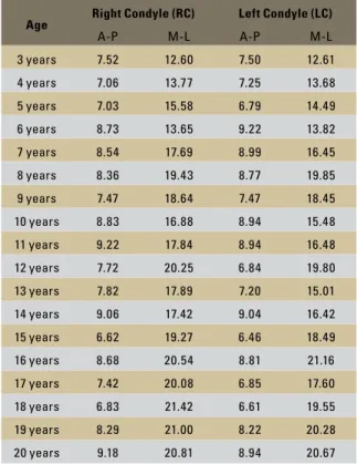
TABLE 1 - Condylar linear measurements (mm) in relation to age.
Lateral: (RC) P=0.322; (LC) P=0.294 / Frontal: (RC) P=0.909; (LC) P=0.856.
separately for right and left condyles in lateral and frontal sections. Differences for right and left condyles in lateral dimensions were tested using Mann-Whitney test and for frontal dimension a non-paired Student t-test.
RESuLtS
Linear measurements of the mandibular con-dyles on lateral and frontal sections are presented in Table 1. The values for intra-operator reliability were similar with no statistical difference, indicat-ing agreement for the lateral (right, P= 0.322; left, P= 0.294) and the frontal (right, P= 0.909; left, P= 0.856) duplicated measurements.
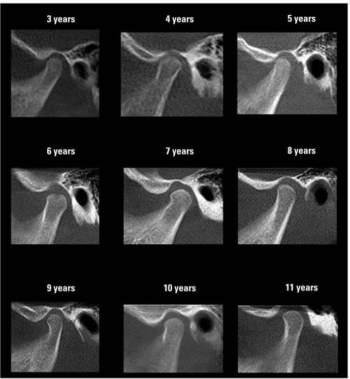
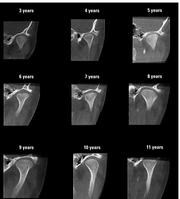
There were no significant differences between right and left mandibular condyles for lateral (P=0.815) and frontal (P=0.374) sections. Figures 2 and 3 show mandibular condyle sequences on CBCT imaging between 3 to 20 years of age and the behavior of morphological changes with time is presented on Figure 4.
dISCuSSIOn
The mandibular condyle is one of the main sites of facial growth, which is expressed in an upward and backward direction.8 The present study did not aim to quantify the participation of condylar growth on total mandibular growth but, instead, assess in a cross-sectional study the local morpho-logical changes of the mandibular condyle during growth using CBCT images. The results showed that the lateral dimension (A-P) seemed to be es-tablished early and to vary a little with age, while the frontal dimension (M-L) increases (Fig 4). Therefore, the mandibular condyle develops by a remodeling process and replaces itself by preserv-ing its lateral dimension and enlargpreserv-ing laterally.
Rodrigues et al21 investigated the diameter of the right and left condyles in subjects aged 13 to 30 years old. All subjects presented Class I maloc-clusion and were evaluated by computed tomog-raphy. Mean sagittal (lateral) dimensions for right and left condyles were, respectively, 9.39 mm and
Age Right Condyle (RC) Left Condyle (LC)
A-P M-L A-P M-L
3 years 7.52 12.60 7.50 12.61
4 years 7.06 13.77 7.25 13.68
5 years 7.03 15.58 6.79 14.49
6 years 8.73 13.65 9.22 13.82
7 years 8.54 17.69 8.99 16.45
8 years 8.36 19.43 8.77 19.85
9 years 7.47 18.64 7.47 18.45
10 years 8.83 16.88 8.94 15.48
11 years 9.22 17.84 8.94 16.48
12 years 7.72 20.25 6.84 19.80
13 years 7.82 17.89 7.20 15.01
14 years 9.06 17.42 9.04 16.42
15 years 6.62 19.27 6.46 18.49
16 years 8.68 20.54 8.81 21.16
17 years 7.42 20.08 6.85 17.60
18 years 6.83 21.42 6.61 19.55
19 years 8.29 21.00 8.22 20.28
20 years 9.18 20.81 8.94 20.67
9.30 mm, and for mediolateral (frontal) 20.62 mm and 20.57 mm with no statistically significant dif-ferences between right and left condyles. The lat-eral dimensions were slightly larger for the same age group when compared to the present study, but the measurements were done on the axial plane.
FIGURE 2 - Sequence of morphological variation of the mandibular condyle in lateral view according to age (3 to 20 years old) (continue).
panoramic radiography to evaluate the purpose of symmetry with contrasting results.15,23 It is known that panoramic radiography is not the most appro-priate method since it produces magnification and distortion in the vertical and horizontal directions.17
Similar studies should be performed with a larger sample to confirm the present data and to correlate them to gender, facial patterns and con-dyle types. The vertical dimensions, shape of man-dibular fossae, articular eminence, and degree of
3 years
6 years
9 years
4 years
7 years
10 years
5 years
8 years
FIGURE 2 - Sequence of morphological variation of the mandibular condyle in lateral view according to age (3 to 20 years old).
inclination of the condyle should be also included with a specific methodology.
CBCT is becoming an important tool in modern dental practice and provides excellent imaging of the osseous components of the TMJ with less radiation
exposure compared to other techniques.1,12 The developed technique showed promising results for condyle measurement and to detect morphological changes during the growth phase in a non-invasive manner using CBCT images in living individuals.
12 years
15 years
18 years
13 years
16 years
19 years
14 years
17 years
FIGURE 3 - Sequence of morphological variation of the mandibular condyle in frontal view according to age (3 to 20 years old) (continue).
3 years
6 years
9 years
4 years
7 years
10 years
5 years
8 years
FIGURE 3 - Sequence of morphological variation of the mandibular condyle in frontal view according to age (3 to 20 years old).
12 years
15 years
18 years
13 years
16 years
19 years
14 years
17 years
A B 21 23
19 17 15 13 11 9
20 19 18 17 16 15 14 13 12 11 10 9 8 7 6 5 4 3 10
8
6
4
2
0
20 19 18 17 16 15 14 13 12 11 10 9 8 7 6 5 4 3
FIGURE 4 - Behavior of mandibular condyle dimensions (in mm) between 3 to 20 years old: Lateral (A) and frontal (B) view.
COnCLuSIOn
The lateral dimension of the mandibular condyle seems to establish itself early because it varied very little with age, while the frontal di-mension increased. Small asymmetries between left and right condyles seem to be common, but with no statistical significance. These prelimi-nary data suggested that CBCT is an useful tool
to measure and to evaluate condylar morphol-ogy during growth.
ACKnOwLEdgMEntS
This study was supported in part by grants from the Nacional Council for Scientific and Technolog-ical Development (CNPq grants #302875/2008-5 and CNPq grants #474642/2009 to C.E.). dimensions (mm) dimensions (mm)
Lateral view Frontal view
Age (years)
Age (years)
1. Alkhader M, Ohbayashi N, Tetsumura A, Nakamura S, Okochi K, Momin MA, et al. Diagnostic performance of magnetic resonance imaging for detecting osseous abnormalities of the temporomandibular joint and its correlation with cone beam computed tomography. Dentomaxillofac Radiol. 2010 Jul;39(5):270-6.
2. Alexiou K, Stamatakis H, Tsiklakis K. Evaluation of the severity of temporomandibular joint osteoarthritic changes related to age using cone beam computed tomography. Dentomaxillofac Radiol. 2009 Mar;38(3):141-7.
3. Berco M, Rigali PH Jr, Miner RM, DeLuca S, Anderson NK, Will LA. Accuracy and reliability of linear cephalometric measurements from cone-beam computed tomography scans of a dry human skull. Am J Orthod Dentofacial Orthop. 2009 Jul;136(1):17.e1-9.
4. Brooks SL, Brand JW, Gibbs SJ, Hollender L, Lurie AG, Omnell KA, et al. Imaging of the temporomandibular joint: a position paper of the American Academy of Oral and Maxillofacial Radiology. Oral Surg Oral Med Oral Pathol Oral Radiol Endod. 1997 May;83(5):609-18.
5. Burke G, Major P, Glover K, Prasad N. Correlations between condylar characteristics and facial morphology in Class II preadolescent patients. Am J Orthod Dentofacial Orthop. 1998 Sep;114(3):328-36.
6. Cimasoni G. Histopathology of the temporomandibular joint following bilateral extractions of molars in the rat. Oral Surg Oral Med Oral Pathol. 1963 May;16:613-21.
7. Chen J, Sorensen KP, Gupta T, Kilts T, Young M, Wadhwa S. Altered functional loading causes differential effects in the subchondral bone and condylar cartilage in the temporomandibular joint from young mice. Osteoarthritis Cartilage. 2009 Mar;17(3):354-61.
8. Enlow DH. Crescimento facial. 3ª ed. São Paulo: Artes Médicas; 1993. p. 88-96.
9. Estrela C, Bueno MR, Azevedo BC, Azevedo JR, Pécora JD. A new periapical index based on cone beam computed tomography. J Endod. 2008;34:1325-31.
10. Estrela C, Bueno MR, Alencar AH, Mattar R, Valladares J Neto,
Azevedo BC, et al. Method to evaluate inlammatory root
resorption by using Cone Beam Computed Tomography. J Endod. 2009 Nov;35(11):1491-7.
11. Garib DG, Raymundo R Junior, Raymundo MV, Raymundo DV,
Ferreira SN. Tomograia computadorizada de feixe cônico
(Cone beam): entendendo este novo método de diagnóstico por imagem com promissora aplicabilidade na Ortodontia. Rev Dental Press Ortod Ortop Facial. 2007;12:139-56.
12. Honey OB, Scarfe WC, Hilgers MJ, Klueber K, Silveira AM, Haskell BS, et al. Accuracy of cone-beam computed tomography imaging of the temporomandibular joint: comparisons with panoramic radiology and linear tomography. Am J Orthod Dentofacial Orthop. 2007 Oct;132(4):429-38. REfEREnCES
Contact address
Carlos Estrela
Rua C-245, Quadra 546, Lote 9, Jardim América CEP: 74.290-200 – Goiânia / GO, Brazil E-mail: [email protected] Submitted: July 2010
Revised and accepted: August 2010
13. Ishibashi H, Takenoshita Y, Ishibashi K, Oka M. Age-related changes in the human mandibular condyle: a morphologic, radiologic and histologic study. J Oral Maxillofac Surg. 1995 Sep;53(9):1016-23.
14. Katsavrias EG, Halazonetis DJ. Condyle and fossa shape in Class II and Class III skeletal patterns: a morphometric tomographic study. Am J Orthod Dentofacial Orthop. 2005 Sep;128(3):337-46.
15. Kilic N, Kiki A, Oktay H. Condylar asymmetry in unilateral posterior crossbite patients. Am J Orthod Dentofacial Orthop. 2008 Mar;133(3):382-7.
16. Kurusu A, Horiuchi M, Soma K. Relationship between occlusal force and mandibular condyle morphology. Angle Orthod. 2009 Nov;79(6):1063-9.
17. Laster WS, Ludlow JB, Bailey LJ, Hershey HG. Accuracy of measurements of mandibular anatomy and prediction of asymmetry in panoramic radiographic images. Dentomaxillofac Radiol. 2005 Nov;34(6):343-9.
18. Ludlow JB, Laster WS, See M, Bailey LJ, Hershey HG. Accuracy of measurements of mandibular anatomy in cone beam computed tomography images. Oral Surg Oral Med Oral Pathol Oral Radiol Endod. 2007 Apr;103(4):534-42.
19. Moreira CR, Sales MA, Lopes PM, Cavalcanti MG. Assessment of linear and angular measurements on three-dimensional cone beam computed tomographic images. Oral Surg Oral Med Oral Pathol Oral Radiol Endod. 2009 Sep;108(3):430-6. 20. Pereira FJ Jr, Lundh H, Westesson PL. Morphologic changes
in the temporomandibular joint in different age groups. An autopsy investigation. Oral Surg Oral Med Oral Pathol. 1994 Sep;78(3):279-87.
21. Rodrigues AF, Fraga MR, Vitral RW. Computed tomography evaluation of the temporomandibular joint in Class I malocclusion patients: condylar symmetry and condyle-fossa relationship. Am J Orthod Dentofacial Orthop. 2009 Aug;136(2):192-8.
22. Schlueter B, Kim KB, Oliver D, Sortiropoulos G. Cone beam computed tomography 3D reconstruction of the mandibular condyle. Angle Orthod. 2008 Sep;78(5):880-8.
23. Uysal T, Sisman Y, Kurt G, Ramoglu SI. Condylar and ramal vertical asymmetry in unilateral and bilateral posterior crossbite patients and a normal occlusion sample. Am J Orthod Dentofacial Orthop. 2009 Jul;136(1):37-43.