Use of miniplates as a method for orthodontic
anchorage: a case report
Fernando Gianzanti Peres1, Luis Eduardo Marques Padovan2, Leandro Eduardo Kluppel2, Gustavo Calvalcanti Albuquerque3, Paulo Cesar Ulson de Souza4, Marcela Claudino5
Introduction: Temporary anchorage devices (TADs) have been developed to be used as direct adjuncts in orthodontic treatment and have facilitated treatment of more complex orthodontic cases, including patients with dental impaction.
Objectives: This clinical case reports the applicability of TADs in the orthodontic treatment of a patient with impacted mandibular second molars. Surgical and orthodontic procedures related to the use of miniplates were also discussed in this study. Conclusions: The use of temporary anchorage devices, such as miniplates, can be suggested as an alternative to treat patients with impacted mandibular second molars.
Keywords: Tooth movement. Orthodontic anchorage procedures. Molar. Tooth, Impacted.
1 MSc in Implantology, Instituto Latino Americano de Pesquisa e Ensino
Odontológico (ILAPEO), Curitiba, Paraná, Brazil.
2 Professor of Implantology, Instituto Latino Americano de Pesquisa e Ensino
Odontológico (ILAPEO), Curitiba, Paraná, Brazil.
3 Professor, Universidade do Estado do Amazonas (UEA), Department of Oral
and Maxillofacial Surgery, Manaus, Amazonas, Brazil.
4 DDS, Instituto Odontológico de Cirurgia e Prótese (IOCP), Bauru, São Paulo,
Brazil.
5 Professor, Universidade Estadual de Ponta Grossa (UEPG), Graduation Course,
Ponta Grossa, Paraná, Brazil.
Submitted: October 20, 2015 - Revised and accepted: June 13, 2016
DOI: http://dx.doi.org/10.1590/2177-6709.21.5.095-102.oar
How to cite this article: Peres FG, Padovan LEM, Kluppel LE, Albuquerque GC, Souza PCU, Claudino M. Use of miniplates as a method for orthodontic anchorage: a case report. Dental Press J Orthod. 2016 Sept-Oct;21(5):95-102. DOI: http://dx.doi.org/10.1590/2177-6709.21.5.095-102.oar
» The authors report no commercial, proprietary or financial interest in the products or companies described in this article.
» Patients displayed in this article previously approved the use of their facial and in-traoral photographs.
Contact address: Marcela Claudino
Rua Carlos Cavalcanti 4748, Uvaranas; CEP: 84030-900 Ponta Grossa/PR Brazil E-mail: [email protected]
Introdução: os dispositivos de ancoragem temporária vêm sendo desenvolvidos para uso como coadjuvantes no trata-mento ortodôntico. Esses dispositivos facilitam o tratatrata-mento ortodôntico de casos mais complexos, incluindo pacientes com dentes impactados. Objetivos: o presente relato de caso reporta a aplicabilidade dos dispositivos de ancoragem temporária no tratamento ortodôntico de um paciente com segundos molares inferiores impactados. Os procedimentos cirúrgicos e ortodônticos relacionados ao uso das miniplacas também são discutidos nesse estudo.Conclusões: o uso de dispositivos de ancoragem temporária, tais como as miniplacas, pode ser sugerido como uma alternativa no tratamento de pacientes com segundos molares inferiores impactados.
Use of miniplates as a method for orthodontic anchorage: a case report
original article
INTRODUCTION
Orthodontic anchorage has been considered relevant in providing satisfactory clinical results. Ini-tially, in conventional treatment, headgear and intra-oral elastics are used to improve anchorage. However, these devices require patient’s compliance, which
can significantly influence treatment.1 In this
con-text, temporary anchorage devices (TADs) have been developed to be used as direct adjuncts in orthodontic treatment, including conventional implants, titanium miniscrews, and bone miniplates. These devices have demonstrated favorable clinical results and have
facil-itated treatment of more complex orthodontic cases.2
Complex cases include patients with dental im-paction. Conceptually, impacted teeth are the re-sult of eruption impairment due to the presence of physical barriers in the tooth eruption pathway or al-tered position of the tooth germ. In a general context, tooth impaction affects about 20% of the population. However, when specific dental groups are assessed, prevalence rates demonstrate significant differences. For example, reduced rates of 0.08% and 0.01% are reported for impaction of mandibular first molars and
second molars, respectively.3 Despite low prevalence,
orthodontic treatment of these cases has a higher de-gree of complexity. Treatment options depend on the degree of tooth tipping, the position of third molars and the desired type of motion. Moreover, this treat-ment can be carried out by means of surgical or
orth-odontic procedures.4
As regards surgical alternatives, surgical reposi-tioning is proposed as a therapeutic method for the treatment of impacted teeth. Although this proce-dure provides an immediate solution, complications, such as endodontic and periodontal impairment,
have been reported.5,6 Orthodontic movement
is also indicated for uprighting impacted molars achieved by means of fixed or removable applianc-es.7,8,9 In this context, the establishment of a distal
force component is difficult because of the presence of impacted third molars, mainly in adolescent
pa-tients.10,11 In these cases, the applicability of
skele-tal anchorage optimizes orthodontic treatment and has advantages, such as decreased overall treatment time and effective anchorage with reduced aesthetic discomfort, when compared to extraoral anchor-age. Occasionally, it allows orthodontic treatment
previously thought to be impossible without surgery to be carried out. These devices also minimize the
need for patient’s compliance.1,12 However,
disad-vantages, such as surgical procedures for placement and removal of devices, as well as inflammation of
surrounding soft tissues, have been reported.13
Based on the relevance of the use of TADs and the higher degree of complexity implied in treating sur-rounding patients with impacted mandibular second molars, the aim of this study was to present a case re-port regarding the applicability of temporary anchor-age devices (TADs) in the orthodontic treatment of a patient with impacted mandibular second molars.
CASE REPORT
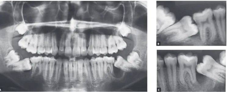
A 14 year-old boy with permanent dentition was referred for treatment after complaining of a need to correct dental position. Evaluation revealed a balanced facial growth pattern, convex profile and passive lip seal. Furthermore, the following were identified: dental alignment, Class I canine rela-tionship and coinciding midlines (Figs 1, 2 and 3). Mandibular second molars were severely mesially tipped and semi-erupted; impacted third molars were also observed (Fig 4). Radiographic exami-nation also revealed the presence of inflammatory cysts in the region of mandibular left second and third molars and in the region of maxillary left sec-ond and third molars. Thus, treatment planning was based on cysts enucleation and uprighting of mandibular second molars (#37 and #47) achieved by means of miniplates.
Treatment was initiated with enucleation of cysts. Subsequently, a miniplate for orthodontic anchorage was installed with an incision in the molar and pre-molar regions on both sides, and with raising of a mu-coperiostal flap in the zygomatic bone region.
The miniplate (Neoortho, Curitiba, Brazil),
22.16 mm in lenght, 1 mm in thickness and
Figure 1 - Initial facial aspect: A) lateral and
B) frontal.
Figure 2 - Initial clinical aspect: A) intraoral right side, B) intraoral frontal aspect, C) intraoral left side, D ) oc-clusal maxillary aspect, and E) occlusal mandibular aspect.
Figure 3 - Initial dental casts: A) Right side aspect, B) frontal aspect, C) left side aspect. B
B
C
C A
A
D E
Use of miniplates as a method for orthodontic anchorage: a case report
original article
Figure 4 - Initial radiographic aspect: panoramic radiograph (A) and periapical radiographs of teeth #46 and #47 (B), and #36 and #37 (C).
Buttons were placed on the buccal surface of man-dibular second molars, followed by placement of ¼ inter-maxillary elastics with a force of 150 g (mouth closed) and approximately 200 g (mouth open). Ater ive months, orthodontic miniplates were removed, since second
molars were in proper position for uprighting with can-tilevers. Ater uprighting, mandibular third molars were extracted, totaling 37 months of treatment (Figs 5, 6, 7, 8, and 9). Ater orthodontic treatment completion, the pa-tient was followed-up for two years (Fig 10).
Figure 5 - Final facial aspect. A) lateral and
B) frontal. C B
A
Figure 6 - Final radiographic aspect.
Figure 7 - Final clinical aspect: A) intraoral right side, (B) intraoral frontal, C) intraoral left side, D) occlusal mandibular aspect, and E) occlusal maxillary aspect.
B
D
C
Use of miniplates as a method for orthodontic anchorage: a case report
original article
Figure 8 - Final dental casts. (A) Right side aspect, (B) frontal aspect, (C) left side aspect, and (D, E) occlusal aspect.
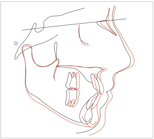
Figure 9 - Superimposition of initial (black) and final (red) cephalometric tracings. B
E
C A
Figure 10 - Radiographic follow-up two years after orthodontic treatment completion.
DISCUSSION
The case described in this study demonstrates a relatively unusual event, since the most common
dental impaction is unilateral.14 In this case, bilateral
impaction was observed in the jaw in a male patient. Regardless of the treatment approach employed for impacted teeth, it is important that treatment be per-formed as soon as possible due to contact with adja-cent teeth, which can result in root resorption, caries
and periodontal diseases.15
For molar uprighting, several options of mechanical approaches are described, namely: cantilevers, tip-back
spring, NiTi wire, among others.7,8,16 In the present
case, the irst approach employed for molar upright-ing was the use of orthodontic elastics supported on miniplates in order to increase molar exposure in the oral cavity. As soon as possible, a cantilever was used to inish the movement. The cantilever is a segment of wire in which one end in inserted in a tube or bracket and the other end has just a point of contact. It was chosen in this case because, as a statistically determined
system,17,18 it generates a predictable force system.16,18,19
The clinical results of the case reported herein demonstrate that the use of miniplates as anchorage devices was an efficient strategy for uprighting im-pacted second molars. The introduction of mini-plate anchorage in Orthodontics was a great progress, since it minimizes the need for patient’s
compli-ance7 and allows for a more predictable orthodontic
movement.20-23 Furthermore, the use of these devices
enables the application of force from the distal side of the impacted molar. This application of force gener-ates a counterclockwise moment, which allows move-ment control, thus promoting rapid disimpaction and
crown distalization.22,24 Despite yielding good
treat-ment results, miniplates are not yet widely used, pos-sibly due to the requirement of surgical procedures with a certain degree of complexity, as well as the need for subsequent orthodontic treatment.
In fact, the majority of case reports describe the
use of screws or microscrews.25-28 However, in the
present case, the use of miniplates was a precise, safe and simple method for skeletal anchorage. Moreover, it does not require complex movements and
involve-ment of several teeth in the process.27 Nevertheless,
disadvantages over conventional devices are reported, including the need for surgical procedures, high cost, difficult cleaning, risk of infection and discomfort
during the first days due to the size of the device28.
Thus, the use of these devices may be indicated for treatment of specific cases.
CONCLUSIONS
This case report demonstrates the applicability of miniplates for uprighting impacted second molars. Thus, the use of these devices may be used as an al-ternative to treat patients with impacted mandibular second molars.
Use of miniplates as a method for orthodontic anchorage: a case report
original article
1. Londa G. The anchorage quality of titanium microplates with short microscrews for orthodontic anchorage applications. J Orofac Orthop. 2005 Jan;66(1):67-77. 2. Takaki T, Tamura N, Yamamoto M, Takano N, Shibahara T, Yasumura T, et al.
Clinical study of temporary anchorage devices for orthodontic treatment: stability of micro/mini-screws and mini-plates: experience with 455 cases. Bull Tokyo Dent Coll. 2010;51(3):151-63.
3. Valmaseda-Castellón E, De-la-Rosa-Gay C, Gay-Escoda C. Eruption disturbances of the irst and second permanent molars: results of treatment in 43 cases. Am J Orthod Dentofacial Orthop. 1999 Dec;116(6):651-8.
4. Mariano RC, Mariano L de C, de Melo WM. Deep impacted mandibular second molar: a case report. Quintessence Int. 2006 Nov-Dec;37(10):773-6. 5. McAboy CP, Grumet JT, Siegel EB, Iacopino AM. Surgical uprighting and
repositioning of severely impacted mandibular second molars. J Am Dent Assoc. 2003 Nov;134(11):1459-62.
6. Pogrel MA. The surgical uprighting of mandibular second molars. Am J Orthod Dentofacial Orthop. 1995 Aug;108(2):180-3.
7. Aksoy AU, Aras S. Use of nickel titanium coil springs for partially impacted second molars. J Clin Orthod. 1998 Aug;32(8):479-82.
8. Shapira Y, Borell G, Nahlieli O, Kuftinec MM. Uprighting mesially impacted mandibular permanent second molars. Angle Orthod. 1998 Apr;68(2):173-8. 9. Sherwood KH, Burch JG, Thompson WJ. Closing anterior open bites by
intruding molars with titanium miniplate anchorage. Am J Orthod Dentofacial Orthop. 2002 Dec;122(6):593-600.
10. Gazit E, Lieberman M. A mesially impacted mandibular second molar. Treatment considerations and outcome: a case report. Am J Orthod Dentofacial Orthop. 1993 Apr;103(4):374-6.
11. Fontenelle A. Limitations in adult Orthodontics. In: Melsen B. Current controversies in Orthodontics. Chicago: Quintessence; 1991. Cap 5, p. 147-79. 12. Miyahira YI, Maltagliati LA, Siqueira DF, Romano R. Miniplates as skeletal
anchorage for treating mandibular second molar impactions. Am J Orthod Dentofacial Orthop. 2008 July;134(1):145-8.
13. Chen YJ, Chang HH, Lin HY, Lai EH, Hung HC, Yao CC. Stability of miniplates and miniscrews used for orthodontic anchorage: experience with 492 temporary anchorage devices. Clin Oral Implants Res. 2008 Nov;19(11):1188-96.
REFERENCES
14. Wellfelt B, Varpio M. Disturbed eruption of the permanent lower second molar: treatment and results. ASDC J Dent Child. 1988 May-Jun;55(3):183-9. 15. Vedtofte H, Andreasen JO, Kjaer I. Arrested eruption of the permanent lower
second molar. Eur J Orthod. 1999 Feb;21(1):31-40.
16. Sakima T, Martins LP, Sakima MT, Terada HH, Kawakami RY, Ozawa TO. Alternativas mecânicas na verticalização de molares. Sistemas de força liberados pelos aparelhos. Rev Dental Press Ortod Ortop Facial. 1999 Jan-Feb; 4(1):79-100. 17. Melsen B, Fiorelli G, Bergamini A. Uprighting of lower molars. J Clin Orthod. 1996
Nov;30(11):640-5.
18. Melo AC, Duarte da Silva R, Shimizu RH, Campos D, Andrighetto AR. Lower molar uprighting with miniscrew anchorage: direct and indirect anchorage. Int J Orthod Milwaukee. 2013 Fall;24(3):9-14.
19. Romeo DA, Burstone CJ. Tip-back Mechanics. Am J Orthod. 1977 Oct;72(4):414-21.
20. Sherwood KH, Burch J, Thompson W. Intrusion of supererupted molars with titanium miniplate anchorage. Angle Orthod. 2003 Oct;73(5):597-601. 21. Sugawara J, Nishimura M. Minibone plates: the skeletal anchorage system.
Semin Orthod. 2005 Aug;11(2):47-56.
22. Park HS, Kyung HM, Sung JH. A simple method of molar uprighting with micro-implant anchorage. J Clin Orthod. 2002 Oct;36(10):592-6.
23. Melsen B. Overview mini-implants: where are we? J Clin Orthod. 2005 Sept;39(9):539-47.
24. Giancotti A, Arcuri C, Barlattani A. Treatment of ectopic mandibular second molar with titanium miniscrews. Am J Orthod Dentofacial Orthop. 2004 July;126(1):113-7.
25. Giancotti A, Muzzi F, Santini F, Arcuri C. Miniscrew treatment of ectopic mandibular molars. J Clin Orthod. 2003 July;37(7):380-3.
26. Kanomi R. Mini-implant for orthodontic anchorage. J Clin Orthod. 1997 Nov;31(11):763-7.
27. Majourau A, Norton LA. Uprighting impacted second molars with segmented springs. Am J Orthod Dentofacial Orthop. 1995 Mar;107(3):235-8.